Abstract
Sweeteners are generally used in food and pharmaceutical formulations, yet their natural conditioning beyond taste isn't well understood. This study explores the remedial eventuality of four extensively used sweeteners, sucrose, lactose, mannitol, and dextrose, focusing on their cytotoxic, antioxidant, and antibacterial properties. Cytotoxicity was assessed using the MTT assay in a mammalian macrophage cell line at colourful attention (0.2 – 6). Both lactose and dextrose enhanced cell viability by 25 – 30 at lower boluses, indicating cytoprotective goods. In discrepancy, mannitol significantly reduced cell viability by over 40 at advanced attention, suggesting cytotoxicity. The antioxidant capacity of the sweeteners was estimated with the DPPH and potassium permanganate assays, revealing that sucrose displayed the loftiest radical-scavenging activity (up to 80), showcasing notable redox-modulating potential. In terms of antibacterial exertion, assessed through the slice prolixity system, lactose demonstrated the most significant inhibition against Escherichia coli, producing the largest inhibition zone measuring 15 ± 1 mm. These findings indicate that common sweeteners retain distinct bioactivities that impact cell survival, oxidative balance, and microbial inhibition. The results emphasise the significance of recognising sweeteners not solely for their taste but also for their implicit remedial, nutraceutical, and preservative roles. This exploration opens avenues for further disquisition into the broader operations of sweeteners in health and drug.
|
Published in
|
Cell Biology (Volume 14, Issue 1)
|
|
DOI
|
10.11648/j.cb.20261401.11
|
|
Page(s)
|
1-19 |
|
Creative Commons
|

This is an Open Access article, distributed under the terms of the Creative Commons Attribution 4.0 International License (http://creativecommons.org/licenses/by/4.0/), which permits unrestricted use, distribution and reproduction in any medium or format, provided the original work is properly cited.
|
|
Copyright
|
Copyright © The Author(s), 2026. Published by Science Publishing Group
|
Keywords
Sweeteners, Cytotoxicity, Antioxidant Activity, Antibacterial Assay, Mtt Assay, Dpph Assay
1. Introduction
The global consumption of sweeteners—both natural sugars and synthetic sugar substitutes—has increased significantly in recent years, prompting growing scientific and public interest in their broader impact on human health. Traditionally, compounds such as sucrose, lactose, dextrose, and mannitol were regarded primarily as sources of sweetness and energy. However, with increasing awareness of metabolic disorders, oxidative stress, and antibiotic resistance, these compounds are now being re-examined for their potential biological activities beyond taste and caloric contribution. Sweeteners are now widely found in processed foods, beverages, nutraceuticals, and pharmaceutical preparations, underscoring the need to understand their physiological and cellular effects more comprehensively
| [1] | Medeleanu, M. L., Sanchez, S. P., Cătunescu, G. M., & Cerezo, A. B. (2024). Risk assessment of food additives including dietary exposure. EFSA journal. European Food Safety Authority, 22(Suppl 1), e221110.
https://doi.org/10.2903/j.efsa.2024.e221110 |
[1]
. This study investigates the functional bioactivities and therapeutic potential of four common sweeteners—sucrose, lactose, mannitol, and dextrose. By systematically evaluating their cytotoxicity via MTT assay, redox properties through DPPH radical scavenging and KMno4 reduction, and antibacterial efficacy using the disc diffusion method, this research explores the multifaceted roles of these compounds beyond basic nutrition. The findings aim to identify potential applications for these sweeteners as functional ingredients in the pharmaceutical, nutraceutical, and food science industries.
The unique chemical and metabolic characteristics of each sweetener determine how it behaves in biological systems. In the food business, sucrose—a disaccharide made up of glucose and fructose—continues to be the standard for sweetness. Although it is a quick source of energy, consuming too much of it has been connected to metabolic diseases, obesity, and insulin resistance
| [2] | Grigsby A, Herron J, Hunter BR. Does the addition of dextrose to IV crystalloid therapy provide clinical benefit in acute dehydration? A systematic review and meta-analysis. CJEM. 2019; 21(5): 638-645. https://doi.org/10.1017/cem.2018.500 |
[2]
. Due to its low glycemic index and osmotic diuretic properties, mannitol, a polyol or sugar alcohol, is often used in diabetic-friendly products. It is also used in medicine to lower intracranial and intraocular pressure
| [3] | Stockwell, B. R., Friedmann Angeli, J. P., Bayir, H., Bush, A. I., Conrad, M., Dixon, S. J., Fulda, S., Gascón, S., Hatzios, S. K., Kagan, V. E., Noel, K., Jiang, X., Linkermann, A., Murphy, M. E., Overholtzer, M., Oyagi, A., Pagnussat, G. C., Park, J., Ran, Q., Rosenfeld, C. S., … Zhang, D. D. (2017). Ferroptosis: A Regulated Cell Death Nexus Linking Metabolism, Redox Biology, and Disease. Cell, 171(2), 273–285.
https://doi.org/10.1016/j.cell.2017.09.021 |
[3]
. The natural sugar in milk, lactose, is vital for baby nourishment but can cause lactose intolerance in adults, which can upset the balance of gut microbes and gut health. Dextrose, also known as D-glucose, is a common ingredient in pharmaceutical formulations that replenishes hydration and maintains metabolic balance. It is the principal energy substrate for cells
| [4] | Gibson, S., Gunn, P., Wittekind, A., & Cottrell, R. (2013). The effects of sucrose on metabolic health: a systematic review of human intervention studies in healthy adults. Critical reviews in food science and nutrition, 53(6), 591–614.
https://doi.org/10.1080/10408398.2012.691574 |
[4]
.
In addition to their industrial and nutritional significance, these sweeteners might have minor effects on microbial and cellular functions. Although the evidence is still sparse and frequently contradictory, recent research indicates that certain sweeteners may influence oxidative stress pathways or possibly prevent bacterial growth. Evaluating the antioxidant potential of dietary compounds has attracted a lot of scientific attention because oxidative stress, which is a state of imbalance between reactive oxygen species (ROS) and antioxidant defences, is a major contributor to the onset of chronic diseases like diabetes, cancer, and cardiovascular disorders
. For example, sugar alcohols such as mannitol have shown a modest ability to scavenge free radicals, but there are currently few systematic comparisons between various sweeteners
| [6] | Ruiz-Ojeda, F. J., Plaza-Díaz, J., Sáez-Lara, M. J., & Gil, A. (2019). Effects of Sweeteners on the Gut Microbiota: A Review of Experimental Studies and Clinical Trials. Advances in nutrition (Bethesda, Md.), 10(suppl_1), S31–S48.
https://doi.org/10.1093/advances/nmy037 |
[6]
.
Additionally, microbial infections remain a significant global health concern, made worse by the quick emergence of strains that are resistant to antibiotics. Finding unconventional substances having antibacterial properties, such as those present in food, may offer safer and more long-lasting infection management methods. According to preliminary observations, some sweeteners may change the dynamics of bacterial development or disrupt microbial metabolism, indicating that they may have a direct or indirect impact on modifying microbial habitats
| [7] | Gill, P. A., Inniss, S., Kumagai, T., Rahman, F. Z., & Smith, A. M. (2022). The Role of Diet and Gut Microbiota in Regulating Gastrointestinal and Inflammatory Disease. Frontiers in immunology, 13, 866059.
https://doi.org/10.3389/fimmu.2022.866059 |
[7]
.
This work fills these gaps by assessing the cytotoxic, antioxidant, and antibacterial properties of four popular sweeteners using a multi-assay methodology. A sensitive colourimetric technique based on mitochondrial enzyme activity, the MTT assay, is used to gauge these substances' potential for cytotoxicity. With the help of the potassium permanganate reduction assay and the DPPH radical scavenging assay, which together offer complementary insights into redox behaviour and free radical neutralisation, antioxidant capacity is assessed. The disc diffusion approach is also used to evaluate the antibacterial properties of specific pathogenic microorganisms
| [8] | Ahmad, Z., Rauf, A., Orhan, I. E., Mubarak, M. S., Akram, Z., Islam, M. R., Imran, M., Edis, Z., Kondapavuluri, B. K., Thangavelu, L., & Thiruvengadam, M. (2025). Antioxidant Potential of Polyphenolic Compounds, Sources, Extraction, Purification and Characterization Techniques: A Focused Review. Food science & nutrition, 13(12), e71259.
https://doi.org/10.1002/fsn3.71259 |
[8]
.
Although sweeteners are indispensable in modern food and pharmaceutical industries, their broader biological and therapeutic properties remain poorly characterised. Most research has traditionally focused on their metabolic roles or sensory functions, leaving significant gaps in understanding how they may influence cellular health, oxidative defence mechanisms, or microbial behaviour. Considering their extensive daily consumption and biochemical diversity, it is essential to assess these compounds in an integrated manner. In this context, our study evaluated cytotoxicity, antioxidant potential, and antibacterial activity of commonly used sweeteners, providing new insights into their health implications and possible therapeutic relevance. Among the sugars tested, sucrose demonstrated the highest antioxidant activity. This superior potential likely arises from a combination of factors: chemically, sucrose is a non-reducing sugar that interacts differently with reactive oxygen species compared to reducing sugars like glucose or lactose; physically, its osmotic properties may help stabilise cells and prevent oxidative damage by regulating water balance; and biologically, sucrose metabolism provides energy that can activate cellular defence mechanisms against oxidative stress. Together, these chemical, physical, and metabolic effects highlight the complex ways in which sugars can modulate oxidative stress, underscoring the importance of evaluating sweeteners beyond their conventional roles
| [9] | Clemente-Suárez, V. J., Redondo-Flórez, L., Beltrán-Velasco, A. I., Yáñez-Sepúlveda, R., Rubio-Zarapuz, A., Martín-Rodríguez, A., Navarro-Jimenez, E., & Tornero-Aguilera, J. F. (2025). Human Digestive Physiology and Evolutionary Diet: A Metabolomic Perspective on Carnivorous and Scavenger Adaptations. Metabolites, 15(7), 453.
https://doi.org/10.3390/metabo15070453 |
[9]
.
Sweeteners are omnipresent in modern foods and pharmaceuticals, yet their broader biological and therapeutic effects remain largely unexplored. While most studies focus on metabolic or sensory roles, little is known about how these compounds influence cellular health, oxidative stress, and microbial behaviour. This study addresses this critical gap by taking a holistic approach, simultaneously evaluating cytotoxicity, antioxidant potential, and antibacterial activity of commonly used sweeteners, including sucrose, glucose, lactose, and mannitol. The novelty of this work lies in its integrated, multi-dimensional perspective—examining chemical structure, osmotic properties, and metabolic effects together to understand how sugars modulate biological systems. Notably, sucrose displayed the highest antioxidant activity, likely due to the combined influence of its non-reducing structure, cellular stabilisation through osmotic effects, and energy-driven activation of defence mechanisms. By systematically comparing multiple sweeteners and revealing functional roles beyond sweetness, this study provides unprecedented insights into their therapeutic relevance, safety, and potential applications in food technology, pharmacology, and preventive healthcare. This integrated evaluation not only bridges existing knowledge gaps but also sets a foundation for future research into the health-promoting and functional properties of sweeteners.
2. Literature Review
Sweeteners, historically used as flavour enhancers and preservatives in food and pharmaceuticals, are gaining attention in scientific research for their potential biological activity. While traditionally assessed for taste and metabolism, recent studies focus on their effects on cellular health, oxidative stress, and antimicrobial properties. This review examines the cytotoxic, antioxidant, and antibacterial activities of common sweeteners like sucrose, mannitol, lactose, and dextrose
.
2.1. Cytotoxic Effects and Cell Viability: The Role of MTT Assay
The MTT (3-(4,5-dimethylthiazol-2-yl)-2,5-diphenyltetrazolium Bromide) test is a common method for assessing cell proliferation and viability. It quantifies the metabolic activity of viable cells by measuring the reduction of MTT to formazan by mitochondrial dehydrogenase enzymes. This method is frequently used in the toxicological screening of both synthetic and natural compounds
| [11] | Sengupta S, Banerjee S, Nayek S, Das P, Chakraborty D, Mukherjee A, et al. A brief comparative study of the natural sources (lemons) based on protein, vitamin C, their antibacterial, anthelminthic, and cell viability on immune cells. Int J Herb Med. 2023; 11(5): Article 883.
https://doi.org/10.22271/flora.2023.v11.i5a.883 |
[11]
.
The MTT assay remains valid for assessing cytotoxicity across various concentrations. Regarding sweeteners, evidence on their effects on cell viability is limited but growing. For instance, sugar alcohols like mannitol show low cytotoxicity at moderate levels but can inhibit cell function at high levels due to osmotic stress. Sucrose, typically seen as inert in cytotoxicity studies, has produced inconsistent results depending on cell type and exposure duration
.
2.2. Antibacterial Properties of Common Sweeteners
The evaluation of sweeteners for antibacterial activity is relatively recent but has significant implications for both food preservation and therapeutic applications. The disc diffusion method, as described by Bauer, remains a widely accepted technique for testing antimicrobial activity against pathogens such as Escherichia coli and Staphylococcus aureus.
Sugars can influence microbial growth both directly and indirectly. Hyperosmotic environments created by high concentrations of sugars like sucrose and dextrose inhibit bacterial proliferation by disrupting cell membrane function and dehydrating cells. Polyols like mannitol have been studied for their antimicrobial synergy when used with antibiotics, although their standalone antibacterial effect remains modest
| [13] | Sengupta S, Pradhan A, Biswas S, Bhattacharya M. Exploring the antimicrobial properties of lemon: A comparative analysis of peel, seed, and pulp. Microbiol Res J Int. 2024; 34: 10–24. https://doi.org/10.9734/mrji/2024/v34i91477 |
[13]
.
Lactose, a disaccharide prevalent in dairy products, shows limited antibacterial activity but may influence the growth of probiotic bacteria, which can indirectly inhibit pathogenic strains. Emerging research suggests that sugar-mediated bacterial inhibition may also involve interference with quorum-sensing pathways and biofilm formation.
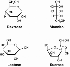
Figure 1. Structural Representation of Common Sweeteners: Dextrose, Mannitol, Lactose, and Sucrose.
2.3. Integration of Antioxidant and Antibacterial Activities in Health Applications
There is increasing interest in compounds with both antioxidant and antibacterial properties for wound healing and functional foods. Polyol-based sweeteners like mannitol protect against oxidative stress and modulate gut microbiota. While dextrose and glucose can show short-term antioxidant effects, prolonged exposure leads to advanced glycation end products (AGEs) that may cause inflammation. Therefore, evaluating both antioxidant and antibacterial effects is crucial for understanding the health impacts of sweeteners.
| [14] | Sengupta S, Khatun R, Kumari A, Rai R, Dutta S, Mondal T, et al. Nutritional, antioxidant, and antimicrobial properties of citrus peels: A sustainable valorization approach. Microbiol Res J Int. 2024; 34: 149–61.
https://doi.org/10.9734/mrji/2024/v34i121517 |
[14]
.
3. Materials and Methods
3.1. Preparation of Sweetener Solutions
This study involved four sweeteners: sucrose, lactose, mannitol, and dextrose. Stock solutions were prepared in sterile distilled water at five concentrations corresponding to 40%, 10%, 5%, 2.5%, and 1.25% (w/v). For consistency across all assays, these concentrations were converted into micromolar (µM) based on molecular weight (
Table 1). After weighing, the sweeteners were dissolved in 2 mL of sterile distilled water, filtered through 0.22 µm syringe filters to ensure sterility, and stored at 4°C until use.
3.2. MTT Cell Viability Assay
Mammalian macrophage cell lines (1 × 10⁴ cells/well) were seeded in 100 µL of complete culture medium in 96-well plates and incubated for 48 hours at 37°C with 5% CO₂ to promote cell adhesion
| [15] | Ghasemi M, Turnbull T, Sebastian S, Kempson I. The MTT assay: Utility, limitations, pitfalls, and interpretation in bulk and single-cell analysis. Int J Mol Sci. 2021; 22: 12827.
https://doi.org/10.3390/ijms222312827 |
[15]
. After incubation, the media was removed, and 100 µL of sweetener solutions at various concentrations were added to the wells (in triplicate). Cells were cultured for an additional 24 hours, with untreated cells serving as a negative control
| [16] | Optimization of some parametric values of MTT for the determination of human melanoma (SK-Mel-30) cell viability. Int J Life Sci Biotechnol. 2022; 1: 9–20.
https://doi.org/10.38001/ijlsb.991615 |
[16]
. Following sweetener exposure, each well received 10 µL of MTT solution (5 mg/mL in PBS). Live cells converted MTT to purple formazan crystals by incubating for four hours at 37°C
| [17] | Basit A, Ahmad S, Khan KUR, Aati HY, Sherif AE, Ovatlarnporn C, et al. Evaluation of the anti-inflammatory, antioxidant, and cytotoxic potential of Cardamine amara L. (Brassicaceae): A comprehensive biochemical, toxicological, and in silico computational study. Front Chem. 2023; 10: 1077581.
https://doi.org/10.3389/fchem2022.1077581 |
[17]
. The media was removed, and 100 µL of DMSO was added to dissolve the crystals. Plates were shaken for ten minutes at room temperature
| [18] | Buranaamnuay K. The MTT assay for measuring the viability of spermatozoa: A variety of assay protocols. Open Vet J. 2021; 11(2): 251–69. https://doi.org/10.5455/OVJ |
[18]
. Absorbance was measured at 570 nm using a microplate reader. Relative cell viability was calculated with the formula:
Cell viability (%) = [(Absorbance of treated cells) / (Absorbance of control)] x 100.
Results were presented as mean ± standard deviation, indicating cell metabolic activity
| [19] | Popova AA, Reischl M, Kazenmaier D, Cui H, Amberger T, Levkin PA. Simple assessment of viability in 2D and 3D cell microarrays using single-step digital imaging. SLAS Technol. 2022; 27(1): 44–53.
https://doi.org/10.1016/j.slast.2021.10.017 |
[19]
.
3.3. Antioxidant Assay
3.3.1. DPPH Radical Scavenging Assay
The DPPH test assesses the antioxidant capacity to neutralise free radicals. A 0.1 mM DPPH stock solution was prepared in 100% ethanol and stored in the dark. One mL of each sweetener solution was mixed with 1 mL of DPPH in test tubes, with a control using ethanol. After incubating for 30 minutes at room temperature in the dark, absorbance was measured at 517 nm
| [20] | Gulcin I, Alwasel S. DPPH Radical Scavenging Assay. Processes. 2023; 11: 2248. https://doi.org/10.3390/pr11082248 |
| [21] | Rana MS, Rayhan NMA, Emon MSH, Islam MT, Rathry K, Hasan MM, et al. Antioxidant activity of Schiff base ligands using the DPPH scavenging assay: an updated review. RSC Adv. 2024; 14(45): 33094–123.
HYPERLINK https://doi.org/10.1039/d4ra04375h |
[20, 21]
.
3.3.2. Potassium Permanganate (KMnO₄) Assay
The KMnO₄ assay uses potassium permanganate, a strong oxidising agent that appears purple and has an absorption maximum at 525 nm. Antioxidants reduce the purple colour, with a decrease in absorbance indicating antioxidant capacity
| [22] | Alonso-Salinas R, López-Miranda S, González-Báidez A, Pérez-López AJ, Noguera-Artiaga L, Núñez-Delicado E, et al. Effect of potassium permanganate, ultraviolet radiation and titanium oxide as ethylene scavengers on preservation of postharvest quality and sensory attributes of broccoli stored with tomatoes. Foods (Basel). 2023; 12(12): 2418.
https://doi.org/10.3390/foods12122418 |
[22]
. A 0.1% KMnO₄ solution was prepared and stored in amber bottles. In the assay, 1 mL of sweetener solution was mixed with 1 mL of KMnO₄, while the control used purified water. After incubating in the dark for 10 minutes, absorbance was measured at 525 nm. A decrease in absorbance compared to the control indicated antioxidant activity, with larger declines reflecting higher activity. Each experiment was repeated three times for accuracy
.
3.4. Antibacterial Assay
A standardised bacterial suspension is spread on Mueller-Hinton Agar (MHA) plates, where wells are created using a sterile tool. Various concentrations of test substances, like sweeteners (sucrose, lactose, dextrose, and mannitol), along with negative (sterile water) and positive (ampicillin) controls, are added to the wells
. After incubating at 37°C for 24 hours, antibacterial activity is assessed by measuring the zone of inhibition (ZOI) around each well. The diameter of the ZOI (zone of inhibition) is measured in millimetres, and average values are calculated from triplicate readings. This method is suitable for both Gram-positive and Gram-negative bacteria, including
Staphylococcus aureus, G. bacillus, Enterobacter cloacae, and Escherichia coli
| [25] | Hossain T. Methods for screening and evaluation of antimicrobial activity: A review of protocols, advantages, and limitations. Eur J Microbiol Immunol. 2024.
https://doi.org/10.1556/1886.2024.00035 |
[25]
.
4. Result and Discussion
4.1. MTT Cell Viability Assay
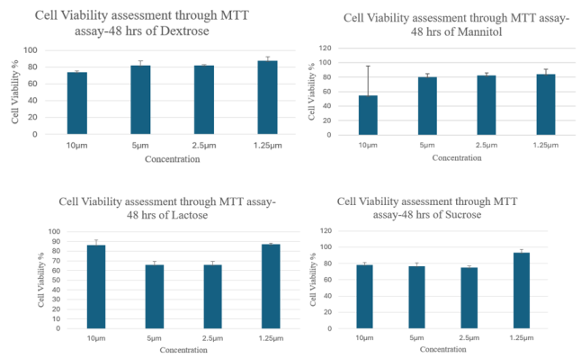
The MTT assay results after 48 hours indicate that Lactose, Sucrose, Mannitol, and Dextrose influence cell viability in a concentration-dependent manner, but their effects vary significantly:
Figure 2. Comparative analysis of cell viability (%) of cells treated with dextrose, mannitol, lactose, and sucrose at different concentrations using MTT assay after 48 hours.
The MTT assay results after 48 hours indicated varying effects of sweeteners on cell viability. Dextrose showed the highest viability (~88%) at 1.25µM, decreasing to ~75% at 10 µM, highlighting a concentration-dependent decline likely due to osmotic stress. Mannitol had the lowest viability (~55%) at 10 µM, with the highest at 1.25 µM (~85%), indicating toxicity at higher doses. Lactose displayed a non-linear response, peaking at 1.25 µM (~88%) but dropping to ~67% at 2.5–5 µM, possibly due to limited metabolism. Sucrose achieved the highest viability (~92%) at 1.25, remaining stable (~75–78%) from 2.5 to 10µM, suggesting mild dose-dependence with lower concentrations enhancing viability.
Table 1. Comparative Analysis Across Sugars (MTT Assay at 48 hrs).
Sugar | 10µM Viability | 1.25µM Viability | Dose-Response Trend |
Dextrose | ~75% | ~88% | Moderate increase with lower dose |
Mannitol | ~55% | ~85% | Sharp increase with lower dose |
Lactose | ~85% | ~88% | Non-linear/ biphasic |
Sucrose | ~85% | ~92% | Steady, increasing with lower dose |
The MTT assay results indicate that lower sugar concentrations support greater cell viability, while higher concentrations can be inhibitory due to factors like osmotic stress and metabolic overload. Excessive sugars can lead to cellular dehydration and hinder function. Non-metabolizable sugars like mannitol contribute to osmotic effects without providing energy, while cell responses vary based on sugar type and enzymatic capacity. Sucrose and dextrose showed high viability at low concentrations, making them suitable as energy supplements. However, mannitol may be cytotoxic at higher doses, so it's best to use low concentrations (1.25µM) to preserve cell health. The choice of sugar and its concentration are crucial for maintaining cell viability in vitro.
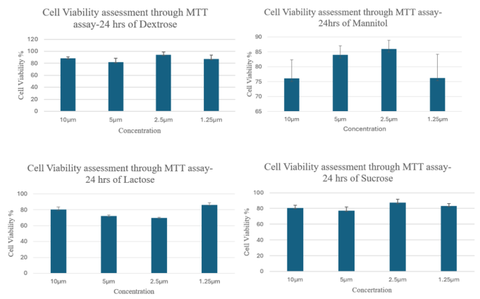
The MTT assay results after 24 hours indicate that Lactose, Sucrose, Mannitol, and Dextrose influence cell viability in a concentration-dependent manner, but their effects vary significantly:
Figure 3. Comparative analysis of cell viability (%) of cells treated with dextrose, mannitol, lactose, and sucrose at different concentrations using MTT assay after 24 hours.
In this experiment, four sugars—Dextrose, Mannitol, Lactose, and Sucrose—were evaluated for their effects on cell viability after 24 hours using the MTT assay at concentrations of 10, 5, 2.5, and 1.25 µM. Dextrose showed the highest viability (~93%) at 2.5µM, with 10 and 1.25µM also maintaining high viability (~88-90%). The 5µM concentration resulted in the lowest viability (~83%). Mannitol peaked at ~86% viability at 2.5µM, while 10 and 1.25µM saw reduced viability (~75-76%). Lactose had the highest viability (~85%) at 1.25µM but dropped to ~70% at mid-range concentrations. Sucrose showed consistent viability (~80-90%), peaking at 2.5µM (~90%) without any cytotoxic effects. Overall, the results demonstrated concentration-dependent effects of each sugar on cell viability.
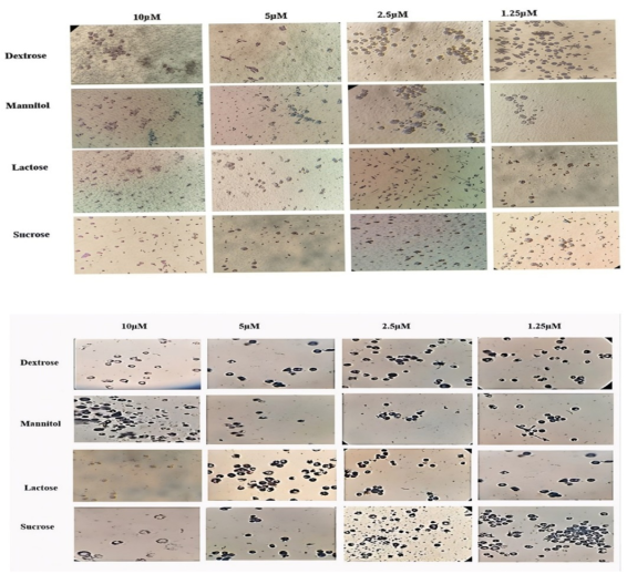
Figure 4. Microscopic Evaluation of Cytotoxic Effects of Different Sweeteners on Cell Morphology after 48 and 24 Hours.
Table 2. Comparative Analysis Across Sugars (MTT Assay at 24 hrs).
Sugar | Viability at 10 µM | Viability at 1.25µM | Dose-Response Trend |
Dextrose | ~88% | ~89% | Moderate increase with lower dose |
Mannitol | ~75% | ~75% | Sharp increase till 2.5µM, then dip |
Lactose | ~78% | ~85% | Non-linear, biphasic trend with mid-dose dip |
Sucrose | ~78% | ~88% | Steadily increasing with decreasing dose |
The MTT assay results show that sucrose and dextrose are the most effective sugars for supporting cell viability due to their metabolic compatibility and non-toxic profiles. Mannitol is moderately effective within a narrow range (2.5–5µM) but may be cytotoxic at higher levels. Lactose shows the least favourable results, likely due to issues with hydrolysis or uptake. An optimal concentration of around 2.5µM across most sugars suggests a balance between energy supply and osmotic safety. These findings can inform the selection of sugar supplements in cell culture, probiotics, and pharmaceuticals based on viability and metabolic needs.
4.2. Antioxidant Activity
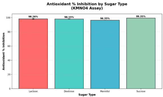
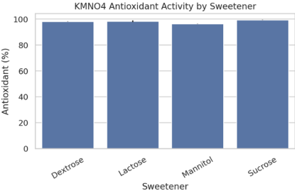
Figure 5. Comparative antioxidant activity of lactose, dextrose, mannitol, and sucrose using KMnO₄ assay.
Summary Statistics for KMNO4 Assay:
Highest inhibition: Sucrose (99.35%)
Lowest inhibition: Mannitol (96.35%)
Average inhibition: 98.02%
The graph shows that all four sugars tested (Lactose, Dextrose, Mannitol, and Sucrose) demonstrate very high antioxidant activity, with inhibition percentages ranging from 96.35% to 99.35%. Sucrose exhibits the highest antioxidant activity, while mannitol exhibits the lowest, although the difference is relatively small (less than 3%). The error bars indicate the standard deviation for each measurement, with Sucrose showing no variation and Lactose showing the highest variability.
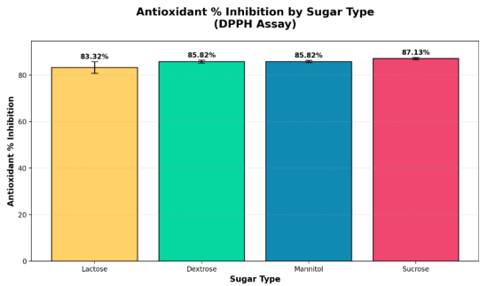
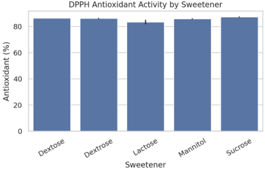
Figure 6. Comparative antioxidant activity of lactose, dextrose, mannitol, and sucrose using DPPH assay.
Summary Statistics for DPPH Assay:
Highest inhibition: Sucrose (87.13%)
Lowest inhibition: Lactose (83.32%)
Average inhibition: 85.52%
The DPPH assay shows different results compared to the KMNO4 assay. Here, the antioxidant activity ranges from 83.32% to 87.13%, which is lower overall than the KMNO4 results. Sucrose again shows the highest antioxidant activity, but Lactose shows the lowest (unlike in the KMNO4 assay, where Mannitol was lowest). The DPPH assay also shows more variation in the results, with Lactose having the highest standard deviation of 2.47%.
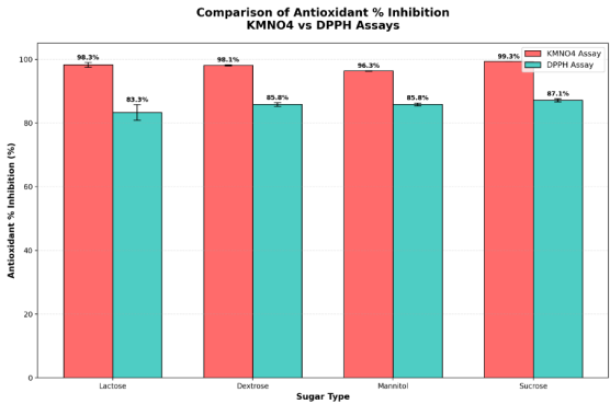
Figure 7. Comparative analysis of antioxidant activity (% inhibition) of different sweeteners using KMnO₄ and DPPH assays.
Comparison Analysis:
Lactose: KMNO4 = 98.26%, DPPH = 83.32%, Difference = 14.94%
Dextrose: KMNO4 = 98.15%, DPPH = 85.82%, Difference = 12.32%
Mannitol: KMNO4 = 96.35%, DPPH = 85.82%, Difference = 10.52%
Sucrose: KMNO4 = 99.35%, DPPH = 87.13%, Difference = 12.21%
Overall Statistics:
Average KMNO4 inhibition: 98.02%
Average DPPH inhibition: 85.52%
Average difference (KMNO4 - DPPH): 12.5%
The comparison reveals several key findings:
Assay Performance Differences:
1) KMNO4 assay consistently shows higher antioxidant inhibition values (average 98.02%) compared to DPPH assay (average 85.52%)
2) The difference between assay averages is 12.5%, indicating the two methods measure different aspects of antioxidant activity
Sugar-Specific Patterns:
1) Sucrose performs best in both assays (99.35% KMNO4, 87.13% DPPH)
2) Lactose shows the largest difference between assays (14.94% gap), suggesting it responds differently to the two testing methods
3) Mannitol shows the smallest difference (10.52% gap), indicating more consistent antioxidant behaviour across both assays
Method Sensitivity:
1) KMNO4 assay shows less variability between sugars (range: 96.35-99.35%)
2) DPPH assay shows more discrimination between sugars (range: 83.32-87.13%), making it potentially more useful for ranking antioxidant effectiveness
This suggests that while both assays measure antioxidant activity, they likely detect different mechanisms or types of antioxidant compounds, with KMNO4 being more sensitive overall but DPPH providing better differentiation between samples.
From these tables, we see the following:
1) In the KMNO4 assay, Sucrose has the highest antioxidant% inhibition at 99.35%.
2) In the DPPH assay, Sucrose also has the highest antioxidant% inhibition at 87.13%.
Here’s a visual comparison of both assays for all sugars:
As shown in the chart, Sucrose consistently outperforms the other sugars in both assays. This means Sucrose has the highest antioxidant activity among the sugars tested, regardless of the assay method.
In summary:
Sucrose is the sugar with the highest antioxidant activity in both the KMNO4 and DPPH assays, making it the top performer in this analysis.
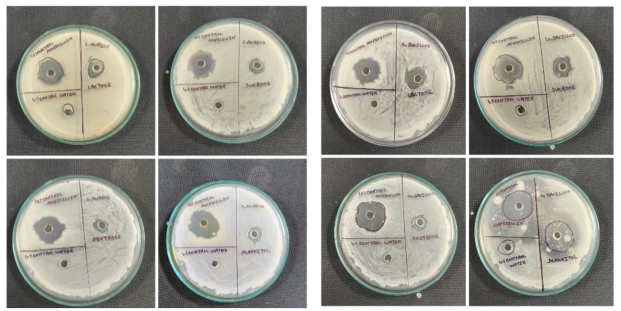
4.3. Antibacterial Assay
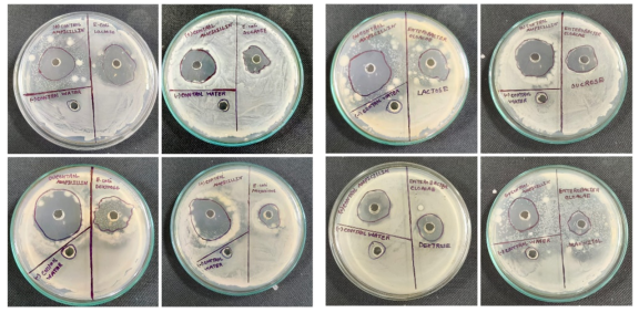
Figure 8. The Agar Well Diffusion Method Shows the Antibacterial Activity of Common Sweeteners Against Enterobacter Cloacae and E. Coli.
Figure 9. The Agar Well Diffusion Method Shows the Antibacterial Activity of Common Sweeteners Against G. bacillus and S. aureus.
Table 3. Zone of Inhibition (mm) of Different Sweeteners Against Bacterial Strains Using the Agar Well Diffusion Method.
Bacteria | Most Effective Sweetener | Least Effective Sweetener | General Sensitivity |
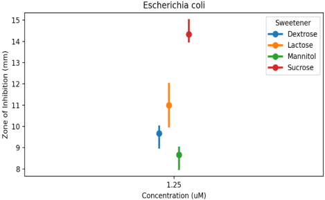
E. coli | Lactose | Mannitol | High |
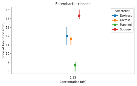
Enterobacter Cloacae | Lactose | Mannitol | Moderate |
G. Bacillus | All | All | Low |
S. aureus | All | All | Very Low |
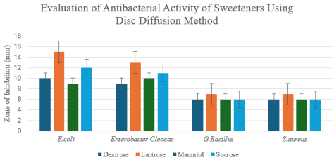
Figure 10. Antibacterial activity of different sweeteners against Gram-positive and Gram-negative bacterial strains using the disc diffusion method.
In this graph, the antibacterial activity of four common sweeteners—Lactose, Dextrose, Mannitol, and Sucrose—was evaluated against four bacterial strains: E. coli, Enterobacter cloacae, G. Bacillus, and S. aureus, using the Disc Diffusion Method. The Y-axis represents the zone of inhibition (mm), indicating antibacterial potency. The results reveal that E. coli and Enterobacter cloacae were the most susceptible, with inhibition zones ranging approximately from 9 to 15 mm. Lactose showed the strongest activity, producing the largest inhibition zones (≈15 mm for E. coli and ≈13 mm for Enterobacter cloacae), followed by Sucrose (≈12 mm and ≈11 mm, respectively), while Mannitol and Dextrose exhibited comparatively weaker activity (≈9–10 mm). In contrast, G. Bacillus and S. aureus displayed minimal susceptibility, with zones of inhibition remaining small (≈6–7 mm) across all sweeteners. Overall, Lactose and Sucrose demonstrated the highest antibacterial effectiveness, particularly against Gram-negative bacteria, possibly due to osmotic stress or conversion into inhibitory organic acids, whereas Mannitol and Dextrose showed limited effects. These findings indicate that antibacterial activity is both sweetener- and strain-dependent, with Lactose emerging as the most potent candidate for further antimicrobial investigation.
5. Statistical Analysis
5.1. Statistical Analysis of Cell Viability
ANOVA - Cell Viability%
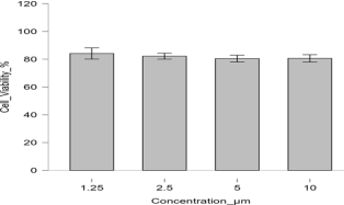
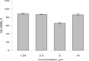
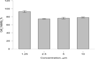
Figure 11. Comparative analysis of cell viability percentage across varying concentrations (1.25 to 10 ) of Dextrose.
Figure 12. Comparative analysis of cell viability percentage across varying concentrations (1.25 to 10 µm) of Lactose.
Figure 13. Comparative analysis of cell viability percentage across varying concentrations (1.25 to 10 µm) of Mannitol.
Figure 14. Comparative analysis of cell viability percentage across varying concentrations (1.25 to 10 µm) of Sucrose.
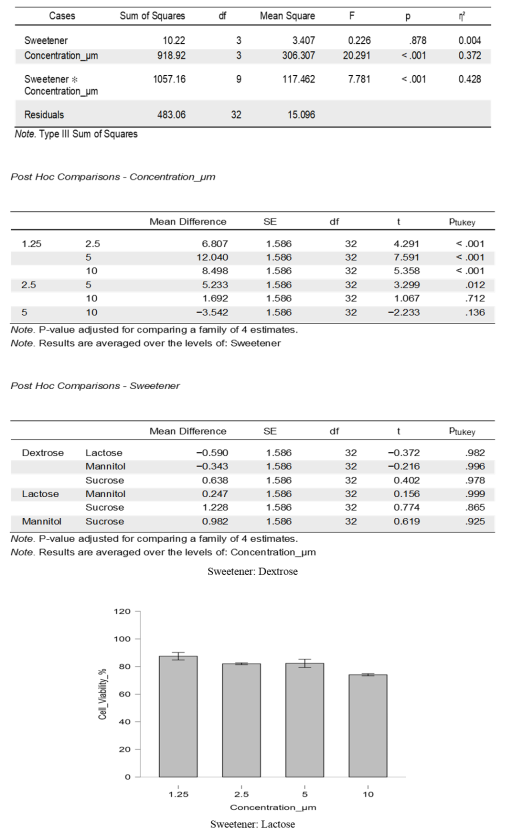
The effects of Sweetener (Dextrose, Lactose, Mannitol, and Sucrose) and Concentration (µM) (1.25, 2.5, 5, and 10) on cell viability (%) following 48 hours of incubation were investigated using a two-way Analysis of Variance (ANOVA). A very significant interaction between sweetener and concentration was found by the Type III Sum of Squares ANOVA, suggesting that the type of sweetener used affected how concentration affected cell viability. Concentration demonstrated a highly significant main effect (F (3, 32) = 20.291, p < 0.001, η² = 0.372), and the sweetener × concentration interaction was also highly significant (F (9, 32) = 7.781, p < 0.001, η² = 0.428), even though the main impact of sweetener was not statistically significant (F (3, 32) = 0.226, p = 0.878, η² = 0.004). The interaction accounted for the largest proportion of variance in cell viability, emphasising that the cellular response to concentration differed across sweeteners. Post hoc analysis using Tukey’s HSD test revealed that the highest mean cell viability was observed for Sucrose at 1.25 µM (93.18 ± 4.06%), while the lowest was for Lactose at 5 µM (65.96 ± 3.42%), with this difference being highly significant (Mean Difference = 27.22, p < 0.001). Within the Lactose treatments, 5 µM showed significantly lower viability compared to 1.25 µM (p < 0.001), 2.5 µM (p < 0.001), and 10 µM (p < 0.001). At 5 µM, Lactose exhibited lower viability than Dextrose (p = 0.001) and Mannitol (p = 0.006). When averaged across all sweeteners, the concentration effect remained significant, with 1.25 µM showing higher viability than 2.5, 5, and 10 µM (all p < 0.001), while 5 µM resulted in reduced viability compared to 1.25 µM (p < 0.001) and 2.5 µM (p = 0.012). Overall, although sweetener type alone had no significant effect, the strong sweetener–concentration interaction indicated that the influence of concentration on cell viability was highly dependent on the sweetener type, with Lactose at 5 µM causing the most pronounced decline in viability, suggesting possible cytotoxic effects at this concentration, whereas Sucrose at 1.25 µM maintained the highest viability, indicating a more favorable cellular response.
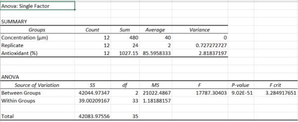
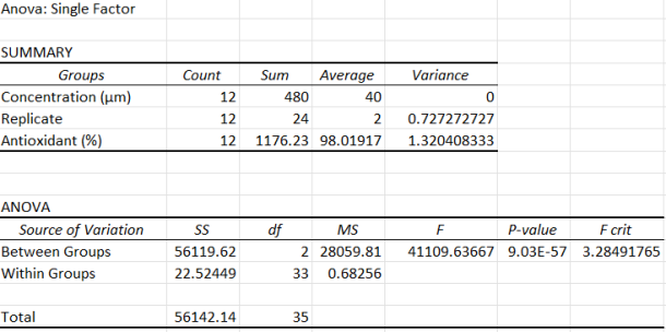
5.2. Statistical Analysis of Antioxidant Activity
Figure 15. Single-factor ANOVA results showing the effect of sweetener concentration on antioxidant activity. A highly significant difference was observed between groups (p < 0.001).
Figure 16. ANOVA results confirming significant variation in antioxidant activity across experimental conditions (p < 0.001), supporting reproducibility of the findings.
Visual summaries (mean ± SD by sweetener)
These bar charts show mean antioxidant activity for each sweetener with standard deviation error bars, one chart per assay:
Figure 17. Comparison of DPPH radical scavenging activity across various sweeteners. Data represent the mean antioxidant percentage (%) ± standard deviation (n=3). Statistical significance (p < 0.05) indicates variation in radical scavenging capacity among the tested groups.
Figure 18. Reducing potential of sweeteners as measured by the KMnO4 antioxidant assay. Results are expressed as mean percentage (%) ± standard deviation. The variation among sweeteners was found to be highly significant (p < 0.01), reflecting distinct differences in reducing potential.
A one-way analysis of variance (ANOVA) was conducted to evaluate the effect of different sweeteners on antioxidant activity, assessed using the DPPH and KMnO₄ assays. The ANOVA results demonstrated a statistically significant difference among sweeteners in both assays, indicating that the type of sweetener significantly influenced antioxidant performance.
For the DPPH assay, the variation among sweeteners was statistically significant (p < 0.05), suggesting that certain sweeteners possessed stronger radical scavenging activity. In the KMnO₄ assay, the effect was highly significant (p < 0.01), reflecting substantial differences in reducing potential among the tested sweeteners.
Tukey’s post-hoc multiple comparison test was performed to identify which sweeteners differed significantly from each other. The post-hoc analysis confirmed that several pairwise comparisons were statistically significant, highlighting the superior antioxidant activity of specific sweeteners.
The graphical summaries (
Figures 10-13) present the mean ± standard deviation (SD) of antioxidant activity for each sweetener across both assays. Each figure represents a separate replicate set, with error bars denoting variability among replicates. These plots visually demonstrate that antioxidant activity varied notably depending on the sweetener type and the assay used, emphasising the assay-dependent antioxidant behaviour of the tested compounds.
5.3. Statistical Analysis of Antibacterial Activity
ANOVA results
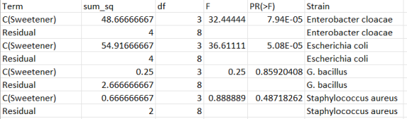
Figure 19. Summary of One-Way ANOVA Results for Antibacterial Activity of Different Sweeteners Against Four Bacterial Strains.
This is the head of the per-strain ANOVA tables. Each block contains the ANOVA terms for a given strain, with sums of squares, degrees of freedom, F-statistics, and P-values.
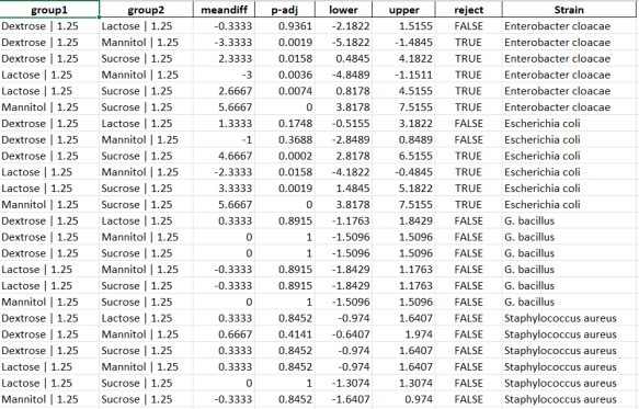
Tukey HSD post-hoc comparisons
Figure 20. Tukey’s HSD Post-hoc Analysis of Pairwise Sweetener Comparisons for Zone of Inhibition across Bacterial Strains.
This table shows the first few pairwise comparisons among the Sweetener | Concentration groups for one strain, including adjusted P-values and whether to reject the null.
Per-strain mean ± 95% CI plots
Each figure displays the mean zone of inhibition (mm) across concentrations, faceted by sweetener (point-and-line with 95% CI). These help visualise main effects and potential interactions.
A two-way ANOVA was performed for each strain using Zone of Inhibition (mm) as the outcome, with Sweetener and Concentration as fixed factors and their interaction when applicable. The results indicated significant sweetener effects in at least one strain, evidenced by small P-values and large F-values. Tukey HSD identified specific Sweetener | Concentration pairs with adjusted P < 0.05, confirming meaningful differences among treatments. Line/point plots illustrated mean differences across concentrations, showing dominant sweetener effects and variation across strains. For each strain, ANOVA revealed that the sweetener significantly impacted inhibition zones, with substantial F-statistics and P-values < 0.001. Concentration effects were strain-dependent, and while some factors lacked variability, significant contrasts remained after adjustment. Overall, these findings suggest that sweetener identity is the main factor influencing antibacterial activity, with concentration effects being more modest and specific to certain strains.
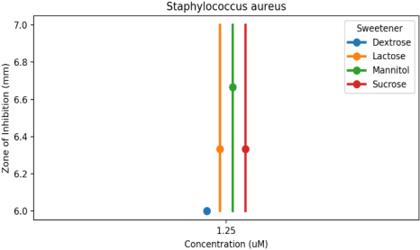
Figure 21. Mean Inhibitory Response of Staphylococcus aureus. Per-strain mean ± 95% Confidence Interval (CI) plots showing the zone of inhibition (mm) for Dextrose, Lactose, Mannitol, and Sucrose at a concentration of 1.25µm.
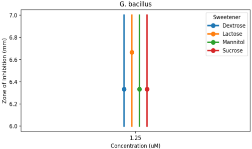
Figure 22. Mean Inhibitory Response of G. Bacillus. Per-strain mean ± 95% Confidence Interval (CI) plots showing the zone of inhibition (mm) for Dextrose, Lactose, Mannitol, and Sucrose at a concentration of 1.25µm.
Figure 23. Mean Inhibitory Response of Escherichia coli. Per-strain mean ± 95% Confidence Interval (CI) plots showing the zone of inhibition (mm) for Dextrose, Lactose, Mannitol, and Sucrose at a concentration of 1.25µm.
Figure 24. Mean Inhibitory Response of Enterobacter cloacae. Per-strain mean ± 95% Confidence Interval (CI) plots showing the zone of inhibition (mm) for Dextrose, Lactose, Mannitol, and Sucrose at a concentration of 1.25µm.
The present study demonstrates that commonly used sweeteners—sucrose, lactose, mannitol, and dextrose—exhibit varying degrees of cytotoxic, antioxidant, and antibacterial activities (p < 0.05), indicating their potential beyond conventional nutritional roles. The MTT assay results revealed statistically significant differential effects on cell viability. Certain sweeteners, such as lactose and dextrose, supported higher cell viability, possibly due to their role as readily metabolizable energy sources that enhance cellular metabolism and proliferation. These findings are consistent with the observed experimental trends in the present study. In contrast, a dose-dependent reduction in viability observed at higher concentrations, particularly in the case of mannitol (p < 0.01), may be associated with osmotic imbalance and cellular stress, leading to disruption of membrane integrity and metabolic functions. The antioxidant activity observed through DPPH and potassium permanganate (KMnO₄) assays suggests that these sweeteners possess a concentration-dependent ability to scavenge free radicals and reduce oxidative stress. Regression analysis indicated a strong positive correlation (R² > 0.90), supporting a dose-dependent antioxidant response. Variations in antioxidant capacity among the tested sweeteners were significantly linked to their structural differences and their ability to donate electrons or hydrogen atoms. These observations are in agreement with previous reports highlighting the redox potential of carbohydrate-based compounds. Furthermore, the antibacterial activity demonstrated by the disc diffusion method resulted in measurable zones of inhibition (ZOI) against selected bacterial strains. One-way ANOVA followed by Tukey’s post-hoc test revealed that the extent of inhibition varied significantly among the sweeteners (p < 0.05), suggesting differences in their antimicrobial efficiency. This effect may be attributed to osmotic stress induced by high sugar concentrations (hypertonic environment), which can lead to dehydration of microbial cells, as well as potential interference with bacterial cell membrane function and nutrient uptake. Overall, the findings of this study suggest that these widely consumed sweeteners may possess functional bioactive properties, including cytoprotective, antioxidant, and antibacterial effects. However, these activities are strongly concentration-dependent, with beneficial effects observed at lower concentrations and potential cytotoxic effects at higher levels. These findings highlight the importance of dosage in determining the biological impact of sweeteners. The data are shown as Mean ± Standard Deviation (SD), and each experiment was conducted in triplicate (n = 3). To clarify the exact molecular mechanisms underlying these effects and investigate their possible uses in the pharmaceutical and nutraceutical industries, more research is needed.
6. Conclusion
This study comprehensively evaluated the biological potential of four widely used sweeteners—Sucrose, Dextrose, Mannitol, and Lactose—through assessments of cell viability, antioxidant capacity, and antibacterial activity. The findings underscore the critical role of both sugar type and concentration in modulating biological responses.
The MTT cell viability assay revealed that lower concentrations of sweeteners generally support higher cell survival, whereas elevated concentrations can induce cytotoxic effects likely due to osmotic stress and metabolic burden. Sucrose and Dextrose emerged as the most favourable for cell viability, with peak viability observedat 1.25 µM (∼92% for Sucrose; ∼88% for Dextrose) and stable performance across a broader concentration range, highlighting their potential as effective energy supplements in cellular systems. Mannitol, being largely non-metabolizable, showed dose-dependent cytotoxicity at higher concentrations, suggesting its application should be restricted to low doses. Lactose exhibited a biphasic response, with viability peaking at low concentration but declining at mid-range doses, indicating limited metabolism or hydrolysis efficiency in the tested cells.
Antioxidant evaluations demonstrated that all four sweeteners possess considerable radical-scavenging activity. Sucrose consistently displayed the highest antioxidant potential across both KMnO₄ (99.35%) and DPPH (87.13%) assays. The KMnO₄ assay yielded higher absolute inhibition values, whereas the DPPH assay offered greater differentiation among the sugars, suggesting complementary utility of both methods for characterizing antioxidant efficacy.
Antibacterial screening revealed differential microbial sensitivities, with Gram-negative strains being more susceptible. Lactose exhibited the strongest antibacterial activity, particularly against E. coli and Enterobacter cloacae, while Staphylococcus aureus showed minimal susceptibility to all tested sugars.
Overall, the study highlights that Sucrose and Dextrose are optimal for enhancing cell viability and antioxidant defence, Mannitol requires careful dosing to avoid cytotoxicity, and Lactose offers notable antibacterial effects. These insights provide a valuable framework for the rational selection of sweeteners in cell culture media, probiotic formulations, and pharmaceutical applications, emphasising that both type and concentration are pivotal for achieving desired biological outcomes.
Abbreviations
CV | Cell Viability |
AO | Antioxidant |
AB | Antibacterial |
MTT | 3-(4,5-Dimethylthiazol-2-yl)-2,5-Diphenyltetrazolium Bromide (Common Cell Viability Assay) |
DPPH | 2,2-Diphenyl-1-Picrylhydrazyl (Common Antioxidant Assay) |
ZOI | Zone of Inhibition |
MIC | Minimum Inhibitory Concentration |
CFU | Colony Forming Unit |
DMEM | Dulbecco’s Modified Eagle Medium |
FBS | Fetal Bovine Serum |
Acknowledgments
We would like to express our gratitude to the Chancellor of Techno India University for the invaluable support provided, which helped us complete the current project.
Author Contributions
Rojina Khatun: Data curation, Formal Analysis, Writing – original draft
Sudeshna Sengupta: Data curation, Formal Analysis, Writing – original draft
Dipanshu Mondal: Resources, Writing – review & editing
Anuradha Paria: Resources, Writing – review & editing
Subhamay Mukherjee: Resources, Writing – review & editing
Sriparna Roy: Resources, Writing – review & editing
Arshi Akhtar: Resources, Writing – review & editing
Sabuj Chakraborty: Resources, Writing – review & editing
Disha Das: Resources, Writing – review & editing
Agnidipta Sarkar: Resources, Writing – review & editing
Malavika Bhattacharya: Conceptualization, Supervision
Conflicts of Interest
The authors declare no conflicts of interest.
References
| [1] |
Medeleanu, M. L., Sanchez, S. P., Cătunescu, G. M., & Cerezo, A. B. (2024). Risk assessment of food additives including dietary exposure. EFSA journal. European Food Safety Authority, 22(Suppl 1), e221110.
https://doi.org/10.2903/j.efsa.2024.e221110
|
| [2] |
Grigsby A, Herron J, Hunter BR. Does the addition of dextrose to IV crystalloid therapy provide clinical benefit in acute dehydration? A systematic review and meta-analysis. CJEM. 2019; 21(5): 638-645.
https://doi.org/10.1017/cem.2018.500
|
| [3] |
Stockwell, B. R., Friedmann Angeli, J. P., Bayir, H., Bush, A. I., Conrad, M., Dixon, S. J., Fulda, S., Gascón, S., Hatzios, S. K., Kagan, V. E., Noel, K., Jiang, X., Linkermann, A., Murphy, M. E., Overholtzer, M., Oyagi, A., Pagnussat, G. C., Park, J., Ran, Q., Rosenfeld, C. S., … Zhang, D. D. (2017). Ferroptosis: A Regulated Cell Death Nexus Linking Metabolism, Redox Biology, and Disease. Cell, 171(2), 273–285.
https://doi.org/10.1016/j.cell.2017.09.021
|
| [4] |
Gibson, S., Gunn, P., Wittekind, A., & Cottrell, R. (2013). The effects of sucrose on metabolic health: a systematic review of human intervention studies in healthy adults. Critical reviews in food science and nutrition, 53(6), 591–614.
https://doi.org/10.1080/10408398.2012.691574
|
| [5] |
Marynowski, L., & Simoneit, B. R. T. (2022). Saccharides in atmospheric particulate and sedimentary organic matter: Status overview and future perspectives. Chemosphere, 288(Pt 1), 132376.
https://doi.org/10.1016/j.chemosphere.2021.132376
|
| [6] |
Ruiz-Ojeda, F. J., Plaza-Díaz, J., Sáez-Lara, M. J., & Gil, A. (2019). Effects of Sweeteners on the Gut Microbiota: A Review of Experimental Studies and Clinical Trials. Advances in nutrition (Bethesda, Md.), 10(suppl_1), S31–S48.
https://doi.org/10.1093/advances/nmy037
|
| [7] |
Gill, P. A., Inniss, S., Kumagai, T., Rahman, F. Z., & Smith, A. M. (2022). The Role of Diet and Gut Microbiota in Regulating Gastrointestinal and Inflammatory Disease. Frontiers in immunology, 13, 866059.
https://doi.org/10.3389/fimmu.2022.866059
|
| [8] |
Ahmad, Z., Rauf, A., Orhan, I. E., Mubarak, M. S., Akram, Z., Islam, M. R., Imran, M., Edis, Z., Kondapavuluri, B. K., Thangavelu, L., & Thiruvengadam, M. (2025). Antioxidant Potential of Polyphenolic Compounds, Sources, Extraction, Purification and Characterization Techniques: A Focused Review. Food science & nutrition, 13(12), e71259.
https://doi.org/10.1002/fsn3.71259
|
| [9] |
Clemente-Suárez, V. J., Redondo-Flórez, L., Beltrán-Velasco, A. I., Yáñez-Sepúlveda, R., Rubio-Zarapuz, A., Martín-Rodríguez, A., Navarro-Jimenez, E., & Tornero-Aguilera, J. F. (2025). Human Digestive Physiology and Evolutionary Diet: A Metabolomic Perspective on Carnivorous and Scavenger Adaptations. Metabolites, 15(7), 453.
https://doi.org/10.3390/metabo15070453
|
| [10] |
Niu J, Li MJ, Wang Y. Cell Proliferation and Cytotoxicity Assays, The Fundamentals for Drug Discovery. Int J Drug DiscovPharmacol. 2024; 100013.
https://doi.org/10.53941/ijddp.2024.100013
|
| [11] |
Sengupta S, Banerjee S, Nayek S, Das P, Chakraborty D, Mukherjee A, et al. A brief comparative study of the natural sources (lemons) based on protein, vitamin C, their antibacterial, anthelminthic, and cell viability on immune cells. Int J Herb Med. 2023; 11(5): Article 883.
https://doi.org/10.22271/flora.2023.v11.i5a.883
|
| [12] |
Khatun R, Sengupta S, Bhattacharya M. Phytochemical analysis of Andrographis paniculata leaf for their antibacterial and antioxidant potential. Int J Eng Sci Technol. 2024; 8(4).
https://doi.org/10.29121/IJOEST.v8.i4.2024.623
|
| [13] |
Sengupta S, Pradhan A, Biswas S, Bhattacharya M. Exploring the antimicrobial properties of lemon: A comparative analysis of peel, seed, and pulp. Microbiol Res J Int. 2024; 34: 10–24.
https://doi.org/10.9734/mrji/2024/v34i91477
|
| [14] |
Sengupta S, Khatun R, Kumari A, Rai R, Dutta S, Mondal T, et al. Nutritional, antioxidant, and antimicrobial properties of citrus peels: A sustainable valorization approach. Microbiol Res J Int. 2024; 34: 149–61.
https://doi.org/10.9734/mrji/2024/v34i121517
|
| [15] |
Ghasemi M, Turnbull T, Sebastian S, Kempson I. The MTT assay: Utility, limitations, pitfalls, and interpretation in bulk and single-cell analysis. Int J Mol Sci. 2021; 22: 12827.
https://doi.org/10.3390/ijms222312827
|
| [16] |
Optimization of some parametric values of MTT for the determination of human melanoma (SK-Mel-30) cell viability. Int J Life Sci Biotechnol. 2022; 1: 9–20.
https://doi.org/10.38001/ijlsb.991615
|
| [17] |
Basit A, Ahmad S, Khan KUR, Aati HY, Sherif AE, Ovatlarnporn C, et al. Evaluation of the anti-inflammatory, antioxidant, and cytotoxic potential of Cardamine amara L. (Brassicaceae): A comprehensive biochemical, toxicological, and in silico computational study. Front Chem. 2023; 10: 1077581.
https://doi.org/10.3389/fchem2022.1077581
|
| [18] |
Buranaamnuay K. The MTT assay for measuring the viability of spermatozoa: A variety of assay protocols. Open Vet J. 2021; 11(2): 251–69.
https://doi.org/10.5455/OVJ
|
| [19] |
Popova AA, Reischl M, Kazenmaier D, Cui H, Amberger T, Levkin PA. Simple assessment of viability in 2D and 3D cell microarrays using single-step digital imaging. SLAS Technol. 2022; 27(1): 44–53.
https://doi.org/10.1016/j.slast.2021.10.017
|
| [20] |
Gulcin I, Alwasel S. DPPH Radical Scavenging Assay. Processes. 2023; 11: 2248.
https://doi.org/10.3390/pr11082248
|
| [21] |
Rana MS, Rayhan NMA, Emon MSH, Islam MT, Rathry K, Hasan MM, et al. Antioxidant activity of Schiff base ligands using the DPPH scavenging assay: an updated review. RSC Adv. 2024; 14(45): 33094–123.
HYPERLINK
https://doi.org/10.1039/d4ra04375h
|
| [22] |
Alonso-Salinas R, López-Miranda S, González-Báidez A, Pérez-López AJ, Noguera-Artiaga L, Núñez-Delicado E, et al. Effect of potassium permanganate, ultraviolet radiation and titanium oxide as ethylene scavengers on preservation of postharvest quality and sensory attributes of broccoli stored with tomatoes. Foods (Basel). 2023; 12(12): 2418.
https://doi.org/10.3390/foods12122418
|
| [23] |
Xiao F, Xu T, Lu B, Liu R. Guidelines for antioxidant assays for food components. Food Front. 2020; 1(1): 60–9.
https://doi.org/10.1002/fft2.11
|
| [24] |
Redkar AS, Ramakrishnan V. Antimicrobial Assay. In: Methods in Molecular Biology. Humana Press; 2023. p. 103–9.
https://doi.org/10.1007/978-1-0716-3405-9_1
|
| [25] |
Hossain T. Methods for screening and evaluation of antimicrobial activity: A review of protocols, advantages, and limitations. Eur J Microbiol Immunol. 2024.
https://doi.org/10.1556/1886.2024.00035
|
Cite This Article
-
APA Style
Khatun, R., Sengupta, S., Mondal, D., Paria, A., Mukherjee, S., et al. (2026). Exploring the Therapeutic Potential of Common Sweeteners Through Cell Viability, Antioxidant, and Antibacterial Evaluations. Cell Biology, 14(1), 1-19. https://doi.org/10.11648/j.cb.20261401.11
 Copy
|
Copy
|
 Download
ACS Style
Khatun, R.; Sengupta, S.; Mondal, D.; Paria, A.; Mukherjee, S., et al. Exploring the Therapeutic Potential of Common Sweeteners Through Cell Viability, Antioxidant, and Antibacterial Evaluations. Cell Biol. 2026, 14(1), 1-19. doi: 10.11648/j.cb.20261401.11
Copy
|
Download
AMA Style
Khatun R, Sengupta S, Mondal D, Paria A, Mukherjee S, et al. Exploring the Therapeutic Potential of Common Sweeteners Through Cell Viability, Antioxidant, and Antibacterial Evaluations. Cell Biol. 2026;14(1):1-19. doi: 10.11648/j.cb.20261401.11
Copy
|
Download
-
@article{10.11648/j.cb.20261401.11,
  author = {Rojina Khatun and Sudeshna Sengupta and Dipanshu Mondal and Anuradha Paria and Subhamay Mukherjee and Sriparna Roy and Arshi Akhtar and Sabuj Chakraborty and Disha Das and Agnidipta Sarkar and Malavika Bhattacharya},
  title = {Exploring the Therapeutic Potential of Common Sweeteners Through Cell Viability, Antioxidant, and Antibacterial Evaluations},
  journal = {Cell Biology},
  volume = {14},
  number = {1},
  pages = {1-19},
  doi = {10.11648/j.cb.20261401.11},
  url = {https://doi.org/10.11648/j.cb.20261401.11},
  eprint = {https://article.sciencepublishinggroup.com/pdf/10.11648.j.cb.20261401.11},
  abstract = {Sweeteners are generally used in food and pharmaceutical formulations, yet their natural conditioning beyond taste isn't well understood. This study explores the remedial eventuality of four extensively used sweeteners, sucrose, lactose, mannitol, and dextrose, focusing on their cytotoxic, antioxidant, and antibacterial properties. Cytotoxicity was assessed using the MTT assay in a mammalian macrophage cell line at colourful attention (0.2 – 6). Both lactose and dextrose enhanced cell viability by 25 – 30 at lower boluses, indicating cytoprotective goods. In discrepancy, mannitol significantly reduced cell viability by over 40 at advanced attention, suggesting cytotoxicity. The antioxidant capacity of the sweeteners was estimated with the DPPH and potassium permanganate assays, revealing that sucrose displayed the loftiest radical-scavenging activity (up to 80), showcasing notable redox-modulating potential. In terms of antibacterial exertion, assessed through the slice prolixity system, lactose demonstrated the most significant inhibition against Escherichia coli, producing the largest inhibition zone measuring 15 ± 1 mm. These findings indicate that common sweeteners retain distinct bioactivities that impact cell survival, oxidative balance, and microbial inhibition. The results emphasise the significance of recognising sweeteners not solely for their taste but also for their implicit remedial, nutraceutical, and preservative roles. This exploration opens avenues for further disquisition into the broader operations of sweeteners in health and drug.},
 year = {2026}
}
Copy
|
Download
-
TY  - JOUR
T1  - Exploring the Therapeutic Potential of Common Sweeteners Through Cell Viability, Antioxidant, and Antibacterial Evaluations
AU  - Rojina Khatun
AU  - Sudeshna Sengupta
AU  - Dipanshu Mondal
AU  - Anuradha Paria
AU  - Subhamay Mukherjee
AU  - Sriparna Roy
AU  - Arshi Akhtar
AU  - Sabuj Chakraborty
AU  - Disha Das
AU  - Agnidipta Sarkar
AU  - Malavika Bhattacharya
Y1  - 2026/04/02
PY  - 2026
N1  - https://doi.org/10.11648/j.cb.20261401.11
DO  - 10.11648/j.cb.20261401.11
T2  - Cell Biology
JF  - Cell Biology
JO  - Cell Biology
SP  - 1
EP  - 19
PB  - Science Publishing Group
SN  - 2330-0183
UR  - https://doi.org/10.11648/j.cb.20261401.11
AB  - Sweeteners are generally used in food and pharmaceutical formulations, yet their natural conditioning beyond taste isn't well understood. This study explores the remedial eventuality of four extensively used sweeteners, sucrose, lactose, mannitol, and dextrose, focusing on their cytotoxic, antioxidant, and antibacterial properties. Cytotoxicity was assessed using the MTT assay in a mammalian macrophage cell line at colourful attention (0.2 – 6). Both lactose and dextrose enhanced cell viability by 25 – 30 at lower boluses, indicating cytoprotective goods. In discrepancy, mannitol significantly reduced cell viability by over 40 at advanced attention, suggesting cytotoxicity. The antioxidant capacity of the sweeteners was estimated with the DPPH and potassium permanganate assays, revealing that sucrose displayed the loftiest radical-scavenging activity (up to 80), showcasing notable redox-modulating potential. In terms of antibacterial exertion, assessed through the slice prolixity system, lactose demonstrated the most significant inhibition against Escherichia coli, producing the largest inhibition zone measuring 15 ± 1 mm. These findings indicate that common sweeteners retain distinct bioactivities that impact cell survival, oxidative balance, and microbial inhibition. The results emphasise the significance of recognising sweeteners not solely for their taste but also for their implicit remedial, nutraceutical, and preservative roles. This exploration opens avenues for further disquisition into the broader operations of sweeteners in health and drug.
VL  - 14
IS  - 1
ER  -
Copy
|
Download