Background: The injury of intestinal mucosal barrier in the later period of severe acute pancreatitis (SAP) results in flora migration with the endotoxin and bacterium entering blood circulation, final result of bacteremia, multiple organ dysfunction syndrome (MODS), eventually death. poly (ADP-ribose) polymerase-1(PARP-1) is a nuclear DNA-binding protein that has been revealed a wide role in DNA repair, chromatin structure, necrosis, inflammation and immune function. We plan to determine whether PARP-1 and its inhibition affect the injury of intestinal mucosal barrier in SAP. Methods: SAP was induced by 6 hourly intraperitoneal injection of cerulein (50ug/kg), with lipopolysaccharide (10mg/kg) in the last time. At 30 min prior, rats were treated with the PARP inhibition 3-Aminobenzamide (3-AB) or normal saline. 40 rats were randomly separated into 4 groups of a normal control group, a SAP group, a SAP with inhibition group and an inhibition group. After 12 hours, all rats were killed. We compared serum amylase, interleukin-6 (IL-6), celiac morphological changes, pancreatic pathological injury, intestinal histology. Activities of PARP-1, nuclear factor-kappa B (NF-kB), Occludin in the intestine were also examined. Result: The inflammation and injury has been showed seriously in the intestines. with SAP, the expression of PARP-1 and NF-kB also has been shown high level. But with administration of PARP inhibitor, 3-AB, the severity of SAP and pancreatitis-associated intestinal injury was significantly attenuated, the protein expression of PARP-1 and NF-kB was significantly decreased. Conclusion: Our findings indicate that PARP inhibitors played a positive role in the injury of intestinal mucosal barrier in rat model with SAP.
This is an Open Access article, distributed under the terms of the Creative Commons Attribution 4.0 International License (http://creativecommons.org/licenses/by/4.0/), which permits unrestricted use, distribution and reproduction in any medium or format, provided the original work is properly cited.
Severe Acute Pancreatitis, Poly (ADP-ribose) Polymerase, Intestinal Mucosal Barrier, 3-Aminobenzamide
1. Introduction
SAP is the most serious type of this disorder, is associated with high morbidity and mortality. According to present studies, it is a devastating disease with the mortality ranging from less than 10% to as high as 85%. In the late period of this disease, the patient is at risk for intestinal flora translocation and the development of secondary infection of the necrotic tissue, which can result in sepsis and late multisystem organ failure (MOF). However, no approved drugs or therapy are available for the injury of intestinal mucosal barrier in SAP and even the underlying mechanism is still unclear
Zerem E. Treatment of severe acute pancreatitis and its complications [J]. World Journal of Gastroenterology, 2014, 20(38): 13879-13892.
[3]
Virág L, Szabó C. The therapeutic potential of poly (ADP-ribose) polymerase inhibitors [J]. Pharmacological Reviews, 2002, 54(54): 375-429.
[1-3]
.
PARP is a multifunctional protein modifying enzyme family in eukaryotic cells. It functions as DNA damage sensor and signaling molecule binding to both single- and double- stranded DNA breaks. PARP-1 has the strongest activity, accounting for more than 90% of the total activity of the family. NF-kB is a key transcription factor in the regulation of various proteins, and PARP has been shown to act as coactivator in the NF-kB-mediated transcription. There is no consensus in the literature regarding weather the modulation of NF-kB-mediated transcription by PARP is dependent on the catalytic activity of the enzyme or, alternatively, on its physical presence. PARP participates in inflammatory responses by regulating NF-kB-dependent gene expression, such as TNF-a, IL-1B, IL-6. Which make the inflammation more serious
[4]
Rosado M M, Bennici E, Novelli F, et al. Beyond DNA repair, the immunological role of PARP-1 and its siblings [J]. Immunology, 2013, 139(4): 428-437.
[5]
Lin Z S, Ku C F, Guan Y F, et al. Dihydro-Resveratrol Ameliorates Lung Injury in Rats with Cerulein-Induced Acute Pancreatitis. [J]. Phytotherapy Research, 2016, 30(4): 663-670.
In the present study, we conducted a comprehensive analysis of intestinal barrier in a rat model of SAP and examined the potential effects of PARP inhibition in intestinal barrier.
2. Material and Method
2.1. Ethics Statement
All experiments were in accordance with the guidelines defined by the hospital affiliated to Qingdao University, and the study was approved by the Animal Care and ethics committee of Qingdao Universty.
2.2. Method
2.2.1. Animal and Meterial
Wistar rats (male, 6-8 weeks old, 300-400g) were purchased from the Experimental Animal Center of Affiliated Hospital of Qingdao University and housed in it. Adult male rats received a standard diet and water. Cerulein, lipopolysaccharide, 3-Aminobenzamide was abtained from MedChem Express Company. PARP-1 Anti-body, NF-kB Anti-body, Occludin Anti-body was abtained from Abcam Company.
2.2.2. Severe Acute Pancreatitis Rat Model Establishment
40 rats were randomly separated into 4 groups (n=10) of a normal control group, a SAP group, a 3-AB group (SAP with inhibition group) and 3-AB-CON group (an inhibition group). SAP model was induced by 6 hourly intraperitoneal injection of cerulein (50ug/kg), with lipopolysaccharide (10mg/kg) in the last time. Control group was treated with the same volume of normal saline. At 30 min prior, 3-AB group and 3-AB-CON group rats were treated with the PARP inhibition 3-Aminobenzamide (3-AB) before the SAP model or normal model. After 12 hours, all rats were sacrificed to harvest heart blood, pancreas tissue and intestines tissue for index detection.
2.2.3. Amylase and Interleukin-6(IL-6) Levels in Serum
Blood was taken from the heart, and sat quietly for 30 minutes, then centrifuged for 15 min at 3000 rpm and serum fractions were obtained. The level of serum amylase (AMY) was measured by automatic biochemical instrument, the level of serum interleukin-6 was measured by enzyme linked immunosorbent assay (ELISA).
2.2.4. Pancreatic and Intestinal Histopathology
Fresh pancreas and intestine were dissected and fixed in 10% formaldehyde for histopathological examination and were embedded to paraffin. Pancreas and intestine were stained with hematoxylin/eosin (H&E) for morphology. According to the pathological grading standard of pancreas proposed by Schmidt et al, the pathological grading of pancreas was performed according to pathological changes such as edema, acinar necrosis, bleeding and fat necrosis, inflammation and vascular inflammatory cell infiltration. According to the pathological scoring criteria of the intestinal mucosa proposed by Chiu et al, Grade 0: The epithelial cells of the enteral mucosa were arranged and the villi were normal. Grade I: The epithelial cell structure of intestinal mucosa is basically normal, but the epithelium of the upper epithelium of the intestinal mucosa is widened. Grade II: Intestinal mucosa epithelial cells were disordered, the subvilli were significantly expanded or the epithelium and lamina were dissected. Grade III: Intestinal mucosa epithelium is further disturbed, and the villi epithelium is large and flake off. Grade IV: Epidermis of the intestinal mucosa was significantly damaged, and the villi were completely exfoliated or completely damaged. Grade V: Fracture and rupture of submucosa and inherent membrane of intestinal mucosa were found, bleeding and ulcer were found, and damage degree of intestinal mucosa was observed in each group.
2.2.5. PARP-1 and NF-kB Protein Expression in Intestinal Tissue
Rat ileum tissues were taken and weighed, protein was extracted, protein concentration measured by BCA method, SDS-PAGE gel electrophoresis, protein were transferred onto membranes and blocked with a buffer containing 3% skimmed milk, then get primary antigen antibody reaction at 4℃ overnight, secondary antibody were added for two hours at room temperature. Images scanned for subsequent analysis, and Image J Image analysis software was used to analyze the expression level of the target protein by the gray value ratio of the target band and the corresponding internal reference band.
2.2.6. Occludin Protein Expression in Intestinal Tissue
Fresh intestinal tissues were fixed in 10% formaldehyde, followed by paraffin embedding and specimen slicing, the primary anti-body was used for overnight incubation at 4 ℃. Occludin protein expression level was assessed by specific immunohistochemical kits.
2.3. Statistical Analysis
The PRISM7 software was utilized for statistical analyses. All values are expressed as mean ± SD for each experiment and were analyzed by one-way analysis of variance (ANOVA). Inter-group comparison was carried out by t test; post hoc analysis of group pairs was performed by the SNK test. P<0.05 was considered statistically significant.
3. Result
3.1. PARP Inhibition Reduce the Damage of Abdominal Gross Organs
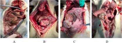
There was no obvious inflammation in the abdominal cavity and no edema and hyperemia in the intestinal tract of rats in the control group (Figure 1A), while the rats in SAP (Figure 1B) and 3-AB groups (Figure 1C) had different degrees of ascites, intestinal edema and hyperemia, and rats in the SAP group were more serious.
Figure 1. Changes in abdominal gross organs in CON (A), SAP (B), 3-AB (C), 3-AB-CON (D) are shown. No obvious change was showed in CON group (A). Acute damage of pancreas and intestines were showed in SAP group (B), with bloody ascites. 3-AB significantly attenuates the damage of abdominal gross organs.
3.2. PARP Inhibition Reduced Pancreatic Enzymes and Histology
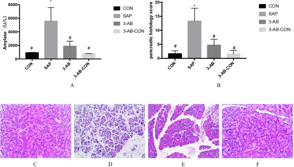
Figure 2. Changes in serum amylase level (A). Values represent mean ± SD,n=10. *p<0.05 vs. CON group, #p<0.05 vs. SAP group. Pancreatic morphological changes of pancreas (C-F) are shown. No histological changes were observed in the pancreas tissue from CON group (C). serious damage of the pancreas was observed in SAP group (D). 3-AB reduced the damage of pancreas significantly (E). Comparison of the pathological score of pancreas (B). Original magnification: x200.
Cerulein infusion increased serum amylase. 3-AB pre-treatment attenuated such increases of observation (Figure 2A). Control rats showed little morphological changes of pancreatic injury (Figure 2C). A substantial increase of pancreatic morphological changes was found in after cerulein infusion,with pancreatic tissue diffuse interlobular space, intercellular space expansion, multi-area cell necrosis, hemorrhage and fat necrosis, multi-lobular inflammation and vascular inflammatory cell infiltration (Figure 2D). Pancreatic damage was attenuated by 3-AB (Figure 2E), as shown histologically and by the scores (Figure 2B).
3.3. PARP Inhibition Attenuated Intestinal Injury and the Expression of Occludin Protein
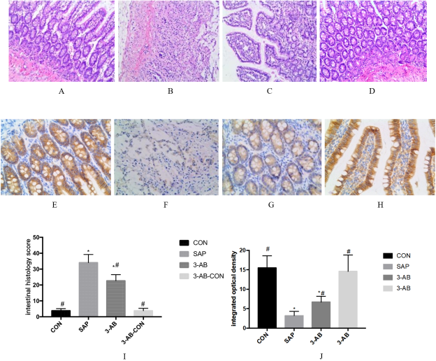
Cerulein infusion caused severe destruction with the epithelial cells of the intestinal mucosa were disordered, the villous morphology was irregular, and inflammatory cells infiltrated in the intestinal mucosa (Figure 3B). 3-AB pre-treatment apparently reduced intestinal histological damages (Figure 3C).
Figure 3. Morphologic changes of rat intestines and immunohistochemical staining for Occludin protein on intestinal mucosal barrier are shown (A-H). No histological changes were observed in the intestinal tissue from CON group (A). 3-AB attenuates the damage of intestines and the expression of Occludin protein (BCFG). Comparison of the pathological score of intestines and expression of Occludin protein on intestinal mucosal barrier (I-J). Original magnification: x200 (A-D) x400 (E-H).
Rat model of acute intestinal injury at 12h after infusion of cerulein to determine the effect 3-AB on Occludin, because of progressive intestinal injury. Occludin expression was assessed by immunohistochemistry: Occludin positive (yellow and brown) granules were mainly found in the intestinal epithelial cell membrance. Occludin levels were significantly lower in the SAP group compared with control group (Figure 3E, F). The decrease of Occludin immunoreactivity caused by infusion of cerulein was attenuated by 3-AB (Figure 3H).
3.4. PARP Inhibition Reduced Intestinal PARP-1, NF-kB and Cytokins Activation
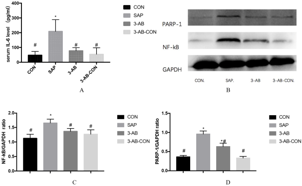
PARP-1 and NF-kB protein levels in intestines were evaluated by Western blot: lower expression was observed in the Control group; meanwhile, the PARP-1 and NF-kB protein was detected in the SAP group and 3-AB group, with the SAP group showing significantly higher amounts compared with the 3-AB group (p<0.05) (Figure 4B-D). cerulein infusion increased the level of IL-6 in serum. 3-AB significantly reduced the level of IL-6 in serum (Figure 4A).
Figure 4. Changes in serum IL-6 level (A). Values represent mean ± SD,n=10. *p<0.05 vs. CON group, #p<0.05 vs. SAP group. Western blot membranes showing PARP-1 and NF-kB protein in intestinal tissue of CON, SAP, 3-AB and 3-AB-CON (B). Treatment of 3-AB reduced the expression of PARP-1, NF-kB protein in intestinal tissue. C and D, quantitation of B. Data are mean ± SD.
4. Discussion
Severe acute pancreatitis accounts for 20-30% of the total cases of acute pancreatitis. It has the characteristics of acute onset, rapid progress, many complications and high mortality. The main manifestations of SAP patients are systemic inflammatory response syndrome (SIRS) and MODS. The mortality rate is about 15%. During the onset of SIRS, inflammatory cells activate, triggering an inflammatory cascade reaction, leading to SIRS
[6]
Pandol SJ, Saluja AK, Imrie CW, et al. Acute pancreatitis: bench to the bedside [J]. Gastroenterology, 2007, 132(3): 1127-1151.
[7]
Beger H G, Rau B, Isenmann R. Natural history of necrotizing pancreatitis. [J]. Pancreatology, 2003, 3(2): 93-101.
[6, 7]
. With the expansion of inflammation, inflammatory factors such as IL-2, IL-6 and TNF can damage remote organs other than pancreas
[8]
Pooran N, Indaram A, Singh P, et al. Cytokines (IL-6, IL-8, TNF): early and reliable predictors of severe acute pancreatitis. [J]. Journal of Clinical Gastroenterology, 2003, 37(3): 263-266.
[8]
, like lung, liver, kidney, intestine and other important organs, and eventually evolve into MODS, leading to death of patients. Small intestine is one of the distant damaged organs. When SAP occurs, microcirculatory disturbance leads to fluid gathering in the third space, blood volume decreases, blood flow redistribution in the whole body, blood supply of visceral vessels, especially intestinal canal, decreases sharply and shows a state of hypoperfusion. Finally, ischemia-reperfusion injury is the main cause of intestinal mucosal barrier damage
[9]
Ammori B J, Leeder P C, King R F, et al. Early increase in intestinal permeability in patients with severe acute pancreatitis: correlation with endotoxemia, organ failure and death [J]. Journal of Gastrointestinal Surgery, 1999, 3(3): 252-262.
[10]
Rahman SH, Ammori BJ, Holmfield J, et al. Intestinal hypoperfusion contributes to gut barrier failure in severe acute pancreatitis. [J]. Journal of Gastrointestinal Surgery, 2003, 7(1): 26-36.
[11]
Capurso G, Zerboni G, Signoretti M, et al. Role of the Gut Barrier in Acute Pancreatitis [J]. Journal of Clinical Gastroenterology, 2012, 46(suppl): S46.
[9-11]
. Intestinal mucosal barrier includes mechanical barrier, immune barrier, chemical barrier, immune barrier and so on. When SAP occurs, the most obvious damage is caused by mechanical barrier, which mainly consists of intestinal epithelial cells and cell-to-cell junctions. Intercellular junctions include tight junctions, adhesion junctions, gap junctions, etc. The main proteins of intestinal epithelial cell-to-cell junctions are Occludin, ZO-1, claudin and so on. Occludin is the main one. One of the functional proteins plays an important role in maintaining normal intestinal mucosal barrier
[12]
Keysami M A, Mohammadpour M. Effect of Bacillus subtilis on Aeromonas hydrophila infection resistance in juvenile freshwater prawn, Macrobrachium rosenbergii (de Man) [J]. Aquaculture International, 2013, 21(3): 553-562.
[13]
Li W, Sun K, Ji Y, et al. Glycine Regulates Expression and Distribution of Claudin-7 and ZO-3 Proteins in Intestinal Porcine Epithelial Cells [J]. Journal of Nutrition, 2016, 146(5): 964-969.
[12, 13]
. Bacterial translocation is the main cause of repeated necrosis of pancreas and sepsis. It is also the second peak period of death in SAP patients
[14]
Fritz S, Hackert T, Hartwig W, et al. Bacterial translocation and infected pancreatic necrosis in acute necrotizing pancreatitis derives from small bowel rather than from colon [J]. American Journal of Surgery, 2010, 200(1): 111-117.
[15]
Simone V. Generoso, Rosana G. Santos, Rosa M. E. Arantes, et al. Protection against increased intestinal permeability and bacterial translocation induced by intestinal obstruction in mice treated with viable and heat-killed Saccharomyces boulardii [J]. European Journal of Nutrition, 2011, 50(4): 261-269.
[14, 15]
.
PARP is a multifunctional protein modifying enzyme family in eukaryotic cells. It is mainly involved in DNA replication and repair, transcription regulation and signal transduction. PARP-1 has the strongest activity, accounting for more than 90% of the total activity of the family. Recent studies have shown that PARP plays an important role in the immune response of pancreatitis, sepsis, multiple injuries and other diseases. In severe infections, PARP overexpression can regulate the entry of multipotent transcription regulator NF-kB into the nucleus, bind to the site of NF-kB, promote the release of downstream cytokines IL-6 and TNF-a, and further activate the release of NF-kB, form positive feedback and release a large number of cytokines. Inflammatory factors and cascade cascade cascade reaction of cytokine network lead to intestinal mucosal barrier damage, ectopic flora, further lead to systemic inflammatory response and multiple organ dysfunction, and even death
[16]
Krishnakumar R, Kraus W L. PARP-1 Regulates Chromatin Structure and Transcription through a KDM5B-Dependent Pathway [J]. Molecular Cell, 2010, 39(5): 736-749.
[17]
Minelli A, Debiasi S, Brecha N C, et al. PARP1 ADP-ribosylates lysine residues of the core histone tails [J]. Nucleic Acids Research, 2010, 38(19): 6350-6362.
[18]
Hassa PO, Hottiger MO. A role of poly (ADP-ribose) polymerase in NF-kappaB transcriptional activation. [J]. Biological Chemistry, 1999, 380(7-8): 953-959.
[16-18]
. Wang Satellite
[19]
Jia Y, Teng Z, Deng W, et al. Poly (ADP-ribose) polymerase inhibition suppresses inflammation and promotes recovery from adrenal injury in a rat model of acute necrotizing pancreatitis: [J]. Bmc Gastroenterology, 2016, 16(1): 81.
[19]
and other studies showed that the content of PARP in the adrenal glands of rats with severe acute pancreatitis was significantly increased, and PARP-1 inhibitor 3-AB could significantly reduce the adrenal tissue damage in severe acute pancreatitis. Mota
[20]
Ruben A Mota, Francisco SánchezBueno, Luis Saenz, et al. Inhibition of poly (ADP-ribose) polymerase attenuates the severity of acute pancreatitis and associated lung injury [J]. Laboratory investigation; a journal of technical methods and pathology, 2005, 85(10): 1250.
[20]
and other studies showed that PARP content in lung tissue increased significantly in rats with severe acute pancreatitis. PARP-1 inhibitor 3-AB could significantly reduce lung injury in severe acute pancreatitis. However, the effect of SAP on intestinal mucosal barrier remains to be further studied and discussed. The results showed that PARP-1 and NF-kB increased significantly in intestinal tissues of SAP rats, while Occludin protein expression in intestinal mucosa decreased significantly. PARP-1 inhibitor 3-AB could significantly reduce pancreatic and intestinal tissue damage and serum amylase levels.
A large number of pro-inflammatory factors released during SAP are the main causes of intestinal mucosal barrier damage. IL-6 and other pro-inflammatory factors activate neutrophils, causing direct damage, deformation and necrosis of intestinal mucosal epithelial cells. At the same time, pro-inflammatory factors promote the release of oxidants and proteolytic enzymes, aggravate the inflammatory reaction of intestinal mucosa, damage of cell tight junction, resulting in increased intestinal mucosal barrier and permeability
[21]
Liu H, Liu Z, Zhao S, et al. Effect of BML-111 on the intestinal mucosal barrier in sepsis and its mechanism of action [J]. Molecular Medicine Reports, 2015, 12(2): 3101-6.
[21]
. The results showed that the serum level of IL-6 was higher in SAP rats, and PARP-1 inhibitor 3-AB could significantly reduce the serum level of IL-6.
In conclusion, the increased expression of PARP in intestinal tissue of SAP rats plays an important role in intestinal mucosal barrier function and disease progression. PARP inhibition can inhibiting the activation of NF-kB and increasing the expression of Occludin protein in intestinal epithelial cells. 3-AB can provide feasible treatment for the prevention and treatment of SAP intestinal mucosal barrier injury.
Abbreviations
PARPP
Poly (ADP-Ribose) Polymerase
SAP
Severe Acute Pancreatitis
Author Contributions
Xinting Pan: Conceptualization, Resources
Yunpeng Zhu: Data curation, Methodology
Yan Liu: Conceptualization, Data curation
Tianjiao Lin: Data curation, Resources, Methodology
Zerem E. Treatment of severe acute pancreatitis and its complications [J]. World Journal of Gastroenterology, 2014, 20(38): 13879-13892.
[3]
Virág L, Szabó C. The therapeutic potential of poly (ADP-ribose) polymerase inhibitors [J]. Pharmacological Reviews, 2002, 54(54): 375-429.
[4]
Rosado M M, Bennici E, Novelli F, et al. Beyond DNA repair, the immunological role of PARP-1 and its siblings [J]. Immunology, 2013, 139(4): 428-437.
[5]
Lin Z S, Ku C F, Guan Y F, et al. Dihydro-Resveratrol Ameliorates Lung Injury in Rats with Cerulein-Induced Acute Pancreatitis. [J]. Phytotherapy Research, 2016, 30(4): 663-670.
Pandol SJ, Saluja AK, Imrie CW, et al. Acute pancreatitis: bench to the bedside [J]. Gastroenterology, 2007, 132(3): 1127-1151.
[7]
Beger H G, Rau B, Isenmann R. Natural history of necrotizing pancreatitis. [J]. Pancreatology, 2003, 3(2): 93-101.
[8]
Pooran N, Indaram A, Singh P, et al. Cytokines (IL-6, IL-8, TNF): early and reliable predictors of severe acute pancreatitis. [J]. Journal of Clinical Gastroenterology, 2003, 37(3): 263-266.
[9]
Ammori B J, Leeder P C, King R F, et al. Early increase in intestinal permeability in patients with severe acute pancreatitis: correlation with endotoxemia, organ failure and death [J]. Journal of Gastrointestinal Surgery, 1999, 3(3): 252-262.
[10]
Rahman SH, Ammori BJ, Holmfield J, et al. Intestinal hypoperfusion contributes to gut barrier failure in severe acute pancreatitis. [J]. Journal of Gastrointestinal Surgery, 2003, 7(1): 26-36.
[11]
Capurso G, Zerboni G, Signoretti M, et al. Role of the Gut Barrier in Acute Pancreatitis [J]. Journal of Clinical Gastroenterology, 2012, 46(suppl): S46.
[12]
Keysami M A, Mohammadpour M. Effect of Bacillus subtilis on Aeromonas hydrophila infection resistance in juvenile freshwater prawn, Macrobrachium rosenbergii (de Man) [J]. Aquaculture International, 2013, 21(3): 553-562.
[13]
Li W, Sun K, Ji Y, et al. Glycine Regulates Expression and Distribution of Claudin-7 and ZO-3 Proteins in Intestinal Porcine Epithelial Cells [J]. Journal of Nutrition, 2016, 146(5): 964-969.
[14]
Fritz S, Hackert T, Hartwig W, et al. Bacterial translocation and infected pancreatic necrosis in acute necrotizing pancreatitis derives from small bowel rather than from colon [J]. American Journal of Surgery, 2010, 200(1): 111-117.
[15]
Simone V. Generoso, Rosana G. Santos, Rosa M. E. Arantes, et al. Protection against increased intestinal permeability and bacterial translocation induced by intestinal obstruction in mice treated with viable and heat-killed Saccharomyces boulardii [J]. European Journal of Nutrition, 2011, 50(4): 261-269.
[16]
Krishnakumar R, Kraus W L. PARP-1 Regulates Chromatin Structure and Transcription through a KDM5B-Dependent Pathway [J]. Molecular Cell, 2010, 39(5): 736-749.
[17]
Minelli A, Debiasi S, Brecha N C, et al. PARP1 ADP-ribosylates lysine residues of the core histone tails [J]. Nucleic Acids Research, 2010, 38(19): 6350-6362.
[18]
Hassa PO, Hottiger MO. A role of poly (ADP-ribose) polymerase in NF-kappaB transcriptional activation. [J]. Biological Chemistry, 1999, 380(7-8): 953-959.
[19]
Jia Y, Teng Z, Deng W, et al. Poly (ADP-ribose) polymerase inhibition suppresses inflammation and promotes recovery from adrenal injury in a rat model of acute necrotizing pancreatitis: [J]. Bmc Gastroenterology, 2016, 16(1): 81.
[20]
Ruben A Mota, Francisco SánchezBueno, Luis Saenz, et al. Inhibition of poly (ADP-ribose) polymerase attenuates the severity of acute pancreatitis and associated lung injury [J]. Laboratory investigation; a journal of technical methods and pathology, 2005, 85(10): 1250.
[21]
Liu H, Liu Z, Zhao S, et al. Effect of BML-111 on the intestinal mucosal barrier in sepsis and its mechanism of action [J]. Molecular Medicine Reports, 2015, 12(2): 3101-6.
Pan, X., Zhu, Y., Liu, Y., Lin, T., Li, A., et al. (2026). Effect of Poly(ADP-ribose) Polymerase Inhibition on the Injury of Intestinal Mucosal Barrier in Rat Model with Severe Acute Pancreatitis. American Journal of Internal Medicine, 14(3), 50-56. https://doi.org/10.11648/j.ajim.20261403.12
Pan, X.; Zhu, Y.; Liu, Y.; Lin, T.; Li, A., et al. Effect of Poly(ADP-ribose) Polymerase Inhibition on the Injury of Intestinal Mucosal Barrier in Rat Model with Severe Acute Pancreatitis. Am. J. Intern. Med.2026, 14(3), 50-56. doi: 10.11648/j.ajim.20261403.12
Pan X, Zhu Y, Liu Y, Lin T, Li A, et al. Effect of Poly(ADP-ribose) Polymerase Inhibition on the Injury of Intestinal Mucosal Barrier in Rat Model with Severe Acute Pancreatitis. Am J Intern Med. 2026;14(3):50-56. doi: 10.11648/j.ajim.20261403.12
@article{10.11648/j.ajim.20261403.12,
  author = {Xinting Pan and Yunpeng Zhu and Yan Liu and Tianjiao Lin and Ainqin Li and Qingyun Zhu},
  title = {Effect of Poly(ADP-ribose) Polymerase Inhibition on the Injury of Intestinal Mucosal Barrier in Rat Model with Severe Acute Pancreatitis},
  journal = {American Journal of Internal Medicine},
  volume = {14},
  number = {3},
  pages = {50-56},
  doi = {10.11648/j.ajim.20261403.12},
  url = {https://doi.org/10.11648/j.ajim.20261403.12},
  eprint = {https://article.sciencepublishinggroup.com/pdf/10.11648.j.ajim.20261403.12},
  abstract = {Background: The injury of intestinal mucosal barrier in the later period of severe acute pancreatitis (SAP) results in flora migration with the endotoxin and bacterium entering blood circulation, final result of bacteremia, multiple organ dysfunction syndrome (MODS), eventually death. poly (ADP-ribose) polymerase-1(PARP-1) is a nuclear DNA-binding protein that has been revealed a wide role in DNA repair, chromatin structure, necrosis, inflammation and immune function. We plan to determine whether PARP-1 and its inhibition affect the injury of intestinal mucosal barrier in SAP. Methods: SAP was induced by 6 hourly intraperitoneal injection of cerulein (50ug/kg), with lipopolysaccharide (10mg/kg) in the last time. At 30 min prior, rats were treated with the PARP inhibition 3-Aminobenzamide (3-AB) or normal saline. 40 rats were randomly separated into 4 groups of a normal control group, a SAP group, a SAP with inhibition group and an inhibition group. After 12 hours, all rats were killed. We compared serum amylase, interleukin-6 (IL-6), celiac morphological changes, pancreatic pathological injury, intestinal histology. Activities of PARP-1, nuclear factor-kappa B (NF-kB), Occludin in the intestine were also examined. Result: The inflammation and injury has been showed seriously in the intestines. with SAP, the expression of PARP-1 and NF-kB also has been shown high level. But with administration of PARP inhibitor, 3-AB, the severity of SAP and pancreatitis-associated intestinal injury was significantly attenuated, the protein expression of PARP-1 and NF-kB was significantly decreased. Conclusion: Our findings indicate that PARP inhibitors played a positive role in the injury of intestinal mucosal barrier in rat model with SAP.},
 year = {2026}
}
TY  - JOUR
T1  - Effect of Poly(ADP-ribose) Polymerase Inhibition on the Injury of Intestinal Mucosal Barrier in Rat Model with Severe Acute Pancreatitis
AU  - Xinting Pan
AU  - Yunpeng Zhu
AU  - Yan Liu
AU  - Tianjiao Lin
AU  - Ainqin Li
AU  - Qingyun Zhu
Y1  - 2026/08/24
PY  - 2026
N1  - https://doi.org/10.11648/j.ajim.20261403.12
DO  - 10.11648/j.ajim.20261403.12
T2  - American Journal of Internal Medicine
JF  - American Journal of Internal Medicine
JO  - American Journal of Internal Medicine
SP  - 50
EP  - 56
PB  - Science Publishing Group
SN  - 2330-4324
UR  - https://doi.org/10.11648/j.ajim.20261403.12
AB  - Background: The injury of intestinal mucosal barrier in the later period of severe acute pancreatitis (SAP) results in flora migration with the endotoxin and bacterium entering blood circulation, final result of bacteremia, multiple organ dysfunction syndrome (MODS), eventually death. poly (ADP-ribose) polymerase-1(PARP-1) is a nuclear DNA-binding protein that has been revealed a wide role in DNA repair, chromatin structure, necrosis, inflammation and immune function. We plan to determine whether PARP-1 and its inhibition affect the injury of intestinal mucosal barrier in SAP. Methods: SAP was induced by 6 hourly intraperitoneal injection of cerulein (50ug/kg), with lipopolysaccharide (10mg/kg) in the last time. At 30 min prior, rats were treated with the PARP inhibition 3-Aminobenzamide (3-AB) or normal saline. 40 rats were randomly separated into 4 groups of a normal control group, a SAP group, a SAP with inhibition group and an inhibition group. After 12 hours, all rats were killed. We compared serum amylase, interleukin-6 (IL-6), celiac morphological changes, pancreatic pathological injury, intestinal histology. Activities of PARP-1, nuclear factor-kappa B (NF-kB), Occludin in the intestine were also examined. Result: The inflammation and injury has been showed seriously in the intestines. with SAP, the expression of PARP-1 and NF-kB also has been shown high level. But with administration of PARP inhibitor, 3-AB, the severity of SAP and pancreatitis-associated intestinal injury was significantly attenuated, the protein expression of PARP-1 and NF-kB was significantly decreased. Conclusion: Our findings indicate that PARP inhibitors played a positive role in the injury of intestinal mucosal barrier in rat model with SAP.
VL  - 14
IS  - 3
ER  -
Pan, X., Zhu, Y., Liu, Y., Lin, T., Li, A., et al. (2026). Effect of Poly(ADP-ribose) Polymerase Inhibition on the Injury of Intestinal Mucosal Barrier in Rat Model with Severe Acute Pancreatitis. American Journal of Internal Medicine, 14(3), 50-56. https://doi.org/10.11648/j.ajim.20261403.12
Pan, X.; Zhu, Y.; Liu, Y.; Lin, T.; Li, A., et al. Effect of Poly(ADP-ribose) Polymerase Inhibition on the Injury of Intestinal Mucosal Barrier in Rat Model with Severe Acute Pancreatitis. Am. J. Intern. Med.2026, 14(3), 50-56. doi: 10.11648/j.ajim.20261403.12
Pan X, Zhu Y, Liu Y, Lin T, Li A, et al. Effect of Poly(ADP-ribose) Polymerase Inhibition on the Injury of Intestinal Mucosal Barrier in Rat Model with Severe Acute Pancreatitis. Am J Intern Med. 2026;14(3):50-56. doi: 10.11648/j.ajim.20261403.12
@article{10.11648/j.ajim.20261403.12,
  author = {Xinting Pan and Yunpeng Zhu and Yan Liu and Tianjiao Lin and Ainqin Li and Qingyun Zhu},
  title = {Effect of Poly(ADP-ribose) Polymerase Inhibition on the Injury of Intestinal Mucosal Barrier in Rat Model with Severe Acute Pancreatitis},
  journal = {American Journal of Internal Medicine},
  volume = {14},
  number = {3},
  pages = {50-56},
  doi = {10.11648/j.ajim.20261403.12},
  url = {https://doi.org/10.11648/j.ajim.20261403.12},
  eprint = {https://article.sciencepublishinggroup.com/pdf/10.11648.j.ajim.20261403.12},
  abstract = {Background: The injury of intestinal mucosal barrier in the later period of severe acute pancreatitis (SAP) results in flora migration with the endotoxin and bacterium entering blood circulation, final result of bacteremia, multiple organ dysfunction syndrome (MODS), eventually death. poly (ADP-ribose) polymerase-1(PARP-1) is a nuclear DNA-binding protein that has been revealed a wide role in DNA repair, chromatin structure, necrosis, inflammation and immune function. We plan to determine whether PARP-1 and its inhibition affect the injury of intestinal mucosal barrier in SAP. Methods: SAP was induced by 6 hourly intraperitoneal injection of cerulein (50ug/kg), with lipopolysaccharide (10mg/kg) in the last time. At 30 min prior, rats were treated with the PARP inhibition 3-Aminobenzamide (3-AB) or normal saline. 40 rats were randomly separated into 4 groups of a normal control group, a SAP group, a SAP with inhibition group and an inhibition group. After 12 hours, all rats were killed. We compared serum amylase, interleukin-6 (IL-6), celiac morphological changes, pancreatic pathological injury, intestinal histology. Activities of PARP-1, nuclear factor-kappa B (NF-kB), Occludin in the intestine were also examined. Result: The inflammation and injury has been showed seriously in the intestines. with SAP, the expression of PARP-1 and NF-kB also has been shown high level. But with administration of PARP inhibitor, 3-AB, the severity of SAP and pancreatitis-associated intestinal injury was significantly attenuated, the protein expression of PARP-1 and NF-kB was significantly decreased. Conclusion: Our findings indicate that PARP inhibitors played a positive role in the injury of intestinal mucosal barrier in rat model with SAP.},
 year = {2026}
}
TY  - JOUR
T1  - Effect of Poly(ADP-ribose) Polymerase Inhibition on the Injury of Intestinal Mucosal Barrier in Rat Model with Severe Acute Pancreatitis
AU  - Xinting Pan
AU  - Yunpeng Zhu
AU  - Yan Liu
AU  - Tianjiao Lin
AU  - Ainqin Li
AU  - Qingyun Zhu
Y1  - 2026/08/24
PY  - 2026
N1  - https://doi.org/10.11648/j.ajim.20261403.12
DO  - 10.11648/j.ajim.20261403.12
T2  - American Journal of Internal Medicine
JF  - American Journal of Internal Medicine
JO  - American Journal of Internal Medicine
SP  - 50
EP  - 56
PB  - Science Publishing Group
SN  - 2330-4324
UR  - https://doi.org/10.11648/j.ajim.20261403.12
AB  - Background: The injury of intestinal mucosal barrier in the later period of severe acute pancreatitis (SAP) results in flora migration with the endotoxin and bacterium entering blood circulation, final result of bacteremia, multiple organ dysfunction syndrome (MODS), eventually death. poly (ADP-ribose) polymerase-1(PARP-1) is a nuclear DNA-binding protein that has been revealed a wide role in DNA repair, chromatin structure, necrosis, inflammation and immune function. We plan to determine whether PARP-1 and its inhibition affect the injury of intestinal mucosal barrier in SAP. Methods: SAP was induced by 6 hourly intraperitoneal injection of cerulein (50ug/kg), with lipopolysaccharide (10mg/kg) in the last time. At 30 min prior, rats were treated with the PARP inhibition 3-Aminobenzamide (3-AB) or normal saline. 40 rats were randomly separated into 4 groups of a normal control group, a SAP group, a SAP with inhibition group and an inhibition group. After 12 hours, all rats were killed. We compared serum amylase, interleukin-6 (IL-6), celiac morphological changes, pancreatic pathological injury, intestinal histology. Activities of PARP-1, nuclear factor-kappa B (NF-kB), Occludin in the intestine were also examined. Result: The inflammation and injury has been showed seriously in the intestines. with SAP, the expression of PARP-1 and NF-kB also has been shown high level. But with administration of PARP inhibitor, 3-AB, the severity of SAP and pancreatitis-associated intestinal injury was significantly attenuated, the protein expression of PARP-1 and NF-kB was significantly decreased. Conclusion: Our findings indicate that PARP inhibitors played a positive role in the injury of intestinal mucosal barrier in rat model with SAP.
VL  - 14
IS  - 3
ER  -





 Share This Article
 Twitter
 Linked In
 Facebook
 Copy
Copy Download