Introduction: Giant emphysematous bulla is defined as an intrapulmonary air collection occupying 30% or more of a hemithorax. Klingman's classification categorizes bullae according to the quality of the underlying lung parenchyma, with type 1 characterized by a giant bulla surrounded by normal lung. Case Report: Female patient, 54 years old, black woman, from an urban area, with no history of smoking, tuberculosis or chronic obstructive pulmonary disease, with a history of controlled arterial hypertension for approximately 10 years, who started about three months ago with slight progressive dyspnea associated with mild exertion, accompanied by chest discomfort. After normal cardiological examinations, chest radiography revealed increased hyperlucency in the right hemithorax. At the general surgery consultation, the respiratory physical examination demonstrated decreased chest expansion, decreased vocal vibrations and decreased vesicular breath sounds in the right hemithorax, with respiratory rate of 22 breaths/minute. Chest radiography confirmed a type 1 emphysematous bulla according to Klingman's classification, and chest computed tomography confirmed the radiological diagnosis. The patient underwent open bullectomy by the Naclerio and Langer technique (1947). The patient remained hospitalized for 14 days due to a postoperative pleural effusion that was promptly treated, with favorable post-operative course. Discussion: This case is particularly relevant as it represents an idiopathic giant emphysematous bulla in a non-smoker female patient, with no history of tuberculosis or chronic obstructive pulmonary disease, resulting inthe first case report of this pathology in Angola. The epidemiological literature review in Africa revealed few reported cases, predominantly associated with spontaneous pneumothorax or other pulmonary comorbidities. Conclusion: Bullectomy by the Naclerio and Langer technique constitutes an effective surgical approach for the treatment of type 1 giant emphysematous bullae, even in patients without classical risk factors. This case represents the first documented report of this pathology in Angola, contributing to the knowledge of the disease's epidemiology in Africa.
This is an Open Access article, distributed under the terms of the Creative Commons Attribution 4.0 International License (http://creativecommons.org/licenses/by/4.0/), which permits unrestricted use, distribution and reproduction in any medium or format, provided the original work is properly cited.
Giant Emphysematous Bulla, Bullectomy, Naclerio and Langer Technique, Klingman Classification, Angola, Africa
1. Introduction
Emphysematous bulla is defined as an intrapulmonary air space resulting from the destruction, dilation and confluence of air spaces distal to the terminal bronchioles, with diameter greater than 1 cm
[1]
Thoracic Key. Bullous Disease of the Lung. 2017. Available at:
. Klingman's classification categorizes bullae according to the quality of the underlying lung parenchyma
[3]
Azzopardi M, Attard D, Catania R, et al. Minimally invasive resection of a marijuana-associated giant bulla: A case report. Malta Medical Journal. 2021; 33(2). Available at:
Type 1: Giant bulla surrounded by normal lung, representing the most favorable form for surgical treatment, as the underlying parenchyma is functional and can reexpand after resection.
Type 2: Bulla surrounded by emphysematous lung with multiple other bullae, less favorable surgical prognosis.
Type 3: Vanishing lung syndrome, the most extreme form, with massive destruction of the parenchyma, not amenable to curative surgical treatment.
The surgical treatment of giant emphysematous bullae was established by Naclerio and Langer in 1947, who described the standard method of bullectomy and plicature without lobectomy
[4]
Verma RK, Nishiki M, Mukai M, et al. Intracavitary Drainage Procedure for Giant Bullae in Compromised Patients. Hiroshima J Med Sci. 1991; 40(4): 115-118.
[4]
. Since then, the technique has been widely adopted and modified, with the development of surgical staplers that facilitated the performance of the procedure by video-assisted thoracoscopic surgery
[5]
Matsumoto T, Omata M, Shidei H, et al. Video-assisted thoracoscopic surgery (VATS) bullectomy with polyglycolic-acid sheet for preventing postoperative residual cavities. Tokyo Women's Medical University Journal. 2022; 6: 2022010.
[5]
. The surgical indications include dyspneic patients with high residual volume and hyperinflation, emergent complications (pneumothorax, infection, hemorrhage), and as a prophylactic measure in bullae occupying more than 50% of a hemithorax
[3]
Azzopardi M, Attard D, Catania R, et al. Minimally invasive resection of a marijuana-associated giant bulla: A case report. Malta Medical Journal. 2021; 33(2). Available at:
. The present case report describes a patient with giant emphysematous bulla type 1 (Klingman) surgically treated by the Naclerio and Langer technique, discussing the diagnostic, therapeutic aspects and the epidemiological relevance of this case as the first documented report in Angola.
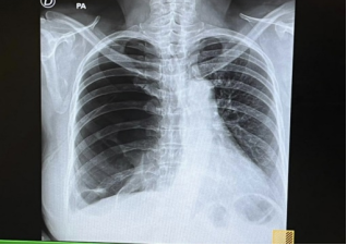
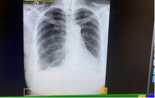
Figure 1. Marked radiotransparency is observed in the right hemithorax with collapsed lung parenchyma.
Female patient, 54 years of age, black woman, urban origin. With personal history of controlled arterial hypertension for approximately 10 years, denies family pathological history, denies smoking, tuberculosis, chronic obstructive pulmonary disease. The patient was apparently in good health until approximately three months before the consultation, when she presented withslight progressive dyspnea, associated with minimal exertion and accompanied by chest discomfort. Given the persistence of symptoms, the patient sought the cardiology services of the General Hospital of Benguela, where she underwent an electrocardiogram and echocardiogram, both without significant alterations. Due to persistence of symptoms, the patient was submitted to a chest radiography, which revealed increased hyperlucency in the right hemithorax, initially interpreted as an emphysematous bulla.
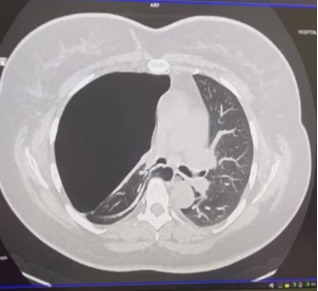
Figure 2. A large homogeneous hypodense air-density area is seen occupying most of the right hemitorax, with absence of pulmonary vascular markings, bounded by the visceral pleural line, and associated compression and retraction of the adjacent lung parenchyma.
Given the radiological finding, the patient was referred to the general surgery services for outpatient evaluation. Physical Examination was done at the General Surgery Consultation: during the physical examination the following was found in the respiratory system: respiratory rate of 22 breaths/minute, decreased chest expansion in the, decreased vocal vibrations and decreased vesicular breaths sounds all in the right hemithorax. The patient was hospitalized and submitted to chest radiography, which confirmed the presence of a type 1 emphysematous bulla according to Klingman's classification
[3]
Azzopardi M, Attard D, Catania R, et al. Minimally invasive resection of a marijuana-associated giant bulla: A case report. Malta Medical Journal. 2021; 33(2). Available at:
, giant bulla surrounded by normal lung, without evidence of associated diffuse emphysema. Chest computed tomography confirmed the previous radiological diagnosis, observing a giant emphysematous bulla of type 1, as already described in the radiography. CT allowed detailed characterization of the lesion, confirming the absence of diffuse emphysema in the adjacent parenchyma and the viability of surgical treatment.
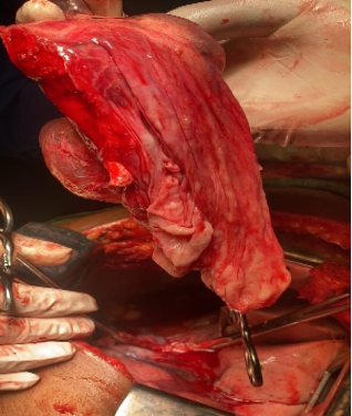
Figure 4. Surgical specimen obtained after bullectomy through a right posterolateral thoracotomy. The specimen demonstrates a giant thin-walled pulmonary bulla arising from the subpleural surface of the lung.
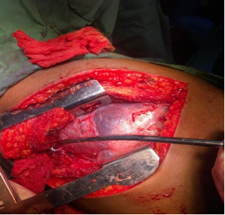
The patient was prepared for elective surgery. Preoperative examinations were performed, which showed no alterations of note. Anesthetic evaluation was performed and informed consent was obtained for the surgical procedure. Intraoperatively, an open bullectomy was performed by the Naclerio and Langer technique (1947)
[4]
Verma RK, Nishiki M, Mukai M, et al. Intracavitary Drainage Procedure for Giant Bullae in Compromised Patients. Hiroshima J Med Sci. 1991; 40(4): 115-118.
[5]
Matsumoto T, Omata M, Shidei H, et al. Video-assisted thoracoscopic surgery (VATS) bullectomy with polyglycolic-acid sheet for preventing postoperative residual cavities. Tokyo Women's Medical University Journal. 2022; 6: 2022010.
[4, 5]
. The Naclerio and Langer technique consists of: thoracic access by thoracotomy at the appropriate intercostal space; longitudinal opening of the bulla; identification and closure of bronchial orifices by "X" sutures (mattress sutures); plicature of the bulla base with "through-and-through" sutures (through-and-through mattress sutures) through healthy lung tissue to obtain hermetic sealing; resection of the bulla wall up to the previous suture line; and imbrication of the lung surface with the visceral pleura, suturing the edges of the resected bulla wall.
The patient remained hospitalized for 14 days, during which she presented as a postoperative complication a pleural effusion, which was promptly treated. After adequate treatment of the complication, the patient presented favorable postoperative course, with progressive recovery of respiratory function and resolution of dyspneic symptoms.
Figure 5. The follow-up chest radiography of the patient on the sixth postoperative day.
3. Discussion
3.1. Epidemiology and Etiology
Giant emphysematous bullae occur predominantly in male patients, smokers, over 50 years of age. However, they can occur in female patients and in non-smokers, as in the reported case
[2]
Siddiqui NA. Bullous Emphysema. StatPearls. Treasure Island (FL): StatPearls Publishing; 2025. Available at:
. The etiology of emphysematous bullae is associated with various factors, including smoking, alpha-1 antitrypsin deficiency, intravenous drug use and, more recently, marijuana use, which appears to have greater potential to cause giant bullae compared to tobacco
[3]
Azzopardi M, Attard D, Catania R, et al. Minimally invasive resection of a marijuana-associated giant bulla: A case report. Malta Medical Journal. 2021; 33(2). Available at:
The epidemiological literature review in Africa revealed few case reports of giant emphysematous bullae. In Nigeria, Anumenechi et al.
[10]
Anumenechi N, Adamu YB, Alioke II, et al. Uniportal Video-Assisted ThoracoscopicBullectomy for Recurrent Spontaneous Pneumothorax: A First Experience Case Report in a Private Hospital. African Journal of Thoracic and Cardiovascular Surgery. 2025; 8(1): 123-128.
[10]
reported a case of bullectomy by uniportal VATS for recurrent spontaneous pneumothorax in a 25-year-old patient with bilateral apical bullae, but without characterization as an isolated giant emphysematous bulla. In South Africa, the African Journal of Emergency Medicine
[8]
African Journal of Emergency Medicine. Please do not put a chest drain in my chest! Vanishing lung syndrome. 2020; 10(4): 193-194.
[8]
published a case of vanishing lung syndrome in a 61-year-old male patient with severe COPD, not representing an idiopathic giant emphysematous bulla. In Saudi Arabia, Hamad et al.
[11]
Hamad AMM, Nosseir MM, Alorainy SM. Unilateral Giant Bullae: Pulmonary Placental Transmogrification Should Be Kept in Mind: Case Reports. J Chest Surg. 2021; 54(4): 329-333.
[11]
described two cases of pulmonary placental transmogrification in non-smoker female patients (one African of 25 years and one Jordanian of 28 years), which presented as unilateral giant bullae, although the histopathological diagnosis differed from the classic giant emphysematous bulla.
3.3. Pathophysiology
Giant emphysematous bullae affect gas exchange in multiple ways: ventilation-perfusion mismatch, by the destruction of alveolar walls that creates a bulla with minimal blood supply; compression of adjacent parenchyma, which has better blood supply, limiting ventilation in these air spaces; and reduction of pulmonary compliance, by the destruction of air space walls that decreases elasticity, increasing lung compliance and leading to preferential air entry into the bulla during ventilation
[3]
Azzopardi M, Attard D, Catania R, et al. Minimally invasive resection of a marijuana-associated giant bulla: A case report. Malta Medical Journal. 2021; 33(2). Available at:
Azzopardi M, Attard D, Catania R, et al. Minimally invasive resection of a marijuana-associated giant bulla: A case report. Malta Medical Journal. 2021; 33(2). Available at:
. Type 1 presents an isolated giant bulla with normal underlying lung and excellent surgical prognosis with complete reexpansion. Type 2 presents multiple bullae with emphysematous lung and moderate prognosis with partial benefit. Type 3 corresponds to vanishing lung syndrome with massive destruction of the parenchyma and reserved prognosis, not amenable to curative surgery. The patient in the reported case presented a type 1 bulla, which justifies the surgical indication and the excellent expected prognosis.
3.5. Surgical Treatment
Bullectomy is indicated in symptomatic patients with bullae occupying more than one third of the hemithorax, or more than 50% as a prophylactic measure
[3]
Azzopardi M, Attard D, Catania R, et al. Minimally invasive resection of a marijuana-associated giant bulla: A case report. Malta Medical Journal. 2021; 33(2). Available at:
. The Naclerio and Langer technique, described in 1947, remains the reference in the treatment of giant emphysematous bullae
[4]
Verma RK, Nishiki M, Mukai M, et al. Intracavitary Drainage Procedure for Giant Bullae in Compromised Patients. Hiroshima J Med Sci. 1991; 40(4): 115-118.
[12]
Li C, Hu X, Jiang G, Jiang YL. Case Report: A case of giant emphysematous bullae successfully treated with percutaneous aspiration and sclerotherapy for staged reduction of pulmonary bullae. Front Med. 2025; 12: 1693942.
[4, 12]
. The modification of the technique with the use of surgical staplers (staplers) and the video-assisted thoracoscopic approach (VATS) has made the procedure less invasive, with reduced morbidity and comparable results to open surgery
[5]
Matsumoto T, Omata M, Shidei H, et al. Video-assisted thoracoscopic surgery (VATS) bullectomy with polyglycolic-acid sheet for preventing postoperative residual cavities. Tokyo Women's Medical University Journal. 2022; 6: 2022010.
[13]
Panagopoulos N, et al. VATS bullectomy and apical pleurectomy for spontaneous pneumothorax in a young patient with Swyer-James-Mc Leod syndrome: case report presentation and literature review focusing on surgically treated cases.
[5, 13]
.
3.6. Postoperative Complications
The postoperative pleural effusion observed in the patient is a recognized complication after bullectomy, occurring in approximately 5-15% of cases. The etiology may be related to pleural irritation by the surgical procedure, inflammatory reaction or small transient bronchopleural fistula. Treatment generally consists of thoracic drainage and, in refractory cases, chemical or mechanical pleurodesis
[7]
Krishnamohan P, Shen KR, Wigle DA, et al. Bullectomy for Symptomatic or Complicated Giant Lung Bullae. Ann Thorac Surg. 2014; 97(2): 425-431.
[7]
.
3.7. Relevance of the Case
The present case presents multiple aspects of clinical and scientific relevance.
3.8. Idiopathic Giant Emphysematous Bulla
The patient does not present any of the classical risk factors for the development of giant emphysematous bullae: absence of smoking, as most cases reported in the literature are associated with chronic smoking
[2]
Siddiqui NA. Bullous Emphysema. StatPearls. Treasure Island (FL): StatPearls Publishing; 2025. Available at:
Adloye D, Basquil C. Estimating the prevalence of COPD in Africa: a systematic review and meta-analysis. J Glob Healyh. 2014; 4(2): 020415.
[2, 14]
; and absence of alpha-1 antitrypsin deficiency, although not formally investigated, the absence of family history and the late age of onset make this etiology less likely. This characterization of "idiopathic" is particularly rare in literature. Garvey et al.
described a similar case of unilateral idiopathic giant emphysematous bulla in a non-smoker female patient, highlighting the rarity of this presentation.
3.9. First Case Report in Angola
After exhaustive review of indexed medical literature, no case reports of giant emphysematous bulla in Angola were identified. This case constitutes, therefore, the first documented report of this pathology in the country.
3.10. Considerations on the Case
The present case highlights the importance of systematic investigation of patients with progressive dyspnea, even when initial cardiological examinations are normal and there are no classical risk factors; the value of respiratory physical examination in the characterization of unilateral thoracic alterations; the need for complementary imaging examinations (chest CT) for diagnostic confirmation and therapeutic planning; the efficacy of bullectomy by the Naclerio and Langer technique in the treatment of type 1 bullae, even in patients without classical risk factors; and the need for surveillance for postoperative complications, such as the pleural effusion observed in this case.
4. Conclusion
Giant emphysematous bulla type 1 (Klingman) in a non-smoker female patient, without history of tuberculosis or chronic obstructive pulmonary disease, represents a rare and idiopathic condition, amenable to surgical treatment with excellent results. The present case illustrates the importance of early diagnosis and the efficacy of open bullectomy by the Naclerio and Langer technique in the definitive treatment of the condition, even in the absence of classical risk factors. The performance of adequate imaging examinations, especially computed tomography, is fundamental for the characterization of the lesion, Klingman classification and adequate therapeutic planning. Surveillance for postoperative complications, such as pleural effusion, is essential for treatment success. This case constitutes the first documented report of giant emphysematous bulla in Angola and one of the few cases of idiopathic giant emphysematous bulla reported in Africa, contributing significantly to the knowledge of the epidemiology of this disease on the African continent.
Abbreviations
VATS
Video-Assisted Thoracic Surgery
CT
Computed Tomography
COPD
Chronic Obstructive Pulmonary Disease
Author Contributions
Maria Nioca: Conceptualization, Methodology, Investigation, Writing – original draft, Supervision, Project Administration, Formal Analysis
Meloise Tchiloia: Conceptualization, Data Curation, Visualization, Project Administration
Eduardo Kedisobua: Resources
Dulcilene De Sousa: Writing – review & editing
Conflicts of Interest
The authors declare no conflict of interest.
References
[1]
Thoracic Key. Bullous Disease of the Lung. 2017. Available at:
Azzopardi M, Attard D, Catania R, et al. Minimally invasive resection of a marijuana-associated giant bulla: A case report. Malta Medical Journal. 2021; 33(2). Available at:
Verma RK, Nishiki M, Mukai M, et al. Intracavitary Drainage Procedure for Giant Bullae in Compromised Patients. Hiroshima J Med Sci. 1991; 40(4): 115-118.
[5]
Matsumoto T, Omata M, Shidei H, et al. Video-assisted thoracoscopic surgery (VATS) bullectomy with polyglycolic-acid sheet for preventing postoperative residual cavities. Tokyo Women's Medical University Journal. 2022; 6: 2022010.
[6]
Marchiori E. Hemitóraxopaco. JornalBrasileiro de Pneumologia. 2017. Available at:
Anumenechi N, Adamu YB, Alioke II, et al. Uniportal Video-Assisted ThoracoscopicBullectomy for Recurrent Spontaneous Pneumothorax: A First Experience Case Report in a Private Hospital. African Journal of Thoracic and Cardiovascular Surgery. 2025; 8(1): 123-128.
[11]
Hamad AMM, Nosseir MM, Alorainy SM. Unilateral Giant Bullae: Pulmonary Placental Transmogrification Should Be Kept in Mind: Case Reports. J Chest Surg. 2021; 54(4): 329-333.
[12]
Li C, Hu X, Jiang G, Jiang YL. Case Report: A case of giant emphysematous bullae successfully treated with percutaneous aspiration and sclerotherapy for staged reduction of pulmonary bullae. Front Med. 2025; 12: 1693942.
[13]
Panagopoulos N, et al. VATS bullectomy and apical pleurectomy for spontaneous pneumothorax in a young patient with Swyer-James-Mc Leod syndrome: case report presentation and literature review focusing on surgically treated cases.
[14]
Adloye D, Basquil C. Estimating the prevalence of COPD in Africa: a systematic review and meta-analysis. J Glob Healyh. 2014; 4(2): 020415.
Nioca, M., Tchiloia, M., Kedisobua, E., Sousa, D. D. (2026). Idiopathic Type I Giant Emphysematous Bulla in a
Non-Smoking Female Patient: The First Reported Case from Angola. Journal of Surgery, 14(4), 76-80. https://doi.org/10.11648/j.js.20261404.12
Nioca, M.; Tchiloia, M.; Kedisobua, E.; Sousa, D. D. Idiopathic Type I Giant Emphysematous Bulla in a
Non-Smoking Female Patient: The First Reported Case from Angola. J. Surg.2026, 14(4), 76-80. doi: 10.11648/j.js.20261404.12
Nioca M, Tchiloia M, Kedisobua E, Sousa DD. Idiopathic Type I Giant Emphysematous Bulla in a
Non-Smoking Female Patient: The First Reported Case from Angola. J Surg. 2026;14(4):76-80. doi: 10.11648/j.js.20261404.12
@article{10.11648/j.js.20261404.12,
  author = {Maria Nioca and Meloise Tchiloia and Eduardo Kedisobua and Dulcilene De Sousa},
  title = {Idiopathic Type I Giant Emphysematous Bulla in a
Non-Smoking Female Patient: The First Reported Case from Angola},
  journal = {Journal of Surgery},
  volume = {14},
  number = {4},
  pages = {76-80},
  doi = {10.11648/j.js.20261404.12},
  url = {https://doi.org/10.11648/j.js.20261404.12},
  eprint = {https://article.sciencepublishinggroup.com/pdf/10.11648.j.js.20261404.12},
  abstract = {Introduction: Giant emphysematous bulla is defined as an intrapulmonary air collection occupying 30% or more of a hemithorax. Klingman's classification categorizes bullae according to the quality of the underlying lung parenchyma, with type 1 characterized by a giant bulla surrounded by normal lung. Case Report: Female patient, 54 years old, black woman, from an urban area, with no history of smoking, tuberculosis or chronic obstructive pulmonary disease, with a history of controlled arterial hypertension for approximately 10 years, who started about three months ago with slight progressive dyspnea associated with mild exertion, accompanied by chest discomfort. After normal cardiological examinations, chest radiography revealed increased hyperlucency in the right hemithorax. At the general surgery consultation, the respiratory physical examination demonstrated decreased chest expansion, decreased vocal vibrations and decreased vesicular breath sounds in the right hemithorax, with respiratory rate of 22 breaths/minute. Chest radiography confirmed a type 1 emphysematous bulla according to Klingman's classification, and chest computed tomography confirmed the radiological diagnosis. The patient underwent open bullectomy by the Naclerio and Langer technique (1947). The patient remained hospitalized for 14 days due to a postoperative pleural effusion that was promptly treated, with favorable post-operative course. Discussion: This case is particularly relevant as it represents an idiopathic giant emphysematous bulla in a non-smoker female patient, with no history of tuberculosis or chronic obstructive pulmonary disease, resulting inthe first case report of this pathology in Angola. The epidemiological literature review in Africa revealed few reported cases, predominantly associated with spontaneous pneumothorax or other pulmonary comorbidities. Conclusion: Bullectomy by the Naclerio and Langer technique constitutes an effective surgical approach for the treatment of type 1 giant emphysematous bullae, even in patients without classical risk factors. This case represents the first documented report of this pathology in Angola, contributing to the knowledge of the disease's epidemiology in Africa.},
 year = {2026}
}
TY  - JOUR
T1  - Idiopathic Type I Giant Emphysematous Bulla in a
Non-Smoking Female Patient: The First Reported Case from Angola
AU  - Maria Nioca
AU  - Meloise Tchiloia
AU  - Eduardo Kedisobua
AU  - Dulcilene De Sousa
Y1  - 2026/08/10
PY  - 2026
N1  - https://doi.org/10.11648/j.js.20261404.12
DO  - 10.11648/j.js.20261404.12
T2  - Journal of Surgery
JF  - Journal of Surgery
JO  - Journal of Surgery
SP  - 76
EP  - 80
PB  - Science Publishing Group
SN  - 2330-0930
UR  - https://doi.org/10.11648/j.js.20261404.12
AB  - Introduction: Giant emphysematous bulla is defined as an intrapulmonary air collection occupying 30% or more of a hemithorax. Klingman's classification categorizes bullae according to the quality of the underlying lung parenchyma, with type 1 characterized by a giant bulla surrounded by normal lung. Case Report: Female patient, 54 years old, black woman, from an urban area, with no history of smoking, tuberculosis or chronic obstructive pulmonary disease, with a history of controlled arterial hypertension for approximately 10 years, who started about three months ago with slight progressive dyspnea associated with mild exertion, accompanied by chest discomfort. After normal cardiological examinations, chest radiography revealed increased hyperlucency in the right hemithorax. At the general surgery consultation, the respiratory physical examination demonstrated decreased chest expansion, decreased vocal vibrations and decreased vesicular breath sounds in the right hemithorax, with respiratory rate of 22 breaths/minute. Chest radiography confirmed a type 1 emphysematous bulla according to Klingman's classification, and chest computed tomography confirmed the radiological diagnosis. The patient underwent open bullectomy by the Naclerio and Langer technique (1947). The patient remained hospitalized for 14 days due to a postoperative pleural effusion that was promptly treated, with favorable post-operative course. Discussion: This case is particularly relevant as it represents an idiopathic giant emphysematous bulla in a non-smoker female patient, with no history of tuberculosis or chronic obstructive pulmonary disease, resulting inthe first case report of this pathology in Angola. The epidemiological literature review in Africa revealed few reported cases, predominantly associated with spontaneous pneumothorax or other pulmonary comorbidities. Conclusion: Bullectomy by the Naclerio and Langer technique constitutes an effective surgical approach for the treatment of type 1 giant emphysematous bullae, even in patients without classical risk factors. This case represents the first documented report of this pathology in Angola, contributing to the knowledge of the disease's epidemiology in Africa.
VL  - 14
IS  - 4
ER  -
Nioca, M., Tchiloia, M., Kedisobua, E., Sousa, D. D. (2026). Idiopathic Type I Giant Emphysematous Bulla in a
Non-Smoking Female Patient: The First Reported Case from Angola. Journal of Surgery, 14(4), 76-80. https://doi.org/10.11648/j.js.20261404.12
Nioca, M.; Tchiloia, M.; Kedisobua, E.; Sousa, D. D. Idiopathic Type I Giant Emphysematous Bulla in a
Non-Smoking Female Patient: The First Reported Case from Angola. J. Surg.2026, 14(4), 76-80. doi: 10.11648/j.js.20261404.12
Nioca M, Tchiloia M, Kedisobua E, Sousa DD. Idiopathic Type I Giant Emphysematous Bulla in a
Non-Smoking Female Patient: The First Reported Case from Angola. J Surg. 2026;14(4):76-80. doi: 10.11648/j.js.20261404.12
@article{10.11648/j.js.20261404.12,
  author = {Maria Nioca and Meloise Tchiloia and Eduardo Kedisobua and Dulcilene De Sousa},
  title = {Idiopathic Type I Giant Emphysematous Bulla in a
Non-Smoking Female Patient: The First Reported Case from Angola},
  journal = {Journal of Surgery},
  volume = {14},
  number = {4},
  pages = {76-80},
  doi = {10.11648/j.js.20261404.12},
  url = {https://doi.org/10.11648/j.js.20261404.12},
  eprint = {https://article.sciencepublishinggroup.com/pdf/10.11648.j.js.20261404.12},
  abstract = {Introduction: Giant emphysematous bulla is defined as an intrapulmonary air collection occupying 30% or more of a hemithorax. Klingman's classification categorizes bullae according to the quality of the underlying lung parenchyma, with type 1 characterized by a giant bulla surrounded by normal lung. Case Report: Female patient, 54 years old, black woman, from an urban area, with no history of smoking, tuberculosis or chronic obstructive pulmonary disease, with a history of controlled arterial hypertension for approximately 10 years, who started about three months ago with slight progressive dyspnea associated with mild exertion, accompanied by chest discomfort. After normal cardiological examinations, chest radiography revealed increased hyperlucency in the right hemithorax. At the general surgery consultation, the respiratory physical examination demonstrated decreased chest expansion, decreased vocal vibrations and decreased vesicular breath sounds in the right hemithorax, with respiratory rate of 22 breaths/minute. Chest radiography confirmed a type 1 emphysematous bulla according to Klingman's classification, and chest computed tomography confirmed the radiological diagnosis. The patient underwent open bullectomy by the Naclerio and Langer technique (1947). The patient remained hospitalized for 14 days due to a postoperative pleural effusion that was promptly treated, with favorable post-operative course. Discussion: This case is particularly relevant as it represents an idiopathic giant emphysematous bulla in a non-smoker female patient, with no history of tuberculosis or chronic obstructive pulmonary disease, resulting inthe first case report of this pathology in Angola. The epidemiological literature review in Africa revealed few reported cases, predominantly associated with spontaneous pneumothorax or other pulmonary comorbidities. Conclusion: Bullectomy by the Naclerio and Langer technique constitutes an effective surgical approach for the treatment of type 1 giant emphysematous bullae, even in patients without classical risk factors. This case represents the first documented report of this pathology in Angola, contributing to the knowledge of the disease's epidemiology in Africa.},
 year = {2026}
}
TY  - JOUR
T1  - Idiopathic Type I Giant Emphysematous Bulla in a
Non-Smoking Female Patient: The First Reported Case from Angola
AU  - Maria Nioca
AU  - Meloise Tchiloia
AU  - Eduardo Kedisobua
AU  - Dulcilene De Sousa
Y1  - 2026/08/10
PY  - 2026
N1  - https://doi.org/10.11648/j.js.20261404.12
DO  - 10.11648/j.js.20261404.12
T2  - Journal of Surgery
JF  - Journal of Surgery
JO  - Journal of Surgery
SP  - 76
EP  - 80
PB  - Science Publishing Group
SN  - 2330-0930
UR  - https://doi.org/10.11648/j.js.20261404.12
AB  - Introduction: Giant emphysematous bulla is defined as an intrapulmonary air collection occupying 30% or more of a hemithorax. Klingman's classification categorizes bullae according to the quality of the underlying lung parenchyma, with type 1 characterized by a giant bulla surrounded by normal lung. Case Report: Female patient, 54 years old, black woman, from an urban area, with no history of smoking, tuberculosis or chronic obstructive pulmonary disease, with a history of controlled arterial hypertension for approximately 10 years, who started about three months ago with slight progressive dyspnea associated with mild exertion, accompanied by chest discomfort. After normal cardiological examinations, chest radiography revealed increased hyperlucency in the right hemithorax. At the general surgery consultation, the respiratory physical examination demonstrated decreased chest expansion, decreased vocal vibrations and decreased vesicular breath sounds in the right hemithorax, with respiratory rate of 22 breaths/minute. Chest radiography confirmed a type 1 emphysematous bulla according to Klingman's classification, and chest computed tomography confirmed the radiological diagnosis. The patient underwent open bullectomy by the Naclerio and Langer technique (1947). The patient remained hospitalized for 14 days due to a postoperative pleural effusion that was promptly treated, with favorable post-operative course. Discussion: This case is particularly relevant as it represents an idiopathic giant emphysematous bulla in a non-smoker female patient, with no history of tuberculosis or chronic obstructive pulmonary disease, resulting inthe first case report of this pathology in Angola. The epidemiological literature review in Africa revealed few reported cases, predominantly associated with spontaneous pneumothorax or other pulmonary comorbidities. Conclusion: Bullectomy by the Naclerio and Langer technique constitutes an effective surgical approach for the treatment of type 1 giant emphysematous bullae, even in patients without classical risk factors. This case represents the first documented report of this pathology in Angola, contributing to the knowledge of the disease's epidemiology in Africa.
VL  - 14
IS  - 4
ER  -





 Share This Article
 Twitter
 Linked In
 Facebook
 Copy
Copy Download