Background: Invasive fungal pyelonephritis is a rare but potentially life-threatening renal infection, predominantly affecting immunocompromised or critically ill individuals. The diagnosis is often delayed due to nonspecific clinical presentation and laboratory findings, which can lead to adverse renal outcomes. Objective: To highlight the diagnostic challenges and emphasize the role of renal biopsy in identifying invasive fungal infections of the kidney. Methods/Case Presentation: We report the case of a 75-year-old male with a history of diabetes mellitus and recent nonsteroidal anti-inflammatory drug–induced acute kidney injury, who presented with persistent renal dysfunction and pancytopenia. Despite supportive management, renal parameters failed to improve, prompting a renal biopsy. Histopathological examination revealed extensive neutrophilic tubulitis and abscess formation. Fungal elements were identified on special stains, confirming invasive fungal infection. Subsequent urine culture grew Candida tropicalis, establishing the etiological diagnosis. Results: The integration of histopathology and microbiological findings enabled timely diagnosis of invasive fungal pyelonephritis in an otherwise diagnostically challenging clinical scenario. Conclusion: This case underscores the importance of considering fungal infections in patients with unexplained renal dysfunction, particularly in the presence of risk factors such as diabetes and recent renal injury. Early renal biopsy and prompt antifungal therapy are crucial to prevent irreversible renal damage and improve patient outcomes.
This is an Open Access article, distributed under the terms of the Creative Commons Attribution 4.0 International License (http://creativecommons.org/licenses/by/4.0/), which permits unrestricted use, distribution and reproduction in any medium or format, provided the original work is properly cited.
Renal dysfunction is a frequently encountered clinical problem with a diverse range of underlying etiologies, including metabolic, autoimmune, vascular, and infectious causes. Among infectious causes, bacterial infections are most common; however, invasive fungal infections of the kidney remain exceedingly rare and often underrecognized, particularly in immunocompetent individuals. The majority of community-acquired urinary tract infections are attributed to Enterobacterales. The isolation of Candida species from urine is typically associated with predisposing factors such as indwelling urinary catheters, diabetes mellitus, and prior exposure to antimicrobial agents; nevertheless, true urinary tract infections caused by Candida species remain uncommon
[1]
Sahara S, Kinoshita T, Takimoto N, et al.: A case of pyelonephritis and bacteremia caused by Candida glabrata in a patient on sodium glucose cotransporter 2 inhibitor, successfully treated with micafungin. J Pharm Health Care Sci. 2025, 11: 65.
. Invasive fungal pyelonephritis represents a potentially life-threatening condition characterized by rapid renal tissue destruction, systemic dissemination, and high morbidity and mortality if not promptly diagnosed and treated
[2]
Pappas PG, Kauffman CA, Andes DR, et al.: Clinical practice guidelines for the management of candidiasis. 2016, 2016: 1-50.
Early detection is critical, as clinical manifestations are often nonspecific, including flank pain, fever, and varying degrees of renal impairment, which can mimic more common bacterial infections
[1]
Sahara S, Kinoshita T, Takimoto N, et al.: A case of pyelonephritis and bacteremia caused by Candida glabrata in a patient on sodium glucose cotransporter 2 inhibitor, successfully treated with micafungin. J Pharm Health Care Sci. 2025, 11: 65.
Mabadi A, Davoudi-Monfared E, Jafarieh A, et al.: Emphysematous pyelonephritis due to Candida albicans in a diabetic patient: a case report. Urol Case Rep. 2025, 59: 102960.
. Conventional laboratory tests and imaging studies may not reliably distinguish fungal from bacterial causes, making histopathological examination via renal biopsy an indispensable diagnostic tool
[2]
Pappas PG, Kauffman CA, Andes DR, et al.: Clinical practice guidelines for the management of candidiasis. 2016, 2016: 1-50.
Yelavarthy YM, Wyawahare M, Toi PC, et al.: Isolated renal mucormycosis masquerading as bacterial pyelonephritis in a diabetic patient: a case report. Cureus. 2025, 17: 96487.
. Timely identification of fungal organisms in renal tissue not only facilitates early initiation of antifungal therapy but also significantly improves patient outcomes by preventing progression to irreversible renal damage or systemic dissemination
[1]
Sahara S, Kinoshita T, Takimoto N, et al.: A case of pyelonephritis and bacteremia caused by Candida glabrata in a patient on sodium glucose cotransporter 2 inhibitor, successfully treated with micafungin. J Pharm Health Care Sci. 2025, 11: 65.
Effective antifungal management is critical to prevent the development of drug-resistant Candida strains and to lower associated mortality. Because most cases of candiduria are asymptomatic, determining the need for antifungal therapy and identifying the optimal treatment strategy can be difficult
[6]
Deng R, Meng X, Li R, et al.: Asymptomatic Candida glabrata urinary tract infection in an immunocompetent young female: a case report. Medicine (Baltimore. 2023, 102: 33798.
Here, we report a case of invasive fungal pyelonephritis diagnosed on renal biopsy, highlighting the diagnostic challenges, the pivotal role of histopathology, and the clinical significance of early recognition in guiding effective management strategies.
2. Clinical Presentation
A 75-year-old male with a history of type 2 diabetes mellitus and hypothyroidism presented for routine follow-up. Three months prior, he experienced acute kidney injury (AKI) attributed to Pyroxicam use, for which he underwent four sessions of dialysis. He also had a history of tuberculosis exposure.
On follow-up, routine laboratory investigations revealed elevated serum creatinine (S. Cr: 4.2 mg/dL). He had persistent pancytopenia, but was otherwise asymptomatic with no overt urinary complaints. Given his history, genitourinary tuberculosis was considered in the differential diagnosis.
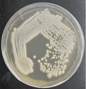
Figure 5. Sabouraud dextrose agar culture showing growth of Candida tropicalis as smooth, creamy, moist, off-white yeast-like colonies.
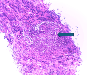
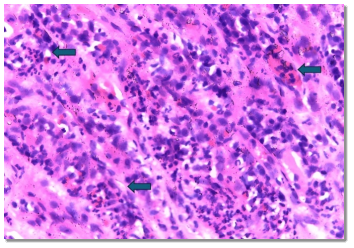
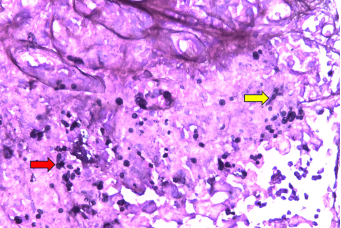
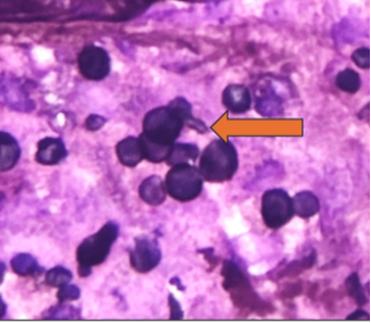
A renal biopsy was performed. Histopathology (H&E) revealed mixed inflammatory infiltrates in the renal medulla, predominantly neutrophils, with interspersed eosinophils, plasma cells, and histiocytes. Extensive neutrophilic tubulitis and focal neutrophilic abscess formation were noted. JMS (Jones' methenamine silver) stain demonstrated budding yeast, a few hyphal forms, and germ tube formation, suggestive of fungal infection.
Subsequent urine culture on Sabouraud Dextrose Agar (SDA) confirmed growth of Candida tropicalis, establishing the diagnosis of Invasive fungal pyelonephritis.
3. Discussion
Invasive fungal pyelonephritis is an uncommon but potentially life-threatening cause of renal dysfunction, most frequently caused by Candida species. Although candiduria is commonly encountered in hospitalized and catheterized patients, true invasive renal parenchymal involvement is rare and often underdiagnosed. The condition predominantly affects individuals with predisposing risk factors such as diabetes mellitus, prior antibiotic exposure, immunosuppression, neutropenia, prolonged hospitalization, and underlying renal disease
[1]
Sahara S, Kinoshita T, Takimoto N, et al.: A case of pyelonephritis and bacteremia caused by Candida glabrata in a patient on sodium glucose cotransporter 2 inhibitor, successfully treated with micafungin. J Pharm Health Care Sci. 2025, 11: 65.
Mabadi A, Davoudi-Monfared E, Jafarieh A, et al.: Emphysematous pyelonephritis due to Candida albicans in a diabetic patient: a case report. Urol Case Rep. 2025, 59: 102960.
. In the present case, advanced age, diabetes mellitus, prior acute kidney injury requiring dialysis, and persistent pancytopenia likely contributed to susceptibility to invasive candidal infection.
Candiduria is rare in healthy individuals but is commonly detected in hospitalized patients, particularly in intensive care units, where multiple predisposing factors coexist. While most cases in critically ill adults represent colonization and do not warrant antifungal therapy, candiduria may occasionally signal disseminated infection. In contrast, neonates in intensive care more frequently exhibit candiduria as a manifestation of invasive disease, often with associated fungal obstruction. Despite its association with increased mortality in ICU patients, candiduria is infrequently attributable to true Candida urinary tract infection
[2]
Pappas PG, Kauffman CA, Andes DR, et al.: Clinical practice guidelines for the management of candidiasis. 2016, 2016: 1-50.
Urinary tract or renal fungal bezoars develop through the aggregation of necrotic tissue, mucous material, and foreign or lithiasic debris, forming an obstructive mass. Although infrequent, these lesions represent a serious complication of fungal urinary tract infections. They most commonly arise either from ascending candiduria or through hematogenous spread of candidiasis, with renal parenchymal involvement occurring in the majority of cases and being associated with substantial mortality
[7]
Villegas KJ, Orhun N, Vera Garces S, et al.: Unilateral renal fungus ball caused by Candida glabrata. Med Mycol Case Rep. 2024, 44: 100649.
The clinical presentation of invasive candidal pyelonephritis is often nonspecific, with patients frequently lacking classic symptoms of urinary tract infection. Many cases present with isolated deterioration of renal function, as seen in our patient, making early clinical recognition challenging. Radiological findings may be inconclusive or mimic bacterial pyelonephritis, renal tuberculosis, or obstructive uropathy. Consequently, the diagnosis is often delayed, leading to progression of renal injury, abscess formation, obstruction due to fungal balls, or systemic dissemination
[1]
Sahara S, Kinoshita T, Takimoto N, et al.: A case of pyelonephritis and bacteremia caused by Candida glabrata in a patient on sodium glucose cotransporter 2 inhibitor, successfully treated with micafungin. J Pharm Health Care Sci. 2025, 11: 65.
Renal biopsy plays a pivotal role in establishing the diagnosis in such diagnostically ambiguous cases. Histopathological identification of fungal elements within renal tissue, particularly when highlighted by special stains such as Gomori methenamine silver, confirms invasive disease and distinguishes it from colonization or contamination
[9]
Pfaller MA, Diekema DJ: Epidemiology of invasive candidiasis: a persistent public health problem. Clin Microbiol Rev. 2007, 20: 133-163.
. In our case, the presence of budding yeast forms with hyphal forms and germ tube formation within areas of neutrophilic tubulitis and abscess formation provided definitive evidence of invasive fungal pyelonephritis. Subsequent urine culture isolating Candida tropicalis further corroborated the histological diagnosis. Notably, Candida tropicalis has been increasingly recognized as an emerging pathogen in invasive candidiasis and is associated with higher virulence and antifungal resistance compared to Candida albicans
[9]
Pfaller MA, Diekema DJ: Epidemiology of invasive candidiasis: a persistent public health problem. Clin Microbiol Rev. 2007, 20: 133-163.
Early diagnosis is crucial, as prompt initiation of appropriate antifungal therapy can significantly improve outcomes and prevent irreversible renal damage. Delay in recognition and treatment has been associated with poor prognosis, including progression to chronic kidney disease, sepsis, and increased mortality
. This case underscores the importance of maintaining a high index of suspicion for invasive fungal infections in high-risk patients presenting with unexplained renal dysfunction and highlights the indispensable role of renal biopsy in guiding timely and targeted therapy.
4. Conclusion
Invasive candidal pyelonephritis, though rare, should be considered in high-risk patients presenting with persistent or unexplained renal dysfunction, particularly in the setting of diabetes and recent renal injury. This case demonstrates that clinical and routine laboratory findings may be nonspecific, leading to diagnostic delay. Renal biopsy played a pivotal role by revealing characteristic inflammatory changes and fungal elements, which, in conjunction with urine culture confirming Candida tropicalis, established the diagnosis. Early recognition through a combination of histopathological and microbiological evaluation is essential, as timely initiation of antifungal therapy can prevent irreversible renal damage and improve clinical outcomes.
Abbreviations
AKI
Acute kidney Injury
H&E
Hematoxylin & Eosin
JMS
Jones' Methenamine Silver Stain
SDA
Sabouraud Dextrose Agar
Acknowledgments
The authors would like to acknowledge the contributions of the technical and laboratory staff involved in specimen processing and microbiological culture, whose support was essential for the completion of this work.
Author Contributions
Shemeera Saidalavi Vellolipadikkal: Conceptualization, Data curation, Investigation, Supervision, Writing – original draft, Writing – review & editing
Sahara S, Kinoshita T, Takimoto N, et al.: A case of pyelonephritis and bacteremia caused by Candida glabrata in a patient on sodium glucose cotransporter 2 inhibitor, successfully treated with micafungin. J Pharm Health Care Sci. 2025, 11: 65.
Mabadi A, Davoudi-Monfared E, Jafarieh A, et al.: Emphysematous pyelonephritis due to Candida albicans in a diabetic patient: a case report. Urol Case Rep. 2025, 59: 102960.
Yelavarthy YM, Wyawahare M, Toi PC, et al.: Isolated renal mucormycosis masquerading as bacterial pyelonephritis in a diabetic patient: a case report. Cureus. 2025, 17: 96487.
Deng R, Meng X, Li R, et al.: Asymptomatic Candida glabrata urinary tract infection in an immunocompetent young female: a case report. Medicine (Baltimore. 2023, 102: 33798.
Vellolipadikkal, S. S., Thomas, M. (2026). Renal Biopsy Revealing an Unusual Pathogen in a Case of Renal Dysfunction: A Case Report. International Journal of Medical Case Reports, 5(3), 24-27. https://doi.org/10.11648/j.ijmcr.20260503.11
Vellolipadikkal, S. S.; Thomas, M. Renal Biopsy Revealing an Unusual Pathogen in a Case of Renal Dysfunction: A Case Report. Int. J. Med. Case Rep.2026, 5(3), 24-27. doi: 10.11648/j.ijmcr.20260503.11
Vellolipadikkal SS, Thomas M. Renal Biopsy Revealing an Unusual Pathogen in a Case of Renal Dysfunction: A Case Report. Int J Med Case Rep. 2026;5(3):24-27. doi: 10.11648/j.ijmcr.20260503.11
@article{10.11648/j.ijmcr.20260503.11,
  author = {Shemeera Saidalavi Vellolipadikkal and Meeta Thomas},
  title = {Renal Biopsy Revealing an Unusual Pathogen in a Case of Renal Dysfunction: A Case Report},
  journal = {International Journal of Medical Case Reports},
  volume = {5},
  number = {3},
  pages = {24-27},
  doi = {10.11648/j.ijmcr.20260503.11},
  url = {https://doi.org/10.11648/j.ijmcr.20260503.11},
  eprint = {https://article.sciencepublishinggroup.com/pdf/10.11648.j.ijmcr.20260503.11},
  abstract = {Background: Invasive fungal pyelonephritis is a rare but potentially life-threatening renal infection, predominantly affecting immunocompromised or critically ill individuals. The diagnosis is often delayed due to nonspecific clinical presentation and laboratory findings, which can lead to adverse renal outcomes. Objective: To highlight the diagnostic challenges and emphasize the role of renal biopsy in identifying invasive fungal infections of the kidney. Methods/Case Presentation: We report the case of a 75-year-old male with a history of diabetes mellitus and recent nonsteroidal anti-inflammatory drug–induced acute kidney injury, who presented with persistent renal dysfunction and pancytopenia. Despite supportive management, renal parameters failed to improve, prompting a renal biopsy. Histopathological examination revealed extensive neutrophilic tubulitis and abscess formation. Fungal elements were identified on special stains, confirming invasive fungal infection. Subsequent urine culture grew Candida tropicalis, establishing the etiological diagnosis. Results: The integration of histopathology and microbiological findings enabled timely diagnosis of invasive fungal pyelonephritis in an otherwise diagnostically challenging clinical scenario. Conclusion: This case underscores the importance of considering fungal infections in patients with unexplained renal dysfunction, particularly in the presence of risk factors such as diabetes and recent renal injury. Early renal biopsy and prompt antifungal therapy are crucial to prevent irreversible renal damage and improve patient outcomes.},
 year = {2026}
}
TY  - JOUR
T1  - Renal Biopsy Revealing an Unusual Pathogen in a Case of Renal Dysfunction: A Case Report
AU  - Shemeera Saidalavi Vellolipadikkal
AU  - Meeta Thomas
Y1  - 2026/07/22
PY  - 2026
N1  - https://doi.org/10.11648/j.ijmcr.20260503.11
DO  - 10.11648/j.ijmcr.20260503.11
T2  - International Journal of Medical Case Reports
JF  - International Journal of Medical Case Reports
JO  - International Journal of Medical Case Reports
SP  - 24
EP  - 27
PB  - Science Publishing Group
SN  - 2994-7049
UR  - https://doi.org/10.11648/j.ijmcr.20260503.11
AB  - Background: Invasive fungal pyelonephritis is a rare but potentially life-threatening renal infection, predominantly affecting immunocompromised or critically ill individuals. The diagnosis is often delayed due to nonspecific clinical presentation and laboratory findings, which can lead to adverse renal outcomes. Objective: To highlight the diagnostic challenges and emphasize the role of renal biopsy in identifying invasive fungal infections of the kidney. Methods/Case Presentation: We report the case of a 75-year-old male with a history of diabetes mellitus and recent nonsteroidal anti-inflammatory drug–induced acute kidney injury, who presented with persistent renal dysfunction and pancytopenia. Despite supportive management, renal parameters failed to improve, prompting a renal biopsy. Histopathological examination revealed extensive neutrophilic tubulitis and abscess formation. Fungal elements were identified on special stains, confirming invasive fungal infection. Subsequent urine culture grew Candida tropicalis, establishing the etiological diagnosis. Results: The integration of histopathology and microbiological findings enabled timely diagnosis of invasive fungal pyelonephritis in an otherwise diagnostically challenging clinical scenario. Conclusion: This case underscores the importance of considering fungal infections in patients with unexplained renal dysfunction, particularly in the presence of risk factors such as diabetes and recent renal injury. Early renal biopsy and prompt antifungal therapy are crucial to prevent irreversible renal damage and improve patient outcomes.
VL  - 5
IS  - 3
ER  -
Vellolipadikkal, S. S., Thomas, M. (2026). Renal Biopsy Revealing an Unusual Pathogen in a Case of Renal Dysfunction: A Case Report. International Journal of Medical Case Reports, 5(3), 24-27. https://doi.org/10.11648/j.ijmcr.20260503.11
Vellolipadikkal, S. S.; Thomas, M. Renal Biopsy Revealing an Unusual Pathogen in a Case of Renal Dysfunction: A Case Report. Int. J. Med. Case Rep.2026, 5(3), 24-27. doi: 10.11648/j.ijmcr.20260503.11
Vellolipadikkal SS, Thomas M. Renal Biopsy Revealing an Unusual Pathogen in a Case of Renal Dysfunction: A Case Report. Int J Med Case Rep. 2026;5(3):24-27. doi: 10.11648/j.ijmcr.20260503.11
@article{10.11648/j.ijmcr.20260503.11,
  author = {Shemeera Saidalavi Vellolipadikkal and Meeta Thomas},
  title = {Renal Biopsy Revealing an Unusual Pathogen in a Case of Renal Dysfunction: A Case Report},
  journal = {International Journal of Medical Case Reports},
  volume = {5},
  number = {3},
  pages = {24-27},
  doi = {10.11648/j.ijmcr.20260503.11},
  url = {https://doi.org/10.11648/j.ijmcr.20260503.11},
  eprint = {https://article.sciencepublishinggroup.com/pdf/10.11648.j.ijmcr.20260503.11},
  abstract = {Background: Invasive fungal pyelonephritis is a rare but potentially life-threatening renal infection, predominantly affecting immunocompromised or critically ill individuals. The diagnosis is often delayed due to nonspecific clinical presentation and laboratory findings, which can lead to adverse renal outcomes. Objective: To highlight the diagnostic challenges and emphasize the role of renal biopsy in identifying invasive fungal infections of the kidney. Methods/Case Presentation: We report the case of a 75-year-old male with a history of diabetes mellitus and recent nonsteroidal anti-inflammatory drug–induced acute kidney injury, who presented with persistent renal dysfunction and pancytopenia. Despite supportive management, renal parameters failed to improve, prompting a renal biopsy. Histopathological examination revealed extensive neutrophilic tubulitis and abscess formation. Fungal elements were identified on special stains, confirming invasive fungal infection. Subsequent urine culture grew Candida tropicalis, establishing the etiological diagnosis. Results: The integration of histopathology and microbiological findings enabled timely diagnosis of invasive fungal pyelonephritis in an otherwise diagnostically challenging clinical scenario. Conclusion: This case underscores the importance of considering fungal infections in patients with unexplained renal dysfunction, particularly in the presence of risk factors such as diabetes and recent renal injury. Early renal biopsy and prompt antifungal therapy are crucial to prevent irreversible renal damage and improve patient outcomes.},
 year = {2026}
}
TY  - JOUR
T1  - Renal Biopsy Revealing an Unusual Pathogen in a Case of Renal Dysfunction: A Case Report
AU  - Shemeera Saidalavi Vellolipadikkal
AU  - Meeta Thomas
Y1  - 2026/07/22
PY  - 2026
N1  - https://doi.org/10.11648/j.ijmcr.20260503.11
DO  - 10.11648/j.ijmcr.20260503.11
T2  - International Journal of Medical Case Reports
JF  - International Journal of Medical Case Reports
JO  - International Journal of Medical Case Reports
SP  - 24
EP  - 27
PB  - Science Publishing Group
SN  - 2994-7049
UR  - https://doi.org/10.11648/j.ijmcr.20260503.11
AB  - Background: Invasive fungal pyelonephritis is a rare but potentially life-threatening renal infection, predominantly affecting immunocompromised or critically ill individuals. The diagnosis is often delayed due to nonspecific clinical presentation and laboratory findings, which can lead to adverse renal outcomes. Objective: To highlight the diagnostic challenges and emphasize the role of renal biopsy in identifying invasive fungal infections of the kidney. Methods/Case Presentation: We report the case of a 75-year-old male with a history of diabetes mellitus and recent nonsteroidal anti-inflammatory drug–induced acute kidney injury, who presented with persistent renal dysfunction and pancytopenia. Despite supportive management, renal parameters failed to improve, prompting a renal biopsy. Histopathological examination revealed extensive neutrophilic tubulitis and abscess formation. Fungal elements were identified on special stains, confirming invasive fungal infection. Subsequent urine culture grew Candida tropicalis, establishing the etiological diagnosis. Results: The integration of histopathology and microbiological findings enabled timely diagnosis of invasive fungal pyelonephritis in an otherwise diagnostically challenging clinical scenario. Conclusion: This case underscores the importance of considering fungal infections in patients with unexplained renal dysfunction, particularly in the presence of risk factors such as diabetes and recent renal injury. Early renal biopsy and prompt antifungal therapy are crucial to prevent irreversible renal damage and improve patient outcomes.
VL  - 5
IS  - 3
ER  -





 Share This Article
 Twitter
 Linked In
 Facebook
 Copy
Copy Download