A 48-year-old man with a history of schizophrenia, bipolar disorder, spinal cord lipoma resection, retinal artery occlusion, alcohol use disorder, and methamphetamine use presented to the hospital after being found unresponsive at his house. On initial evaluation, he was inattentive, but oriented to self, time, and location, and was able to follow commands, speak fluently in short sentences, and had no focal deficits, aside from chronic lower extremity weakness and sensory loss related to his spinal cord lipoma. He was treated for a urinary tract infection however continued to have neurologic decline. An extensive neurological workup was conducted, which included multiple CT scans, three days of electroencephalogram (EEG), lumbar puncture and Magnetic Resonance Imaging (MRI) brain with and without contrast. Brain MRI showed diffusion weighted imaging (DWI) hyperintensities along the cortical ribbon of the temporal, parietal, and frontal lobes with intrinsic T1 hyperintense changes but no true contrast enhancement, as well as prominent medial temporal lobe DWI and T2/FLAIR hyperintensities. He was found to have a positive serum RPR at a titer of 1:64, positive serum treponemal antibody, and positive CSF VDRL at a titer of 1:8. CSF had a lymphocytic pleocytosis. Following a full course of intravenous Penicillin G 24 million units daily for 2 weeks for treatment of neurosyphilis, the patient showed signs of improvement, specifically in cognitive domains of attention and comprehension. However, one week later, his mentation worsened again. He developed agitation, mood swings, confabulation, and intermittent unresponsiveness. A second lumbar puncture revealed elevated 14-3-3 protein levels (41,897), T-Tau >20,000, and a positive RT-QuIC test, consistent with prion disease. A repeat brain MRI showed progressive cortical ribboning (figure), also consistent with a diagnosis of CJD. In practice, when faced with rapidly progressive dementia, clinicians should consider treatable etiologies such as neurosyphilis first but remain vigilant for co-pathologies if deterioration persists.
This is an Open Access article, distributed under the terms of the Creative Commons Attribution 4.0 International License (http://creativecommons.org/licenses/by/4.0/), which permits unrestricted use, distribution and reproduction in any medium or format, provided the original work is properly cited.
Neurosyphilis can mimic many conditions that cause rapidly progressive dementia (RPD), including CJD. While there are documented cases of patients initially suspected to have CJD but later found to have neurosyphilis on autopsy, reports of patients diagnosed with neurosyphilis who were ultimately confirmed to have CJD are rare. Markers such as 14-3-3 protein and T-Tau, previously used for CJD diagnosis, lack specificity as they can be elevated in various pathologies, including both CJD and neurosyphilis
[1]
Jang J-W, Park JH, Eo YJ, Kim SH, Choi KH, Yi S, Park YH, Kim S. Neurosyphilis mimicking Creutzfeldt-Jakob disease. Dement Neurocognitive Disord (2016) 15: 170–173.
[1]
. The advent of RT-QuIC testing in 2010 has significantly improved diagnostic accuracy for CJD with a sensitivity of 96% and specificity of 98%. However, false positives can occur in up to 3% of cases of neurosyphilis
[2]
Hermann P, Schmitz M, Cramm M, Goebel S, Bunck T, Schütte-Schmidt J, Schulz-Schaeffer W, Stadelmann C, Matschke J, Glatzel M, et al. Application of real-time quaking-induced conversion in Creutzfeldt-Jakob disease surveillance. J Neurol (2023) 270: 2149–2161.
[2]
.
To date, there are no documented cases of co-occurring neurosyphilis and CJD; only misdiagnoses and mimickers
[3]
Mead S, Rudge P. CJD mimics and chameleons. Pract Neurol (2017) 17: 113–121.
[3]
. Although neurosyphilis can present with rapidly progressive dementia and acute psychiatric symptoms, it is often not considered on the differential in patients presenting with these symptoms
[4]
Stefani A, Riello M, Rossini F, Mariotto S, Fenzi F, Gambina G, Zanusso G, Monaco S. Neurosyphilis manifesting with rapidly progressive dementia: report of three cases. Neurol Sci (2013) 34: 2027–2030.
[4]
. The rates of newly diagnosed syphilis have been growing over the last decade. In the United States, there has been a 78.9% increase in syphilis diagnoses between 2018 and 2022 without any significant change in diagnostic testing modalities
[5]
Horberg M, Thompson M, Agwu A, Colasanti J, Haddad M, Jain M, McComsey G, Radix A, Rakhmanina N, Short WR, et al. Primary Care Guidance for providers who care for persons with human immunodeficiency virus: 2024 update by the HIV Medicine Association of the Infectious Diseases Society of America. Clin Infect Dis (2024)
While syphilis is on the rise, CJD remains a very rare diagnosis with approximately 1 to 2 cases per million per year globally
[7]
Uttley L, Carroll C, Wong R, Hilton DA, Stevenson M. Creutzfeldt-Jakob disease: a systematic review of global incidence, prevalence, infectivity, and incubation. Lancet Infect Dis (2020) 20: e2–e10.
[8]
Gao L-P, Tian T-T, Xiao K, Chen C, Zhou W, Liang D-L, Cao R-D, Shi Q, Dong X-P. Updated global epidemiology atlas of human prion diseases. Front Public Health (2024) 12: 1411489.
[7, 8]
. The incidence of CJD has remained stable in recent years, suggesting that there has not been an increase in the rate of false positive RT-QuIC
[9]
Guidon AC, Amato AA. COVID-19 and neuromuscular disorders. Neurology. 2020.
Jones SM, Lazar EB, Porter AL, Prusinski CC, Brier MR, Bucelli RC, et al. Real-time quaking-induced conversion assays for prions: applying a sensitive but imperfect test in clinical practice. Eur J Neurol. 2023; 30(7): 1854-1860.
. This case represents the first reported instance of a patient with true simultaneous diagnoses of CJD and neurosyphilis and highlights the challenges associated with the clinical evaluation of patients with multiple neuroinfectious diseases.
2. Case Report
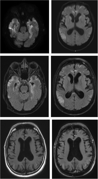
A 48-year-old man with a history of schizophrenia, bipolar disorder, spinal cord lipoma resection, retinal artery occlusion, alcohol use disorder, and methamphetamine use presented to the hospital after being found unresponsive at his house. On initial evaluation, he was inattentive, but oriented to self, time, and location, and was able to follow commands, speak fluently in short sentences, and had no focal deficits, aside from chronic lower extremity weakness and sensory loss related to his spinal cord lipoma. Diagnostic testing revealed that he had both pneumonia and a urinary tract infection (UTI), and he was treated with ceftriaxone and azithromycin. However, his memory, attention, abstraction and executive processing declined despite antimicrobial treatment. An extensive neurological workup was conducted, which included multiple CT scans, three days of electroencephalogram (EEG), lumbar puncture and MRI brain with and without contrast. EEG showed diffuse slowing with no epileptiform discharges or concerning patterns. Brain MRI showed diffusion weighted imaging (DWI) hyperintensities along the cortical ribbon of the temporal, parietal, and frontal lobes with intrinsic T1 hyperintense changes, unrelated to prior ischemic insults or metabolic abnormalities, but no true contrast enhancement, as well as prominent medial temporal lobe DWI and T2/FLAIR hyperintensities (figure). Relevant testing results included a cerebrospinal fluid (CSF) white blood cell count of 21 cells/µL with 90% lymphocytes, CSF protein of 33 mg/dL, CSF glucose of 76 mg/dL (serum glucose 196) negative serum HIV and hepatitis B PCR, positive serum hepatitis C, positive serum RPR at a titer of 1:64, positive serum treponemal antibody, and positive CSF VDRL at a titer of 1:8. The patient was subsequently noted to have Argyll Robertson pupils, all of which were consistent with neurosyphilis, and he was treated with high-dose penicillin.
Following a full course of intravenous Penicillin G 24 million units daily for 2 weeks for treatment of neurosyphilis, the patient showed signs of improvement, specifically in cognitive domains of attention and comprehension. However, one week later, his mentation worsened again. He developed agitation, mood swings, confabulation, and intermittent unresponsiveness. A second lumbar puncture revealed elevated 14-3-3 protein levels (41,897), T-Tau >20,000, and a positive RT-QuIC test, consistent with prion disease. A repeat brain MRI showed progressive cortical ribboning (figure), also consistent with a diagnosis of CJD. A brain biopsy was considered, given the competing diagnoses of CJD and neurosyphilis, but was deferred given that he had transient improvement in his symptoms following a course of antibiotics, confirming active neurosyphilis. His co-existing CJD led to non-sustained improvement in symptoms indicating the high likelihood of prion disease with risk to the patient and surgical team.
The patient was discharged to long-term care but continued to deteriorate. Follow-up examination in the clinic 8 weeks after discharge revealed further cognitive decline, as well as new onset of spontaneous and startle myoclonus, increased tone in the lower extremities leading to contractures, a jaw jerk reflex, and primitive reflexes such as palmar grasp. Based on his clinical progression despite adequate neurosyphilis treatment, and the CSF results and neuroimaging, a diagnosis of CJD was made.
3. Conclusion
This case underscores the importance of considering overlapping diagnoses in patients presenting with RPD
[11]
Funayama M, Kuramochi S, Kudo S. Neurosyphilis initially misdiagnosed as behavioral variant frontotemporal dementia: life-changing differential diagnosis. J Alzheimers Dis Rep. 2023; 7(1): 1077-1083.
Liu X, Sun Y, Zhang X, Liu P, Zhang K, Yu L, Su Y, Yuan Y, Ke Q, Peng G. Prevalence and outcomes of rapidly progressive dementia: a retrospective cohort study in a neurologic unit in China. BMC Geriatr. 2023; 23: 142.
. Given the better prognosis associated with neurosyphilis compared to CJD
[14]
Roberts MC, Emsley RA. Cognitive change after treatment for neurosyphilis: correlation with CSF laboratory measures. Gen Hosp Psychiatry. 1995; 17(4): 305-309.
, there was hope that this patient would improve in the weeks following neurosyphilis treatment, suggesting that he had neurosyphilis without concomitant CJD. His serum and CSF testing for neurosyphilis, as well as early improvement following appropriate antibiotic treatment, was diagnostic of neurosyphilis. However, his subsequent clinical decline, including characteristic features of CJD such as startle myoclonus, as well as imaging findings of DWI hyperintense changes along multiple regions of the cortical ribbon, and positive CSF RT-QuIC with elevated CSF 14-3-3 and T-Tau levels, were all diagnostic of co-existing CJD, and precluded the need for brain biopsy.
In practice, when faced with rapidly progressive dementia, clinicians should consider treatable etiologies such as neurosyphilis first but remain vigilant for co-pathologies if deterioration persists. This case highlights the need for meticulous diagnostic evaluation when a patient's neurological symptoms and deficits continue to progress despite appropriate treatment for the suspected condition. Early recognition of dual pathologies is critical for appropriate management and prognostic discussions with patients and their families, as well as avoidance of interventions that may put both patients and healthcare providers at risk.
Figure depicting MRI with cortical diffusion restriction in multiple areas of the brain (row 1) as well as FLARI hyperintensities in similar brain regions (row 2) and intrinsic T1 hyperintensities (row 3 image 1) without contrast enhancement (row 3 image 2).
Table 1. Comparison of typical CSF findings in CJD and Neurosyphilis.
CSF Finding
CJD
Neuro-Syphilis
Glucose
Normal
Normal or mildly decreased
Protein
Normal or Elevated
Elevated
Cell Counts
Elevated >5 or >20 if concomitant HIV
Elevated >5 or >20 if concomitant HIV
VDRL
Negative
Positive
14-3-3
Elevated
Normal or Elevated
RTQuic
Positive
Negative
Abbreviations
EEG
Electroencephalogram
DWI
Diffusion Weighted Imaging
CJD
Creutzfeldt-Jakob Disease
RPD
Rapidly Progressive Dementia
MRI
Magnetic Resonance Imaging
UTI
Urinary Tract Infection
CSF
Cerebrospinal Fluid
Author Contributions
Emma Frost: Conceptualization, Writing – original draft, Writing – review & editing
Melody Lee Yu: Conceptualization, Writing – review & editing
Marina Santos De Sousa: Writing – review & editing
Olga Thon: Conceptualization, Supervision, Writing – review & editing
Conflicts of Interest
The authors declare no conflicts of interest.
References
[1]
Jang J-W, Park JH, Eo YJ, Kim SH, Choi KH, Yi S, Park YH, Kim S. Neurosyphilis mimicking Creutzfeldt-Jakob disease. Dement Neurocognitive Disord (2016) 15: 170–173.
[2]
Hermann P, Schmitz M, Cramm M, Goebel S, Bunck T, Schütte-Schmidt J, Schulz-Schaeffer W, Stadelmann C, Matschke J, Glatzel M, et al. Application of real-time quaking-induced conversion in Creutzfeldt-Jakob disease surveillance. J Neurol (2023) 270: 2149–2161.
[3]
Mead S, Rudge P. CJD mimics and chameleons. Pract Neurol (2017) 17: 113–121.
[4]
Stefani A, Riello M, Rossini F, Mariotto S, Fenzi F, Gambina G, Zanusso G, Monaco S. Neurosyphilis manifesting with rapidly progressive dementia: report of three cases. Neurol Sci (2013) 34: 2027–2030.
[5]
Horberg M, Thompson M, Agwu A, Colasanti J, Haddad M, Jain M, McComsey G, Radix A, Rakhmanina N, Short WR, et al. Primary Care Guidance for providers who care for persons with human immunodeficiency virus: 2024 update by the HIV Medicine Association of the Infectious Diseases Society of America. Clin Infect Dis (2024)
Uttley L, Carroll C, Wong R, Hilton DA, Stevenson M. Creutzfeldt-Jakob disease: a systematic review of global incidence, prevalence, infectivity, and incubation. Lancet Infect Dis (2020) 20: e2–e10.
[8]
Gao L-P, Tian T-T, Xiao K, Chen C, Zhou W, Liang D-L, Cao R-D, Shi Q, Dong X-P. Updated global epidemiology atlas of human prion diseases. Front Public Health (2024) 12: 1411489.
[9]
Guidon AC, Amato AA. COVID-19 and neuromuscular disorders. Neurology. 2020.
Jones SM, Lazar EB, Porter AL, Prusinski CC, Brier MR, Bucelli RC, et al. Real-time quaking-induced conversion assays for prions: applying a sensitive but imperfect test in clinical practice. Eur J Neurol. 2023; 30(7): 1854-1860.
Liu X, Sun Y, Zhang X, Liu P, Zhang K, Yu L, Su Y, Yuan Y, Ke Q, Peng G. Prevalence and outcomes of rapidly progressive dementia: a retrospective cohort study in a neurologic unit in China. BMC Geriatr. 2023; 23: 142.
Roberts MC, Emsley RA. Cognitive change after treatment for neurosyphilis: correlation with CSF laboratory measures. Gen Hosp Psychiatry. 1995; 17(4): 305-309.
Frost, E., Yu, M. L., Sousa, M. S. D., Bhatti, K., Elmandouh, O., et al. (2026). A Case of Creutzfeldt-Jakob Disease with Concomitant Neurosyphilis. International Journal of Medical Case Reports, 5(2), 15-18. https://doi.org/10.11648/j.ijmcr.20260502.11
Frost, E.; Yu, M. L.; Sousa, M. S. D.; Bhatti, K.; Elmandouh, O., et al. A Case of Creutzfeldt-Jakob Disease with Concomitant Neurosyphilis. Int. J. Med. Case Rep.2026, 5(2), 15-18. doi: 10.11648/j.ijmcr.20260502.11
Frost E, Yu ML, Sousa MSD, Bhatti K, Elmandouh O, et al. A Case of Creutzfeldt-Jakob Disease with Concomitant Neurosyphilis. Int J Med Case Rep. 2026;5(2):15-18. doi: 10.11648/j.ijmcr.20260502.11
@article{10.11648/j.ijmcr.20260502.11,
  author = {Emma Frost and Melody Lee Yu and Marina Santos De Sousa and Karandeep Bhatti and Omar Elmandouh and Courtney Curran and Pratit Patel and Raquel Nahra and Jesse Thon and Olga Thon},
  title = {A Case of Creutzfeldt-Jakob Disease with Concomitant Neurosyphilis},
  journal = {International Journal of Medical Case Reports},
  volume = {5},
  number = {2},
  pages = {15-18},
  doi = {10.11648/j.ijmcr.20260502.11},
  url = {https://doi.org/10.11648/j.ijmcr.20260502.11},
  eprint = {https://article.sciencepublishinggroup.com/pdf/10.11648.j.ijmcr.20260502.11},
  abstract = {A 48-year-old man with a history of schizophrenia, bipolar disorder, spinal cord lipoma resection, retinal artery occlusion, alcohol use disorder, and methamphetamine use presented to the hospital after being found unresponsive at his house. On initial evaluation, he was inattentive, but oriented to self, time, and location, and was able to follow commands, speak fluently in short sentences, and had no focal deficits, aside from chronic lower extremity weakness and sensory loss related to his spinal cord lipoma. He was treated for a urinary tract infection however continued to have neurologic decline. An extensive neurological workup was conducted, which included multiple CT scans, three days of electroencephalogram (EEG), lumbar puncture and Magnetic Resonance Imaging (MRI) brain with and without contrast. Brain MRI showed diffusion weighted imaging (DWI) hyperintensities along the cortical ribbon of the temporal, parietal, and frontal lobes with intrinsic T1 hyperintense changes but no true contrast enhancement, as well as prominent medial temporal lobe DWI and T2/FLAIR hyperintensities. He was found to have a positive serum RPR at a titer of 1:64, positive serum treponemal antibody, and positive CSF VDRL at a titer of 1:8. CSF had a lymphocytic pleocytosis. Following a full course of intravenous Penicillin G 24 million units daily for 2 weeks for treatment of neurosyphilis, the patient showed signs of improvement, specifically in cognitive domains of attention and comprehension. However, one week later, his mentation worsened again. He developed agitation, mood swings, confabulation, and intermittent unresponsiveness. A second lumbar puncture revealed elevated 14-3-3 protein levels (41,897), T-Tau >20,000, and a positive RT-QuIC test, consistent with prion disease. A repeat brain MRI showed progressive cortical ribboning (figure), also consistent with a diagnosis of CJD. In practice, when faced with rapidly progressive dementia, clinicians should consider treatable etiologies such as neurosyphilis first but remain vigilant for co-pathologies if deterioration persists.},
 year = {2026}
}
TY  - JOUR
T1  - A Case of Creutzfeldt-Jakob Disease with Concomitant Neurosyphilis
AU  - Emma Frost
AU  - Melody Lee Yu
AU  - Marina Santos De Sousa
AU  - Karandeep Bhatti
AU  - Omar Elmandouh
AU  - Courtney Curran
AU  - Pratit Patel
AU  - Raquel Nahra
AU  - Jesse Thon
AU  - Olga Thon
Y1  - 2026/07/22
PY  - 2026
N1  - https://doi.org/10.11648/j.ijmcr.20260502.11
DO  - 10.11648/j.ijmcr.20260502.11
T2  - International Journal of Medical Case Reports
JF  - International Journal of Medical Case Reports
JO  - International Journal of Medical Case Reports
SP  - 15
EP  - 18
PB  - Science Publishing Group
SN  - 2994-7049
UR  - https://doi.org/10.11648/j.ijmcr.20260502.11
AB  - A 48-year-old man with a history of schizophrenia, bipolar disorder, spinal cord lipoma resection, retinal artery occlusion, alcohol use disorder, and methamphetamine use presented to the hospital after being found unresponsive at his house. On initial evaluation, he was inattentive, but oriented to self, time, and location, and was able to follow commands, speak fluently in short sentences, and had no focal deficits, aside from chronic lower extremity weakness and sensory loss related to his spinal cord lipoma. He was treated for a urinary tract infection however continued to have neurologic decline. An extensive neurological workup was conducted, which included multiple CT scans, three days of electroencephalogram (EEG), lumbar puncture and Magnetic Resonance Imaging (MRI) brain with and without contrast. Brain MRI showed diffusion weighted imaging (DWI) hyperintensities along the cortical ribbon of the temporal, parietal, and frontal lobes with intrinsic T1 hyperintense changes but no true contrast enhancement, as well as prominent medial temporal lobe DWI and T2/FLAIR hyperintensities. He was found to have a positive serum RPR at a titer of 1:64, positive serum treponemal antibody, and positive CSF VDRL at a titer of 1:8. CSF had a lymphocytic pleocytosis. Following a full course of intravenous Penicillin G 24 million units daily for 2 weeks for treatment of neurosyphilis, the patient showed signs of improvement, specifically in cognitive domains of attention and comprehension. However, one week later, his mentation worsened again. He developed agitation, mood swings, confabulation, and intermittent unresponsiveness. A second lumbar puncture revealed elevated 14-3-3 protein levels (41,897), T-Tau >20,000, and a positive RT-QuIC test, consistent with prion disease. A repeat brain MRI showed progressive cortical ribboning (figure), also consistent with a diagnosis of CJD. In practice, when faced with rapidly progressive dementia, clinicians should consider treatable etiologies such as neurosyphilis first but remain vigilant for co-pathologies if deterioration persists.
VL  - 5
IS  - 2
ER  -
Cooper Neurological Institute, Cooper University Hospital, Camden, United States
Marina Santos De Sousa
Cooper Neurological Institute, Cooper University Hospital, Camden, United States
Karandeep Bhatti
Cooper Neurological Institute, Cooper University Hospital, Camden, United States
Omar Elmandouh
Cooper Neurological Institute, Cooper University Hospital, Camden, United States
Courtney Curran
Cooper Neurological Institute, Cooper University Hospital, Camden, United States; Cooper Medical School of Rowan University, Camden, United States
Pratit Patel
Cooper Neurological Institute, Cooper University Hospital, Camden, United States; Cooper Medical School of Rowan University, Camden, United States
Raquel Nahra
Cooper Medical School of Rowan University, Camden, United States; Cooper Department of Infectious Disease, Cooper University Hospital, Camden, United States; Cooper Department of Critical Care Medicine, Cooper University Hospital, Camden, United States
Jesse Thon
Cooper Neurological Institute, Cooper University Hospital, Camden, United States; Cooper Medical School of Rowan University, Camden, United States
Olga Thon
Cooper Neurological Institute, Cooper University Hospital, Camden, United States; Cooper Medical School of Rowan University, Camden, United States
Frost, E., Yu, M. L., Sousa, M. S. D., Bhatti, K., Elmandouh, O., et al. (2026). A Case of Creutzfeldt-Jakob Disease with Concomitant Neurosyphilis. International Journal of Medical Case Reports, 5(2), 15-18. https://doi.org/10.11648/j.ijmcr.20260502.11
Frost, E.; Yu, M. L.; Sousa, M. S. D.; Bhatti, K.; Elmandouh, O., et al. A Case of Creutzfeldt-Jakob Disease with Concomitant Neurosyphilis. Int. J. Med. Case Rep.2026, 5(2), 15-18. doi: 10.11648/j.ijmcr.20260502.11
Frost E, Yu ML, Sousa MSD, Bhatti K, Elmandouh O, et al. A Case of Creutzfeldt-Jakob Disease with Concomitant Neurosyphilis. Int J Med Case Rep. 2026;5(2):15-18. doi: 10.11648/j.ijmcr.20260502.11
@article{10.11648/j.ijmcr.20260502.11,
  author = {Emma Frost and Melody Lee Yu and Marina Santos De Sousa and Karandeep Bhatti and Omar Elmandouh and Courtney Curran and Pratit Patel and Raquel Nahra and Jesse Thon and Olga Thon},
  title = {A Case of Creutzfeldt-Jakob Disease with Concomitant Neurosyphilis},
  journal = {International Journal of Medical Case Reports},
  volume = {5},
  number = {2},
  pages = {15-18},
  doi = {10.11648/j.ijmcr.20260502.11},
  url = {https://doi.org/10.11648/j.ijmcr.20260502.11},
  eprint = {https://article.sciencepublishinggroup.com/pdf/10.11648.j.ijmcr.20260502.11},
  abstract = {A 48-year-old man with a history of schizophrenia, bipolar disorder, spinal cord lipoma resection, retinal artery occlusion, alcohol use disorder, and methamphetamine use presented to the hospital after being found unresponsive at his house. On initial evaluation, he was inattentive, but oriented to self, time, and location, and was able to follow commands, speak fluently in short sentences, and had no focal deficits, aside from chronic lower extremity weakness and sensory loss related to his spinal cord lipoma. He was treated for a urinary tract infection however continued to have neurologic decline. An extensive neurological workup was conducted, which included multiple CT scans, three days of electroencephalogram (EEG), lumbar puncture and Magnetic Resonance Imaging (MRI) brain with and without contrast. Brain MRI showed diffusion weighted imaging (DWI) hyperintensities along the cortical ribbon of the temporal, parietal, and frontal lobes with intrinsic T1 hyperintense changes but no true contrast enhancement, as well as prominent medial temporal lobe DWI and T2/FLAIR hyperintensities. He was found to have a positive serum RPR at a titer of 1:64, positive serum treponemal antibody, and positive CSF VDRL at a titer of 1:8. CSF had a lymphocytic pleocytosis. Following a full course of intravenous Penicillin G 24 million units daily for 2 weeks for treatment of neurosyphilis, the patient showed signs of improvement, specifically in cognitive domains of attention and comprehension. However, one week later, his mentation worsened again. He developed agitation, mood swings, confabulation, and intermittent unresponsiveness. A second lumbar puncture revealed elevated 14-3-3 protein levels (41,897), T-Tau >20,000, and a positive RT-QuIC test, consistent with prion disease. A repeat brain MRI showed progressive cortical ribboning (figure), also consistent with a diagnosis of CJD. In practice, when faced with rapidly progressive dementia, clinicians should consider treatable etiologies such as neurosyphilis first but remain vigilant for co-pathologies if deterioration persists.},
 year = {2026}
}
TY  - JOUR
T1  - A Case of Creutzfeldt-Jakob Disease with Concomitant Neurosyphilis
AU  - Emma Frost
AU  - Melody Lee Yu
AU  - Marina Santos De Sousa
AU  - Karandeep Bhatti
AU  - Omar Elmandouh
AU  - Courtney Curran
AU  - Pratit Patel
AU  - Raquel Nahra
AU  - Jesse Thon
AU  - Olga Thon
Y1  - 2026/07/22
PY  - 2026
N1  - https://doi.org/10.11648/j.ijmcr.20260502.11
DO  - 10.11648/j.ijmcr.20260502.11
T2  - International Journal of Medical Case Reports
JF  - International Journal of Medical Case Reports
JO  - International Journal of Medical Case Reports
SP  - 15
EP  - 18
PB  - Science Publishing Group
SN  - 2994-7049
UR  - https://doi.org/10.11648/j.ijmcr.20260502.11
AB  - A 48-year-old man with a history of schizophrenia, bipolar disorder, spinal cord lipoma resection, retinal artery occlusion, alcohol use disorder, and methamphetamine use presented to the hospital after being found unresponsive at his house. On initial evaluation, he was inattentive, but oriented to self, time, and location, and was able to follow commands, speak fluently in short sentences, and had no focal deficits, aside from chronic lower extremity weakness and sensory loss related to his spinal cord lipoma. He was treated for a urinary tract infection however continued to have neurologic decline. An extensive neurological workup was conducted, which included multiple CT scans, three days of electroencephalogram (EEG), lumbar puncture and Magnetic Resonance Imaging (MRI) brain with and without contrast. Brain MRI showed diffusion weighted imaging (DWI) hyperintensities along the cortical ribbon of the temporal, parietal, and frontal lobes with intrinsic T1 hyperintense changes but no true contrast enhancement, as well as prominent medial temporal lobe DWI and T2/FLAIR hyperintensities. He was found to have a positive serum RPR at a titer of 1:64, positive serum treponemal antibody, and positive CSF VDRL at a titer of 1:8. CSF had a lymphocytic pleocytosis. Following a full course of intravenous Penicillin G 24 million units daily for 2 weeks for treatment of neurosyphilis, the patient showed signs of improvement, specifically in cognitive domains of attention and comprehension. However, one week later, his mentation worsened again. He developed agitation, mood swings, confabulation, and intermittent unresponsiveness. A second lumbar puncture revealed elevated 14-3-3 protein levels (41,897), T-Tau >20,000, and a positive RT-QuIC test, consistent with prion disease. A repeat brain MRI showed progressive cortical ribboning (figure), also consistent with a diagnosis of CJD. In practice, when faced with rapidly progressive dementia, clinicians should consider treatable etiologies such as neurosyphilis first but remain vigilant for co-pathologies if deterioration persists.
VL  - 5
IS  - 2
ER  -




 Share This Article
 Twitter
 Linked In
 Facebook
 Copy
Copy Download