Research Article | | Peer-Reviewed

Phytochemical Analysis and Antibacterial Activity of Aloe Vera Leaf Extracts Across Different Leaf Ages

Received: 23 November 2024     Accepted: 4 December 2024     Published: 23 December 2024
Views:       Downloads:
Abstract

The increasing reliance on plant-based healthcare products, including herbal medicines and dietary supplements, emphasizes the global significance of traditional remedies. Aloe Vera with 400 reported species, has been widely used herbal remedy for health practices worldwide. Despite its extensive historical use and therapeutic reputation, recent studies have raised concerns about potential adverse effects, challenging the notion of Aloe Vera as a universally safe functional food material. This study aimed to analyze the phytochemical composition of whole leaf extracts at various maturation stages of Aloe Vera and assess their antibacterial effect against Staphylococcus aureus. The qualitative phytochemical analysis revealed a concentration gradient, with older leaves exhibiting higher concentrations compared to medium and young leaves, suggesting a dynamic maturation-related variation. The antibacterial assay demonstrated age-dependent inhibitory activities, with older leaves displaying the highest, medium leaves following, and young leaves exhibiting the least inhibition. A consistent minimum inhibitory concentration of 12.5 mg/ml was observed across all leaf ages. These findings stress the need for cautious Aloe Vera consumption, especially in rural communities where whole-leaf extraction is prevalent, as recent studies have reported adverse effects and potential health risks associated with certain compounds. Safer alternatives, and regulating consumption practices are recommended, emphasizing sustainable practices to maximize plant benefits and minimize waste.

Published in Journal of Diseases and Medicinal Plants (Volume 10, Issue 4)
DOI 10.11648/j.jdmp.20241004.11
Page(s) 60-68
Creative Commons

This is an Open Access article, distributed under the terms of the Creative Commons Attribution 4.0 International License (http://creativecommons.org/licenses/by/4.0/), which permits unrestricted use, distribution and reproduction in any medium or format, provided the original work is properly cited.

Copyright

Copyright © The Author(s), 2024. Published by Science Publishing Group

Keywords

Aloe Vera, Medicinal Plants, Staphylococcus aureus, MBC, MIC

1. Introduction
There has been an increase in reliance on plant-based products for health care, particularly herbal medicines and dietary supplements . In 2013, the World Health Organization(WHO) acknowledged and reported that over 80% of the global population relies on plant-based medicines to meet their healthcare needs . According to a study , approximately 79% of cancer survivors and 68% of cancer-free adults reported using one or a combination of complementary and alternative medicines including herbal medicines and dietary supplements.
Aloe Vera has been used from prehistoric times to provide health benefits and is one of the most frequently used herbal remedies throughout the globe. There are more than 400 species of Aloe reported but the most widely utilized species is Aloe barbadensis Miller (also known as Kigajji(Ganda) commonly referred to as Aloe Vera). It’s name is derived from two Arabic words; “alloeh” meaning “bitter shiny substance” and “vera” meaning “truth.
Aloe Vera is a “wonder” perennial xerophyte, with a short stem, drought-resisting succulent leaves in which large amounts of water is stored . It belongs to the family Liliaceae, a family reported to have 400 species of Aloe. The useful parts utilized are the whole leaf extract, gel, and the latex . Due to its therapeutic values, the plant has been cultivated globally for over 200 years in areas like Australia, China, United States, India, South Africa, Kenya as well as Uganda .
Aloe Vera has received strong global reputation due to its widespread usage in cosmetics industry to treat sunburns, acne, dandruffs, and healing chronic wounds . The latex has been used as a laxative in the treatment of disorders and ailments such as x-ray and radium burns, and psoriasis, constipation, external and internal ulcers, hyperlipidemia, and diabetes in the United States, Egypt and India . According to a study by , the plant has been utilized in the treatment of gastrointestinal conditions, inflammatory conditions, in formulating of cosmetics, and also as an additive in food and beverages industries. In Kenya, the plant has been used to treat diarrhea and Newcastle disease in poultry birds . For our local communities in Uganda, Aloe Vera has been used in a number of ways as full or adjunct therapy, and Aloe Vera products are used as over-the-counter medications, self-medication, as home remedies, dietary supplements and as treatment for poultry animals .
Despite its extensive use, recent studies have raised concerns about potential adverse effects, challenging the perception of Aloe Vera as a universally safe functional food material . Recently, the reported adverse effects in humans and toxicity, genotoxicity and carcinogenicity in both in vitro and in vivo studies raise questions as to whether the components in Aloe Vera may have tumor-promoting activities in humans . Taking Aloe whole-leaf extract by mouth as seen in most of our rural communities in Uganda, is unsafe at any dose, since it has been reported to cause side effects such as stomach pain, with long term use of large amounts being linked to serious side effects including kidney and heart problems, and hypertensive reactions . According to a study by , Aloin is the most active compound of Aloe vera, it is a type of anthraquinone metabolized by human gut microflora, resulting in the formation of aloe-emodin anthraquinone, later being associated with several harmful effects such as carcinogenicity, genotoxicity, nephrotoxicity, and purgative. Similarly, several alkaloids and polysaccharides present in the plant are reported to cause hepatotoxicity and male infertility, respectively .
Usually, the natives from local communities obtain the whole-leaf extract in cups of approximately 500mL. Basing on the available literature, this practice becomes dangerous to the lives of the natives continuing to carry it out. Additionally, there is a tendency of wasting the leaves of plants available as the people harvesting, pick anything they find. Given that the beneficial properties of Aloe Vera have been addressed comprehensively, and that the components possess medicinal properties, this study aimed at analyzing the phytochemical composition of whole leaf extracts at various stages of maturation and the antibacterial effect of the leaf extracts against a gram-positive bacterium, Staphylococcus aureus. Through this research, we aspired to contribute valuable insights that inform safer and more informed practices regarding the utilization of Aloe Vera for health-related purposes especially in our underserved communities.
2. Materials and Methods
2.1. Study Area
The study was carried out in the biological sciences department's microbiology laboratory at Kyambogo University's Faculty of science. The samples of Aloe vera were obtained from Kyambogo University, at the gardens behind the biological science laboratory.
2.2. Bacterial Sample Collection
The bacterial sample Staphylococcus aureus stock culture, ATCC 43300 was obtained from the microbiology laboratory at Department of biological sciences, Faculty of Science, Kyambogo University, maintained at -800c in glycerol stocks. This was maintained on MHA at 4ºC.
2.3. Plant Collection and Extraction
Due to their widespread distribution in the region, fresh Aloe vera leaves free of disease were obtained. The collected leaves were cleaned with sterile distilled water and tap water to get rid of any dust, then they were dried for a fortnight in the shade. The dried leaves were ground into a fine powder using a clean mortar and pestle. This was then stored in an airtight container and kept at room temperature for further research.
2.3.1. Preparation of Crude Extracts
The extraction of phytochemicals was carried out using the solvent extraction method by maceration. For this, 20g of each powdered Aloe vera leaf extract were macerated in 200 ml of ethanol in a conical flask, covered with aluminum foil, and kept at room temperature while shaking at various intervals for 72 hours to permit full extraction. Whatman filter paper number one was used to filter the extract after 72 hours. The solvent was extracted from the filtrate under reduced pressure, and then it was collected and dried in a hot air oven with a 40°C temperature setting. The crude extract was weighed and the percentage yield of each plant was calculated and then stored at 4ºC.
2.3.2. Media Preparation
The media was prepared while following manufacturer's instructions. 38g of MHA in a liter of distilled water as per the manufacturer’s instructions, stirred to dissolve with a magnetic stirrer (UK type) to ensure complete dissolution, and then autoclaved for 15 minutes at 121 ºC to sterilize. The MHA was then administered sterile plastic Petri dishes (20ml) and allowed to stand and set for an hour in the sterile biosafety cabinet until the media solidified.
2.3.3. Preparation of Bacterial Inoculum Suspension
S. aureus was acquired from Kyambogo University's microbiology lab. The Staphylococcus aureus inoculum was prepared by direct colony method as described by . Discrete colonies were picked up directly from the plate with a sterile wire loop and suspended into sterile 0.85% saline. A single bacterial colony was grown in an MHB medium at 37°C until it reached an optical density of 0.5, which corresponded to 108 CFU, as measured with the aid of a UV spectrophotometer.
2.4. Statistical Investigation
The mean and standard deviation (SD) of all the experimental data were calculated. One-way Anova was used to calculate the experimental result. Data from statistical sources were analyzed using Graph Pad Prism version 10.
2.5. Antimicrobial Activity of Aloe vera
Aloe vera's effect and potency were assessed using the modified Agar well diffusion method described by . Using sterile swabs, 80µL of standardized bacterial suspension with an OD value of 0.5 was seeded over 4mm of MHA. After allowing the surface to dry for 5 minutes, five wells with a diameter of 0.5mm were drawn. On each plate, three wells were filled with approximately 100µL of plant extracts. The two empty wells in each plate were filled with an equal volume of each of the two controls; the positive control, which had a concentration of 5mg/ml Tetracycline; and the negative control(water). The inhibition zone was measured in millimeters using a ruler on all the plates after 24-hour incubation period at 37°C.
2.6. Qualitative Phytochemical Screening
Using the protocols outlined in the phytochemicals (bioactive compounds) present in the ethanol extracts were identified.
2.6.1. Alkaloids (Wagner’s Test)
1% Sulphuric acid was added to a powdered plant extract, which was then left to stand for two hours before being filtered. The filtrate was then added to a test tube along with five drops of Wagner's reagent, which is created by combining 2g of iodine and 3g of potassium iodide in a small amount of distilled water to make 100ml of water. An amorphous brown precipitate that formed suggested the presence of an alkaloid.
2.6.2. Saponins (Froth Test)
2 mg of extract was dissolved in 20 ml of distilled water and boiled for five minutes. To create the froth, 10 ml of the filtrate and 5 ml of distilled water were thoroughly combined. The development of an emulsion after mixing the froth with 3 drops of olive oil confirmed the presence of saponins.
2.6.3. Flavonoids (Shinoda Test)
2ml of an aqueous extract was combined with 5ml of diluted ammonia solution and 2ml of concentrated sulfuric acid in a test tube. The presence of flavonoids was revealed when the solution turned yellow after being left to stand.
2.6.4. Glycosides (Borntrager’s Test)
Individually, 5ml of concentrated hydrochloric acid was used to hydrolyze 2mg of dried plant extract for two hours in a water bath before filtering it through Whatman filter papers. Two ml of filtered hydrolysate were placed in a test tube, along with 3ml of chloroform, and were thoroughly mixed. A 10% ammonia solution was added to a layer of chloroform after it had been separated. The presence of glycosides was indicated by a pink color formation.
2.6.5. Phenolic Substances (Test Using Iron (III) Chloride)
2mL of the extract was added to 2ml of the iron (III) chloride solution. The presence of phenol was established by the formation of a deep bluish-green solution.
2.6.6. Tannins (Ferric Chloride Test)
2mL of an aqueous extract and two drops of a 5% aqueous ferric chloride solution were put in a test tube. Tannins were detected by a bluish-black color that vanishes after adding a few ml of sulfuric acid.
2.7. Determination of the Minimum Inhibitory Concentrations (MICs)
The broth dilution method was used to determine the MIC for the highly active crude extracts . The dried ethanolic extracts of leaves of Aloe vera were used as the test solutions. The MIC was determined by serial diluting the extracts independently to required concentrations (50, 25, 12.5, 6.25, 3.125, 1.5625 and 0.78125mg/ml). Equal volumes of 1ml extracts at the concentration of 50mg/ml was added to 1ml of nutrient broth and serially diluted to obtain exact concentrations of 25, 12.5, 6.25, 3.125, 1.5625 and 0.78125mg/ml in seven (7) different test tubes uniformly mixed by pipetting up and down several times to create a two-fold serial dilution (1:1). The positive control was setup with only plant extract and broth without the inoculum, while the negative control was only the inoculum and broth. To each test tube, 100μl of 1x10⁸cfu/ml of Staphylococcus aureus strain was inoculated with an equal volume of Mueller Hinton Broth (MHB) Medium. All tubes were incubated for 24 hours at 37oC. The MIC is the lowest concentration of antimicrobial agent that completely inhibits growth of the organism in tubes or micro- dilution wells as detected by the unaided eye, as a result, the lowest concentration of the extracts that produced no visible bacterial growth (no turbidity) in the 24 hours when compared with the control test tubes was considered the MIC.
2.8. Determination of the Minimum Bactericidal Concentration (MBCs) of Aloe vera Leaf Extracts
The MBC of each plant extract was determined according to the guidelines by by sub-culturing all of the MIC tubes that failed to exhibit any signs of growth. These were then incubated overnight where the least concentration that did not show any sign of growth on the plate was considered to be the MBC.
3. Results
3.1. Phytochemical Test
The present study was conducted on the Aloe vera leaf extracts at different ages and revealed the presence of photochemical of probable medicinal value. The phytochemicals were qualitatively analyzed and the results are presented in Table 1 below. The results were recorded based on the color intensity of the test as shown in figure 1 below. The phytochemicals were more concentrated in the older leaves followed by the medium and finally the young leaves due to the differences in the color intensity of the test solutions observed.
Table 1. Phytochemicals Present in the Aloe Vera Leaf Extracts.

Phytochemical

Young leaves extract

Medium leaves extract

Old leaves extract

Saponins

++

++

++

Tannins

+

++

++

Phenols

+

++

+++

Alkaloids

+

+

+

Glycosides

_

+

++

Flavonoids

+

+

_

Key: (+) least; (++) represents moderate; and (+++) represents most abundant; (_) Absence.
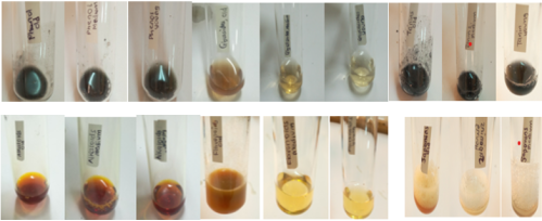
Figure 1. Colour Changes Confirming Presence of Phytochemicals.
3.2. Antibacterial Activity of Different Leaf Ages of Aloe vera Extracts on Staphylococcus aureus Bacteria
The results obtained from the study revealed that the older leaves possessed a higher inhibitory activity of 18.50 ± 0.00 mm followed by the medium leave with 17.50 ± 0.00 mm and finally the young had the least activity of 16.00 ± 0.00 mm and the results were tabulated in the Table 2 below though the results still show that there is no significant difference between the tested Old and Medium ages of the Aloe vera and the positive control (tetracycline) with a p-value ranging from 0.2552 to 0.7557 but significantly different from the Young aged leaves with the p-value of < 0.0001. The results still showed no significant difference between the different leaf ages of the Aloe vera with the p-value ranging between 0.4645 and 0.7557 but all the treatments used were significantly different from the negative control (water) used in the study after Tukey’s multiple comparison tests were run as shown in the Table 3 and Figure 2.
Table 2. Antibacterial Activity of Leaf Extracts against S. aureus showed by zones of inhibition.

Treatments

Inhibition zone (mean ± Sd) mm

Water

0.00 ± 0.00

Tetracycline

19.50 ± 0.00

Young

16.00 ± 0.00

Medium

17.50 ± 0.00

Old

18.50 ± 0.00

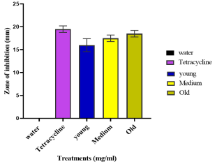
Figure 2. Antibacterial Activity of Leaf Extracts against S. aureus showed by zones of inhibition.
The comparisons of the activities of different parts of the Aloe vera leaves extracts were made to check for significant differences. The results obtained using Tukey's multiple comparisons test were recorded.
Table 3. One-way ANOVA analysis (Tukey’s analysis test).

Tukey's multiple comparisons tests

Mean Diff.

Significant?

Adjusted P Value

water vs. Tetracycline

-19.50

Yes

<0.0001

water vs. young

-16.00

Yes

<0.0001

water vs. Medium

-17.50

Yes

<0.0001

water vs. Old

-18.50

Yes

<0.0001

Tetracycline vs. young

3.500

Yes

0.0427

Tetracycline vs. Medium

2.000

No

0.2552

Tetracycline vs. Old

1.000

No

0.7557

young vs. Medium

-1.500

No

0.4645

young vs. Old

-2.500

No

0.1371

Medium vs. Old

-1.000

No

0.7557

3.3. The Minimum Inhibitory Concentration of Different Leaf Ages of Aloe vera Plant Extracts on Staphylococcus Bacteria
The results obtained after serially diluting the different extracts of different leaf ages of the Aloe vera plant showed that they all exhibited a similar minimum inhibitory concentration of 12.5 mg/ml of the initial concentration which was 50 mg/ml. the results were recorded in the Table 4 and Figure 3.
Table 4. Minimum Inhibitory Concentration Of Aloe Vera Leaf Extracts at Different Ages.

Treatments

MIC (mg/ml)

Young

12.5

Medium

12.5

Old

12.5

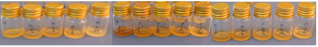
Figure 3. Minimum Inhibitory Concentration Of Aloe Vera Leaf Extracts.
3.4. The Minimum Bactericidal Concentration of Different Leaf Ages of Aloe vera on Staphylococcus aureus Bacteria
All the tested extracts from the different leaf ages of the Aloe vera plant still exhibited a similar MBC of 25 mg/ml on the test organism as shown in Table 5 and Figure 4.
Table 5. Minimum Bactericidal Concentration of leaf extracts of Aloe Vera at Different ages.

Treatments

MBC (mg/ml)

Young

25

Medium

25

Old

25

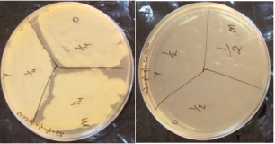
Figure 4. Minimum Bactericidal Concentration Of Aloe Vera Leaf Extracts.
4. Discussion of Results
4.1. Phytochemical Analysis
The phytochemical screening revealed the presence of some active compounds which include saponins, glycosides, flavonoids, alkaloids phenols, and tannins as presented in table 1 above. Secondary metabolites identified in plant material have been reported as having inhibitory action against pathogenic microorganisms . The presence of the above-named secondary metabolites in the various Aloe Vera extracts in different leaf ages could attribute to the inhibitory activities observed in the antimicrobial tests conducted against Staphylococcus aureus.
Saponins and tannins were highest in the old and medium leaf extract of Aloe vera extract when compared to the young leaf extract, while glycosides had the highest intensity in the old leaf extract, least in the medium, and not present in the young leaf extract. Phenols were more abundant in the old leaf extract, moderate in the medium leaf extract and least in the young leaf extract of Aloe vera. Flavonoids are least present in the young and medium leaf extract and absent in the old leaf extract of Aloe vera. Finally, the alkaloids are least present in all the different leaf ages of Aloe vera that is the young, medium, and old leaf extract.
Tannins bind to the cell walls of bacteria, thus inducing bacterial stasis and protease activity . Tannins also interfere with bacterial cell wall development causing disintegration of bacterial colonies thus inhibiting microbial growth through precipitating the microbial protein thus depriving them of nutritional proteins needed for their growth and development . Phenolics and Polyphenol inhibit the growth of microorganisms through enzyme inhibition by the oxidized compounds, possibly through reaction with sulfhydryl groups or through more nonspecific interactions with the proteins .
The current study has also revealed that the amount of the phytochemicals in the extracts varied in the different ages of leaf extracts. The difference in the nature and amount of these phytochemicals in the leaves extract accounts for the difference in their antimicrobial activity. The more the amount of these phytochemicals an extract contains, the higher the antimicrobial activity it possesses . The findings from the study about the present phytochemicals in different leaf ages of the Aloe vera are in line with other researchers who carried out the phytochemical test on the leaves of the plant without considering the difference in ages and found out the present phytochemicals which were, reducing sugar, phenolic compounds, steroids, terpenoids, carbohydrate, amino acid, flavonoids, tannins saponins, glycosides, alkaloids, and glycosides .
There is a slight disagreement in the phytochemicals present whereby we found out that tannins were present when Aloe vera leaves of different ages were extracted with ethanol though according to a study who reported that they were absent when he used the same extracting solvent.
4.2. Antimicrobial Activity
The antimicrobial properties of the leaf extract depend on the type and amount of chemical constituents and exhibit different modes of action depending on the main components present. The antimicrobial activities of Aloe vera leaf extracts at different leaf ages against S. aureus were quantitatively analyzed using inhibition zone diameters, MIC, and MBC values. The older leaves proved to be highly significant for use in the treatment of Staphylococcus infections rather than the medium and young leaves due to the possession of most vital phytochemical concentrations giving the older leaves a chance to exhibit larger inhibition zones through interfering with the growth of the organism. The results are in line with the findings of , who found out that leaf extracts of Aloe Vera have antibacterial activity on a number of bacterial strains and a few fungal strains. As reported by who evaluated the activity of Aloe vera extract against pathogenic bacteria and found that variations in concentrations bring about differences in the inhibition zones. The results are not in line with some researchers who found that the inhibition zone was over 80mm when tested on different fungal diseases such as Penicillium notatum . The results obtained are not consistent with that of who evaluated an antibacterial activity against selected pathogenic organisms of E. coli, Pseudomonas aeruginosa, S. aureus and Bacillus subtilis using ethanol extracts from Aloe vera gel and found out that it exhibited no activity against the bacterial strains.
4.3. Minimum Inhibitory Concentration (MIC)
The MIC obtained from the present study is slightly higher than that recorded by , who tested the gel from Aloe vera leaves on the fungus, gram-positive and negative bacteria of E. coli, Pseudomonas aeruginosa, Staphylococcus aureus, and Candida albicans. This might have happened due to variation in the present chemicals that affected the bacteria. The results obtained from the study agree with those published by , who carried out phytochemical screening and comparative antimicrobial activity of Aloe. vera extracts obtained through the use of methanol solvent. The presence of lower phytochemical concentration or possession of quite similar chemical compositions might have resulted in similar MIC values although there were deviations of phytochemical in different leaf ages but gave equal MIC due to similarities in the mechanism of action.
4.4. Minimum Bactericidal Concentration (MBC)
The MBC obtained from the present study is in line with that recorded by who carried out a comparative antimicrobial test and phytochemical screening of Aloe vera extracts on Bacillus subtilis, Staphylococcus aureus, Proteus mirabilis, and Candida albicans, when methanol extracts were used, similar results were obtained. The findings of this study deviate from those of who evaluated antimicrobial potency of Aloe vera leaves on Staphylococcus aureus bacteria and got different results.
5. Conclusion
The qualitative phytochemical analysis of Aloe vera leaf extracts at various ages revealed a concentration gradient, with older leaves showing higher concentrations followed by medium and young leaves. This observation suggests a dynamic maturation-related variation in the phytochemical composition of Aloe vera.
The antibacterial assay against Staphylococcus aureus showed varying inhibitory activities among different leaf ages. The older leaves had the highest inhibitory activity, followed by medium leaves, and young leaves with the least inhibition. Our findings indicated that there was no significant difference between the inhibitory activities of older and medium-aged leaves, but the young leaves exhibited a significantly lower inhibition zone. This shows a potential age-dependent variation in the antibacterial efficacy of Aloe vera leaf extracts against Staphylococcus aureus.
The serial dilution results indicated a consistent minimum inhibitory concentration of 12.5mg/ml across all leaf ages. Our findings show that, however much variations in phytochemical content and antibacterial activity, the lowest concentration of Aloe vera leaf extracts, at which 99.9% of the final inoculum of Staphylococcus aureus is killed remains consistent for all ages.
6. Recommendations
The variations antibacterial activity across different leaf ages call for further research. In future more work would go deep into the specific phytochemicals for the antibacterial efficacy and also their mode of action. By ensuring that only mature leaves are collected, we would promote sustainable harvesting, to maximize the beneficial properties of the plant while minimizing waste.
Study Limitations
Delays in plant identification and classification due to the country's scarcity of experienced field botanists. Due to a lack of financing, collaborative scientific cooperation for advanced investigations and bioactivity testing were hampered.
Abbreviations

WHO

World Health Organization

OD

Optical Density

MBC

Minimum Bactericidal Concentration

MIC

Minimum Inhibitory Concentration

MHA

Mueller-Hinton Agar

MHB

Mueller-Hinton Broth

CFU

Colony Forming Units

Acknowledgments
Robert, with deep and sincere gratitude, would like to appreciate the efforts to all members (family and at work) who continuously provide support. Isaac would like to dedicate this work to his family and departmental members of Biological Sciences, Kyambogo University for the efforts they have rendered in support of this research work.
Author Contributions
Alule Robert: Conceptualization, Data curation, Formal Analysis, Investigation, Methodology, Software, Visualization, Writing – original draft, Writing – review & editing
Isabirye Isaac: Funding acquisition, Project administration, Resources, Supervision, Validation
Conflicts of Interest
The authors declare no conflicts of interest.
References
[1] A. Robert, I. Isaac, and W. Joel, “Phytochemistry and Antibacterial Activity of Platycerium coronarium (Tanduk Rasa Fern) Leaf Extracts,” J. Dis. Med. Plants, vol. 9, no. 4, pp. 137–147, Nov. 2023,
[2] World Health Organization (WHO), “WHO Traditional Medicine Strategy 2014-2023,” World Heal. Organ., pp. 1–76, 2013.
[3] G. M. John, D. L. Hershman, L. Falci, Z. Shi, W.-Y. Tsai, and H. Greenlee, “Complementary and alternative medicine use among US cancer survivors,” J. Cancer Surviv., vol. 10, no. 5, pp. 850–864, 2016,
[4] U. Nandal and R. L. Bhardwaj, “Aloe vera: A valuable wonder plant for food, medicine and cosmetic use - a review,” Int. J. Pharm. Sci. Rev. Res., vol. 13, pp. 59–67, Mar. 2012.
[5] M. H. Radha and N. P. Laxmipriya, “Evaluation of biological properties and clinical effectiveness of Aloe vera: A systematic review,” J. Tradit. Complement. Med., vol. 5, no. 1, pp. 21–26, 2015,
[6] S. Ishaque, A. Arshad, M. Haider, and F. Fatima, “Biological and Clinical Sciences Research Journal,” Biol. Clin. Sci. Res. J., pp. 1–9, 2021.
[7] E. V. Christaki and P. C. Florou-Paneri, “Aloe vera: A plant for many uses,” J. Food, Agric. Environ., vol. 8, no. 2, pp. 245–249, 2010.
[8] S. Rahman, P. Carter, and N. Bhattarai, “Aloe Vera for Tissue Engineering Applications,” J. Funct. Biomater., vol. 8, no. 1, p. 6, 2017,
[9] G. Anywar, P. Tugume, and E. K. Kakudidi, “A review of Aloe species used in traditional medicine in East Africa,” South African J. Bot., vol. 147, pp. 1027–1041, 2022,
[10] L. K. Keter and P. C. Mutiso, “Ethnobotanical studies of medicinal plants used by Traditional Health Practitioners in the management of diabetes in Lower Eastern Province, Kenya,” J. Ethnopharmacol., vol. 139, no. 1, pp. 74–80, 2012,
[11] P. O. Otieno, “A review on antiplasmodial potential and quantification of aloin and aloe-emodin in aloe vera.,” 2022, Busitema University.
[12] A. Jangra, G. Sharma, S. Sihag, and V. Chhokar, “The dark side of miracle plant-Aloe vera: a review,” Mol. Biol. Rep., vol. 49, no. 6, pp. 5029–5040, 2022,
[13] D. Hekmatpou, F. Mehrabi, K. Rahzani, and A. Aminiyan, “The effect of aloe vera clinical trials on prevention and healing of skin wound: A systematic review,” Iran. J. Med. Sci., vol. 44, no. 1, pp. 1–9, 2019.
[14] X. Guo and N. Mei, “Aloe vera: A review of toxicity and adverse clinical effects,” J. Environ. Sci. Heal. Part C, vol. 34, no. 2, pp. 77–96, Apr. 2016,
[15] A. Manilal et al., “In vitro antibacterial activity of medicinal plants against biofilm-forming methicillin-resistant Staphylococcus aureus: efficacy of Moringa stenopetala and Rosmarinus officinalis extracts,” Heliyon, vol. 6, no. 1, p. e03303, 2020,
[16] L. Damian and S. Paţachia, “Method for Testing the Antimicrobial Character of the Materials and Their Fitting To the Scope,” Bull. Transilv. Univ. Braşov, vol. 7, no. 56, pp. 37–44, 2014, [Online]. Available:
[17] K. S. Banu and L. Cathrine, “General Techniques Involved in Phytochemical Analysis,” Int. J. Adv. Res. Chem. Sci., vol. 2, no. 4, pp. 25–32, 2015, [Online]. Available:
[18] M. A. Wani, S. P. Ceo, U. P. Livestock, O. Prakash, and S. Prasad, “Qualitative phytochemical analysis of various parts of bamboo (Bambusa balcooa) for possible therapeutic usages in bovine reproductive disorders,” ~ 217 ~ J. Pharmacogn. Phytochem., vol. 8, no. 1, pp. 217–221, 2019, [Online]. Available:
[19] M. Balouiri, M. Sadiki, and S. K. Ibnsouda, “Methods for in vitro evaluating antimicrobial activity: A review,” J. Pharm. Anal., vol. 6, no. 2, pp. 71–79, 2016,
[20] M. Benkova, O. Soukup, and J. Marek, “Antimicrobial susceptibility testing: currently used methods and devices and the near future in clinical practice,” J. Appl. Microbiol., vol. 129, no. 4, pp. 806–822, Oct. 2020,
[21] G. C. Omojate, F. O. Enwa, A. O. Jewo, and C. O. Eze, “Mechanisms of Antimicrobial Actions of Phytochemicals against Enteric Pathogens – A Review,” J. Pharm. Chem. Biol. Sci., vol. 2, no. 2, pp. 77–85, 2014.
[22] V. Brezáni and K. Šmejkal, “Secondary metabolites isolated from the genus Eucalyptus,” Chemistry, vol. 1, 2006.
[23] K. Sebei, F. Sakouhi, W. Herchi, M. L. Khouja, and S. Boukhchina, “Chemical composition and antibacterial activities of seven Eucalyptus species essential oils leaves,” Biol. Res., vol. 48, pp. 1–5, 2015,
[24] R. HO and O. PE, “Antimicrobial Potency of Methanolic Leaf Extracts from Selected Medicinal Plants against Staphylococcus aureus,” J. Med. Microbiol. Diagnosis, vol. 5, no. 1, pp. 1–4, 2016,
[25] V. Ikpe, C. S. Eze, P. Mbaoji, and P. E. Joshua, “Phytochemical analysis and antifungi activity of aloe vera leaves,” Bio-Research, vol. 15, no. 1, p. 974, 2019,
[26] K. Thu, Y. Y. Mon, T. A. Khaing, and O. M. Tun, “Study on Phytochemical Properties, Antibacterial Activity and Cytotoxicity of Aloe vera L.,” Int. J. Biotechnol. Bioeng., vol. 7, no. 5, pp. 285–289, 2013.
[27] H. Omer, M. Abakar, S. E. A. Bakhiet, and R. S. M. Abadi, “Antimicrobial activity and minimum inhibitory concentration of Aloe vera sap and leaves using different extracts,” J. Pharmacogn. Phytochem., vol. 6, no. 3, pp. 297–302, 2017.
[28] P. Danish, Q. Ali, M. Hafeez, and A. Malik, “Antifungal and Antibacterial Activity of Aloe Vera Plant Extract,” Biol. Clin. Sci. Res. J., vol. 2020, no. 1, pp. 1–8, 2020,
[29] G. G. Yebpella, M. M. Adeyemi Hassan, C. Hammuel, A. M. Magomya, A. S. Agbaji, and E. M. Okonkwo, “Phtyochemical screening and comparative study of antimicrobial activity of Aloe vera various extracts,” African J. Microbiol. Res., vol. 5, no. 10, pp. 1182–1187, 2011,
Cite This Article
  • APA Style

    Robert, A., Isaac, I. (2024). Phytochemical Analysis and Antibacterial Activity of Aloe Vera Leaf Extracts Across Different Leaf Ages. Journal of Diseases and Medicinal Plants, 10(4), 60-68. https://doi.org/10.11648/j.jdmp.20241004.11

    Copy | Download

    ACS Style

    Robert, A.; Isaac, I. Phytochemical Analysis and Antibacterial Activity of Aloe Vera Leaf Extracts Across Different Leaf Ages. J. Dis. Med. Plants 2024, 10(4), 60-68. doi: 10.11648/j.jdmp.20241004.11

    Copy | Download

    AMA Style

    Robert A, Isaac I. Phytochemical Analysis and Antibacterial Activity of Aloe Vera Leaf Extracts Across Different Leaf Ages. J Dis Med Plants. 2024;10(4):60-68. doi: 10.11648/j.jdmp.20241004.11

    Copy | Download

  • @article{10.11648/j.jdmp.20241004.11,
      author = {Alule Robert and Isabirye Isaac},
      title = {Phytochemical Analysis and Antibacterial Activity of Aloe Vera Leaf Extracts Across Different Leaf Ages
    },
      journal = {Journal of Diseases and Medicinal Plants},
      volume = {10},
      number = {4},
      pages = {60-68},
      doi = {10.11648/j.jdmp.20241004.11},
      url = {https://doi.org/10.11648/j.jdmp.20241004.11},
      eprint = {https://article.sciencepublishinggroup.com/pdf/10.11648.j.jdmp.20241004.11},
      abstract = {The increasing reliance on plant-based healthcare products, including herbal medicines and dietary supplements, emphasizes the global significance of traditional remedies. Aloe Vera with 400 reported species, has been widely used herbal remedy for health practices worldwide. Despite its extensive historical use and therapeutic reputation, recent studies have raised concerns about potential adverse effects, challenging the notion of Aloe Vera as a universally safe functional food material. This study aimed to analyze the phytochemical composition of whole leaf extracts at various maturation stages of Aloe Vera and assess their antibacterial effect against Staphylococcus aureus. The qualitative phytochemical analysis revealed a concentration gradient, with older leaves exhibiting higher concentrations compared to medium and young leaves, suggesting a dynamic maturation-related variation. The antibacterial assay demonstrated age-dependent inhibitory activities, with older leaves displaying the highest, medium leaves following, and young leaves exhibiting the least inhibition. A consistent minimum inhibitory concentration of 12.5 mg/ml was observed across all leaf ages. These findings stress the need for cautious Aloe Vera consumption, especially in rural communities where whole-leaf extraction is prevalent, as recent studies have reported adverse effects and potential health risks associated with certain compounds. Safer alternatives, and regulating consumption practices are recommended, emphasizing sustainable practices to maximize plant benefits and minimize waste.
    },
     year = {2024}
    }
    

    Copy | Download

  • TY  - JOUR
    T1  - Phytochemical Analysis and Antibacterial Activity of Aloe Vera Leaf Extracts Across Different Leaf Ages
    
    AU  - Alule Robert
    AU  - Isabirye Isaac
    Y1  - 2024/12/23
    PY  - 2024
    N1  - https://doi.org/10.11648/j.jdmp.20241004.11
    DO  - 10.11648/j.jdmp.20241004.11
    T2  - Journal of Diseases and Medicinal Plants
    JF  - Journal of Diseases and Medicinal Plants
    JO  - Journal of Diseases and Medicinal Plants
    SP  - 60
    EP  - 68
    PB  - Science Publishing Group
    SN  - 2469-8210
    UR  - https://doi.org/10.11648/j.jdmp.20241004.11
    AB  - The increasing reliance on plant-based healthcare products, including herbal medicines and dietary supplements, emphasizes the global significance of traditional remedies. Aloe Vera with 400 reported species, has been widely used herbal remedy for health practices worldwide. Despite its extensive historical use and therapeutic reputation, recent studies have raised concerns about potential adverse effects, challenging the notion of Aloe Vera as a universally safe functional food material. This study aimed to analyze the phytochemical composition of whole leaf extracts at various maturation stages of Aloe Vera and assess their antibacterial effect against Staphylococcus aureus. The qualitative phytochemical analysis revealed a concentration gradient, with older leaves exhibiting higher concentrations compared to medium and young leaves, suggesting a dynamic maturation-related variation. The antibacterial assay demonstrated age-dependent inhibitory activities, with older leaves displaying the highest, medium leaves following, and young leaves exhibiting the least inhibition. A consistent minimum inhibitory concentration of 12.5 mg/ml was observed across all leaf ages. These findings stress the need for cautious Aloe Vera consumption, especially in rural communities where whole-leaf extraction is prevalent, as recent studies have reported adverse effects and potential health risks associated with certain compounds. Safer alternatives, and regulating consumption practices are recommended, emphasizing sustainable practices to maximize plant benefits and minimize waste.
    
    VL  - 10
    IS  - 4
    ER  - 

    Copy | Download

Author Information
  • Abstract
  • Keywords
  • Document Sections

    1. 1. Introduction
    2. 2. Materials and Methods
    3. 3. Results
    4. 4. Discussion of Results
    5. 5. Conclusion
    6. 6. Recommendations
    Show Full Outline
  • Study Limitations
  • Abbreviations
  • Acknowledgments
  • Author Contributions
  • Conflicts of Interest
  • References
  • Cite This Article
  • Author Information