Abstract
Introduction. Frontal sinus fractures connecting sinus cavity and endocranium without neurological clinical expression or intracranial CT scan injury are "falsely" considered benign especially when no liquorrhea has been objectified. However, because of naso frontal canal presence and the frontal sinus cavity septic nature, these fractures are most often responsible for very serious complications (infections, pneumencephaly) causing sequelae (psychological disorders) most often very disabling even after surgical management. The purpose of this work was to report our therapeutic attitude and evolution of patients. Method. This was a descriptive, analytical retrospective study covering 3 years in the neurosurgery department of the Yalgado Ouedraogo University Hospital in Ouagadougou. All cases of frontal sinus fractures connecting sinus cavity and endocranium with a "workable medical record" were included. Results. We included 47 patients. Average intake rate was 05 days (1 hour - 6.3 months). At admission, 5 patients (10.6%) had febrile meningeal disease. A CT scan made it possible to objectify a compressive pneumencephaly in 9 cases (19.1%), a brain abscess in 2 cases (4.3%). Surgery was performed in 16 patients (34.0%). Incision was made in a frontal skin fold in 10 cases (21.3%). After 3 months and 6 months follow-upf, there were respectively more complications (p=0.0 00017) and more very disabling sequelae (p = 0.016) among non-operated cases. Conclusion. Complications and sequelae were greater among non-operated patients. It is better to cranialize all this types of fractures as quickly as possible through an incision in a frontal skin fold that was the most efficient.
|
Published in
|
International Journal of Neurosurgery (Volume 10, Issue 1)
|
|
DOI
|
10.11648/j.ijn.20261001.11
|
|
Page(s)
|
1-8 |
|
Creative Commons
|

This is an Open Access article, distributed under the terms of the Creative Commons Attribution 4.0 International License (http://creativecommons.org/licenses/by/4.0/), which permits unrestricted use, distribution and reproduction in any medium or format, provided the original work is properly cited.
|
|
Copyright
|
Copyright © The Author(s), 2026. Published by Science Publishing Group
|
Keywords
Fractures, Frontal Sinus, Cranialization, Brain Abscess
1. Introduction
Frontal sinus fractures frequently depressives because of frontal sinus walls thinness. This leads to aesthetic damage and/ or intracranial lesions (meningeal or encephalic)
| [1] | Johnson NR, Roberts MJ. Frontal sinus fracture management: a systematic review and meta-analysis. Int J Oral Maxillofac Surg. 2021 Jan; 50(1): 75-82. |
| [2] | Al-Shami H, Alnemare AK, Mahfoz TB, Salah AM. Traumatic Frontal Sinus Fractures Management: Experience from High-Trauma Centre. Korean J Neurotrauma. 2021 Mar 18; 17(1): 15-24. |
[1, 2]
. Frontal sinus fractures connecting sinus cavity and endocranial without neurological clinical expression or intracranial injury on CT scan are "falsely" considered benign (especially when no cerebrospinal fluid leakage has not been objectified) both by some patients who do not take time to consult immediately and by some surgeons who do not initially operate on them. However, because of the presence of the frontal naso canal (connecting frontal sinus cavity with external environment) and the septic nature of frontal sinus cavity, these fractures are most often providers of very serious intracranial complications (infections, compressive pneumencephaly) causing sequelae (psychological disorders) most often very disabling even after surgical management. The most feared infectious complications are meningitis, mucocele, pyomucocele, brain abscess and empyema
| [3] | Vincent A, Wang W, Shokri T, Gordon E, Inman JC, Ducic Y. Management of Frontal Sinus Fractures. Facial Plast Surg. 2019 Dec; 35(6): 645-650. |
[3]
.
As a result, management of these frontal sinus fractures is still controversial
| [4] | Le P, Martinez R, Black J. Frontal Sinus Fracture Management Meta-analysis: Endoscopic Versus Open Repair. J Craniofac Surg. 2021 Jun 1; 32(4): 1311-1315. |
[4]
. Because of the frequency combination of intracranial lesions, this surgical management is most often done by a multidisciplinary team (neurosurgeon, ORL, maxilo fascial surgeons). Similarly, several approaches have been proposed: open classical surgery (wide uni/bitrage incision; minimal incision in a frontal skin fold); endoscopic surgery
| [1] | Johnson NR, Roberts MJ. Frontal sinus fracture management: a systematic review and meta-analysis. Int J Oral Maxillofac Surg. 2021 Jan; 50(1): 75-82. |
| [4] | Le P, Martinez R, Black J. Frontal Sinus Fracture Management Meta-analysis: Endoscopic Versus Open Repair. J Craniofac Surg. 2021 Jun 1; 32(4): 1311-1315. |
[1, 4]
. To this I add the thorny question of frontal sinus cranialization with or without obliteration
| [5] | Wichova H, Chiu AG, Villwock JA. Does the frontal sinus need to be obliterated following fracture with frontal sinus outflow tract injury? Laryngoscope. 2017 Sep; 127(9): 1967-1969. |
[5]
.
The purpose of this work is to report our therapeutic attitude and the evolution of patients we have taken care for frontal sinus fracture connecting sinus cavity and endocranium.
2. Method
This was a descriptive and analytical study with retrospective collection run over in the neurosurgery department of Yalgado Ouedraogo University Hospital in Ouagadougou. It covered a period of 3 years from January 1, 2013 to December 31, 2015.
Study population was medicals records of patients admitted for head trauma with a head fracture. This was an exhaustive sample that included all cases of closed frontal sinus fracture (without cutaneous opening allowing communication of fracture site with sinus cavity) connecting sinus cavity and endocranium, and having a "usable medical record". Cases with unrecovered or 'unusable' medical records were not included, as were cases with other traumatic injuries more severe than frontal sinus fracture (19 cases of polytrauma, 7 cases of coma).
Exact Fisher test compared qualitative variables with a 95% confidence interval. Any probability value (p) less than 0.05 was considered statistically significant.
3. Results
3.1. Epidemiological Data
We identified 2851 patients with a skull fracture including 47 cases (35.6%) of frontal sinus fracture connecting sinus cavity and endocranium.
Patients average age was 22.4 years with extremes of 3 years and 66 years. Sex ratio was 5.31. Average delay between trauma and admission was 05 days with extremes of 1 hour and 6.3 months. Etiologies were represented by road traffic accidents (30 cases or 6 3.4%), brawls (8 cases or 16.1%), falls from a height (6 cases or 12.3%), landslides / reception of load (3 cases 6.4%) and firearm injuries (1 case or 2.1%).
3.2. Diagnostic Data
Reasons for consultation were represented by the notion of head trauma in all cases; a notion of initial consciousness loss in 18 cases (38.3%): headaches in 16 cases (34.0%); dizziness in 2 cases (4.3%) and epileptic seizures in 2 cases (4.3%).
On head local examination, bone depression was noted in 36 cases (76.6%).
Table 1 shows the distribution of reasons for consultation and local signs.
Table 1. Distribution of reasons for consultation and head local signs.
| Numbers (n=47) | Frequency |
Reason for consultation |
Notion of head trauma | 47 | 100 |
Initial loss of consciousness | 18 | 38,3 |
Alteration of consciousness from the outset | 17 | 36,2 |
Headache | 16 | 34,0 |
Dizziness | 2 | 4,3 |
Seizure | 2 | 4,3 |
Head local examination |
Bone depression | 36 | 76,6 |
Excoriations, scalp | 15 | 31,9 |
Scalp Inflammatory edema | 11 | 23,4 |
Loss of brain matter | 8 | 17,0 |
Rhino-liquorrhea | 19 | 40,4 |
Palpebral edema | 3 | 6,4 |
In 11 cases (23.4%), neurological examination was normal. According to Glasgow coma score scale (GSC), head trauma was mild (GCS 15 to 13) in 33 cases (70.2%) and moderate (GCS 12 to 9) in 14 cases (2 9.8%). Limbs motor deficit was noted in 11 cases (23.4%); agitation in 7 cases (14.9%); aphasia in 5 cases (10.6%); febrile meningeal syndrome in 5 cases (10.6%) and facial paralysis in 1 case (2.1%).
Brain Computed tomography (CT) scan was performed in all patients. Two patients (4.2%) performed skull X-ray that objectified depressive fracture.
The fracture was located in frontal in 32 patients (68.1%), frontoparietal in 12 patients (25.5%); fronto-temporal in 2 patients (4.3%) and fronto-temporo-parietal in 1 patient (2.1%).
Frontal sinus fracture was unilateral in 38 cases (80.9%) and bilateral in 9 cases (19.1%). It was depressive in 44 cases (93.6%) including 35 cases (74.5%) little or no compressive; not depressive in the other cases. In 9 cases (19.1%), depressive fracture was highly compressive and associated with a dural and/or parenchymal lesion. There was pneumencephaly in 23 cases (48.9%) including 13 intradural cases (27.7%). Pneumencephaly was compressive in 9 cases (19.1%). Frontal sinus fracture was isolated with no further associated intracranial injury in 13 patients (27.7%) and associated with minimal intracranial injury in remaining cases. Brain contusion was associated in 25 patients (53.2%).
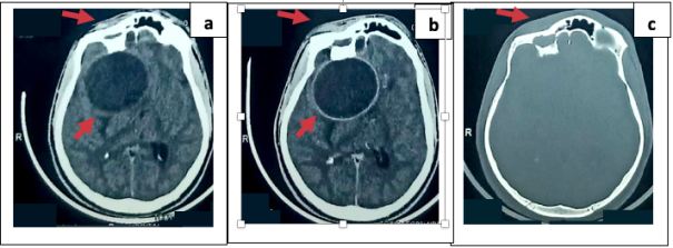
There was an infectious complication of brain abscess type in 2 cases (4.3%). CT scan lesions noted are shown in
Table 2.
Figure 1 shows CT scan images of patient admitted with brain abscess.
Table 2. Distribution of CT scan lesions.
| Numbers (n=47) | Frequency |
Depressive fracture | 44 | 93,6 |
Brain contusion | 25 | 53,2 |
Pneumencephaly | 23 | 48,9 |
Craniocerebral wound | 9 | 19,1 |
Localized brain edema | 7 | 14,9 |
Mass effect on ventricles | 5 | 10,6 |
Small extra dural hematoma | 3 | 6,4 |
Subarachnoid hemorrhage | 2 | 4,3 |
Small intraparenchymal hematoma | 2 | 4,3 |
Small subdural hematoma | 2 | 4,3 |
Brain abscess | 2 | 4,3 |
Figure 1. Cranioencephalic CT scan of a patient admitted complication stage (brain abscess). (a) axial section without contrast product injection showing a roundel image in the right frontal which is enhanced after injection of contrast product (b) and which is opposite a right frontal sinus fracture with intraparenchymal bone fragment (c).
3.3. Therapeutic and Evolutionary Data
Analgesic treatment was instituted in all patients; antibiotics in 17 patients (36.2%); anti-comitials in 2 patients (4.3%).
A surgical indication was retained in 21 patients (44.7%); surgical treatment was conducted in 16 patients (34.0%). Average time from diagnosis to surgical management was 21.4 days, with extremes of 8 hours and 60 days. Incision was made in a frontal skin fold centered on bone depression in 10 cases (21.3%), unitragal in 5 cases (10.6%) and bitragal in 1 case (2.1%). Craniectomy with bone fragments repositioning associated with cranialisation and obliteration of frontal sinus were performed in all patients; dural closure in 4 patients (8.5%), brain abscess evacuation in 2 patients (4.3%).
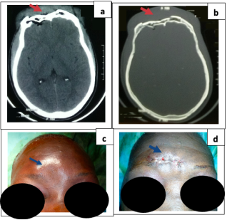
Figure 2 presents CT-scan and clinical images of a right sinus frontal depressive fracture operated through an incision in a frontal skin fold centred on bone depression.
Figure 2. CTscan and clinical images of a right frontal sinus depressive fracture operated through an incision in a frontal skin fold centered on bone depression. Preoperative CT scan images in axial section of parenchymal (a) and bone window (b) showing a compressive right frontal sinus depressive fracture with slight pneumencephaly opposite. Preoperative clinical image showing right mediolateral frontal bone depression (c); immediate postoperative post after depressive fracture removal, cranialization and obliteration of frontal sinus showing barely visible incision made in a frontal skin fold.
Average hospital stay was 8.2 days with extremes of 01 and 26 days. Intra-hospital evolution was favorable (improvement or stationary) in 34 patients, either 72.3%. There were 3 cases (6.4%) of complications such as intracranial infections in non-operated patients. Among operated patients, evolution was considered favorable (stationary or improved clinical condition) in 16 cases and unfavorable (complication or death) in no case; while in non-operated patients, it was favorable in 28 cases and unfavorable in 3 cases. This difference was not statistically significant with p=0.277.
After 3 months post-hospital follow-up, there were 16 cases (34%) of recovery without sequelae including 13 cases (27.7%) among operated cases. Among operated patients, this evolution was favorable (cure without or with slightly disabling sequelae) in 15 cases and unfavorable (infectious complications) in 1 case; while in non-operated patients, it was favorable in 8 cases and unfavorable in 21 cases (infectious complications in all cases associated with compressive pneumencephaly in 9 cases). This difference was statistically significant with p=0.000017.
After 6 months follow-up, post-hospital evolution was marked by 18 cases (38.3%) of recovery without sequelae including 13 cases (27.7%) among operated cases. Among operated patients, this evolution was favourable (cure without or with slightly disabling sequelae) in 15 cases and unfavourable (very disabling sequelae or death) in no case; while in non-operated patients, it was favourable in 18 cases and unfavourable in 9 cases. This difference was statistically significant with p=0.0105.
Table 3 presents intra- and post-hospital evolution of patients.
Table 3. Intra- and post-hospital evolution of patients.
| Operated (n= 16) | Not operated (n=31) | Total (n=47) |
Intra-hospital evolution (average duration = 8.23 days) |
Clinical improvement | 13 | 24 | 37 |
Stationary | 3 | 4 | 7 |
Aggravation / complication | 0 | 3 | 3 |
Post-hospital evolution (average follow-up = 3 months)) |
Healing without sequelae | 13 | 3 | 16 |
Healing with minimally disabling sequelae | 2 | 5 | 7 |
Aggravation / complication | 1 | 21 | 22 |
Lost to follow-up | 0 | 2 | 2 |
Post-hospital evolution (average follow-up = 6 months) |
Healing without sequelae | 13 | 5 | 18 |
Healing with minimally disabling sequelae | 2 | 13 | 15 |
Healing with very disabling sequelae | 0 | 8 | 8 |
Lost to follow-up | 1 | 4 | 5 |
Death | 0 | 1 | 1 |
Still at six months, amont slightly disabling sequelae, there were 05 cases of headache (including 1 operated), 4 cases of non-aesthetic sequelae (all not operated), 4 cases of epilepsy (including 1 operated) and 2 cases of mental disorders (all not operated). Very disabling sequelae were represented by 8 cases of mental disorders (frontal syndrome), all non-operated cases who presented compressive pneumencephaly in the third month of post-hospital follow-up. Despite surgical management of this pneumencephaly, mental disorders persisted in a very disabling way.
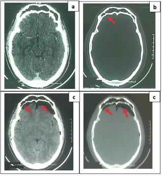
Figure 3 shows CT scans of a non-operated patient who initially had a discrete fracture of right frontal sinus posterior wall which progressed to compressive bifrontal pneumencephaly revealed by significant psychological disorders which persisted despite cranialization of the right frontal sinus.
Figure 3. CT scan images of a non-operated patient who initially had a discrete fracture of right frontal sinus posterior wall (right frontotemporal fracture) that progressed to compressive bifrontal pneumencephaly revealed by significant psychological disorders that persisted despite cranialization and obliteration of the right frontal sinus. Initial axial parenchymal (a) and bony window (b) showing a discrete right frontal sinus posterior wall fracture. Control images in axial section parenchymal (c) and bone window (d) showing a significant compressive bifrontal pneumencephaly complicating the initial right frontal sinus posterior wall fracture.
4. Discussion
Frontal fractures are common
| [6] | Guidah S, Samnuissi S, BA A, Niang EH. Apport de l’imagerie medicale dans le bilan des traumatismes crâniens au Niger. J Afr Imag Med 2012; 1 (4): 4-11. |
| [7] | Bayen E., Jourdan C., Azouvi P, Weiss JJ., Pradat-Diehl P. Prise en charge après lesion cerebrale acquise de type traumatisme crânien. L’Information psychiatrique 2012; 88 (5): 331-7. |
[6, 7]
due to frontal bone protrusion and lack of muscle protection on it. In different series, they occupied the first place of post-traumatic cranial fractures followed by parietal fractures. Thus, 47% of the fracture was frontal and 22.5% parietal
| [8] | Fatigba OH, Savi de Tove KM, Brun L, Babio R, Padonou J. Les lesion de la voûte crânienne chez les motocyclistes dans un centre hospitalier au Benin: à propos de 40 cas. Mali Medical. 2011; 26 (4): 60-4. |
[8]
; 38.9% frontal and 30.6% parietal
| [9] | Kanikomo D, Sogoba Y, Dama M, Coulibaly O, Diarra MS, Thiam AB et al. Etude epidemiologique, clinique et therapeutique des fractures embarrures du crâne au CHU Gabriel Toure. Mali Medical. 2015; 30 (2): 7-12. |
[9]
; 57% frontal and 19.6% parietal
| [10] | Fatigba OH, Savi DE, Tove KM, Hodonou MA, Allode S A. Epidemiologie et prise en charge des embarrures dans un centre hospitalier du Benin. Journal de Neurochirurgie 2012: 16: 10-16. |
[10]
. Frontal sinus fractures often occur against the background of high-kinetic energy trauma. In our series, they occurred most often during traffic accidents and frequently affected young adult males. This finding was made in several other studies. The sex ratio was 4,7
| [8] | Fatigba OH, Savi de Tove KM, Brun L, Babio R, Padonou J. Les lesion de la voûte crânienne chez les motocyclistes dans un centre hospitalier au Benin: à propos de 40 cas. Mali Medical. 2011; 26 (4): 60-4. |
[8]
; 5,2
| [1] | Johnson NR, Roberts MJ. Frontal sinus fracture management: a systematic review and meta-analysis. Int J Oral Maxillofac Surg. 2021 Jan; 50(1): 75-82. |
[1]
; 6,6
| [10] | Fatigba OH, Savi DE, Tove KM, Hodonou MA, Allode S A. Epidemiologie et prise en charge des embarrures dans un centre hospitalier du Benin. Journal de Neurochirurgie 2012: 16: 10-16. |
[10]
; 10,5
| [11] | Firouzbakht PK, Mohiuddin IS, Varman RM, Heinrich MP, Saa L, Cordero J. Analysis of Frontal Sinus Fracture Management and Resource Utilization. J Craniofac Surg. 2020 Nov/Dec; 31(8): 2240-2242. |
[11]
; 13,3
| [9] | Kanikomo D, Sogoba Y, Dama M, Coulibaly O, Diarra MS, Thiam AB et al. Etude epidemiologique, clinique et therapeutique des fractures embarrures du crâne au CHU Gabriel Toure. Mali Medical. 2015; 30 (2): 7-12. |
[9]
. Average age was 17.8 years
| [9] | Kanikomo D, Sogoba Y, Dama M, Coulibaly O, Diarra MS, Thiam AB et al. Etude epidemiologique, clinique et therapeutique des fractures embarrures du crâne au CHU Gabriel Toure. Mali Medical. 2015; 30 (2): 7-12. |
[9]
; 23,1 years
| [10] | Fatigba OH, Savi DE, Tove KM, Hodonou MA, Allode S A. Epidemiologie et prise en charge des embarrures dans un centre hospitalier du Benin. Journal de Neurochirurgie 2012: 16: 10-16. |
[10]
; 24,5 years
| [8] | Fatigba OH, Savi de Tove KM, Brun L, Babio R, Padonou J. Les lesion de la voûte crânienne chez les motocyclistes dans un centre hospitalier au Benin: à propos de 40 cas. Mali Medical. 2011; 26 (4): 60-4. |
[8]
; 36,2 years
| [1] | Johnson NR, Roberts MJ. Frontal sinus fracture management: a systematic review and meta-analysis. Int J Oral Maxillofac Surg. 2021 Jan; 50(1): 75-82. |
[1]
. Road traffic accidents were represented in 50.7% of cases
| [1] | Johnson NR, Roberts MJ. Frontal sinus fracture management: a systematic review and meta-analysis. Int J Oral Maxillofac Surg. 2021 Jan; 50(1): 75-82. |
[1]
.
Frontal sinus fractures were unilateral in 73.9% and bilateral in 26.1%
| [11] | Firouzbakht PK, Mohiuddin IS, Varman RM, Heinrich MP, Saa L, Cordero J. Analysis of Frontal Sinus Fracture Management and Resource Utilization. J Craniofac Surg. 2020 Nov/Dec; 31(8): 2240-2242. |
[11]
. Fracture was associated with intracranial injury in 17.61%
| [2] | Al-Shami H, Alnemare AK, Mahfoz TB, Salah AM. Traumatic Frontal Sinus Fractures Management: Experience from High-Trauma Centre. Korean J Neurotrauma. 2021 Mar 18; 17(1): 15-24. |
[2]
. Cerebrospinal fluid leakage was observed in 32.95% of cases
| [2] | Al-Shami H, Alnemare AK, Mahfoz TB, Salah AM. Traumatic Frontal Sinus Fractures Management: Experience from High-Trauma Centre. Korean J Neurotrauma. 2021 Mar 18; 17(1): 15-24. |
[2]
; 89,1 years
| [12] | Banks C, Grayson J, Cho DY, Woodworth BA. Frontal sinus fractures and cerebrospinal fluid leaks: a change in surgical paradigm. Curr Opin Otolaryngol Head Neck Surg. 2020 Feb; 28(1): 52-60. |
[12]
. Cerebrospinal fluid leakage was found in 40.4% of our cases. This leakage of cerebrospinal fluid is an indirect sign of a dural opening. When this cerebrospinal fluid leakage has not been objectified, frontal sinus fractures connecting sinus cavity and endocranial without neurological clinical expression or intracranial lesion on CT scan are "falsely" considered benign and neglected by both the patient and some surgeons. This could partly explain the long average consultation time (5 days) noted in our series. Indeed, local skin lesions (excoriations / scalp wounds) heal quickly with local care. Similarly, in case of depressive fracture, bone depression could pose a cosmetic problem for some patients but the majority of our patients do not make it a problem. As a result, most often no surgical indication is initially retained for this type of fracture especially when no sign of brain compression is visible on imaging. Yet these fractures are most often responsible for very serious complications
| [4] | Le P, Martinez R, Black J. Frontal Sinus Fracture Management Meta-analysis: Endoscopic Versus Open Repair. J Craniofac Surg. 2021 Jun 1; 32(4): 1311-1315. |
| [11] | Firouzbakht PK, Mohiuddin IS, Varman RM, Heinrich MP, Saa L, Cordero J. Analysis of Frontal Sinus Fracture Management and Resource Utilization. J Craniofac Surg. 2020 Nov/Dec; 31(8): 2240-2242. |
[4, 11]
.
Infections and pneumocephalus causing sequelae (psychological disorders) most often very disabling even after secondary surgical management. The most feared infectious complications are meningitis, mucocele, pyomucocele, brain abscess and empyema
| [3] | Vincent A, Wang W, Shokri T, Gordon E, Inman JC, Ducic Y. Management of Frontal Sinus Fractures. Facial Plast Surg. 2019 Dec; 35(6): 645-650. |
[3]
. These complications are frequent and predictable because the cavities of the frontal sinuses are septic promoting infection and because these cavities are directly in communication with external environment through nasofrontal canal promoting both pneumencephaly and infection. As antibiotic prophylaxis is controversial
| [13] | Hode L, Sogbo DHO, Hounnou MG, Houessou GF, Voyeme A. K. A, Chobli M. Prise en charge des traumatismes cranio-encephaliques chez les enfants à Cotonou. AJNS 2016; 35(1): 55-65. |
| [14] | Samake BM, Coulibaly Y, Diallo A, Tchamko FRD, Sidibe S, Toure A. Traumatisme crânio-encephalique à l’hôpital Gabriel Toure. Mali Medical. 2010; 25 (4): 3-7. |
[13, 14]
; these infections could be prevented by surgery to repair traumatic injuries. However, conservative treatment is the most realized. Rate of surgical indications was low in our series (44.7%) and literature (25%
| [15] | Obayemi A, Losenegger T, Long S, Spielman D, Casiano MF, Reeve G and al. Frontal Sinus Fractures: 10-Year Contemporary Experience at a Level 1 Urban Trauma Center J Craniofac Surg. 2021 Jun 1; 32(4): 1376-1380. |
[15]
; 13%
| [11] | Firouzbakht PK, Mohiuddin IS, Varman RM, Heinrich MP, Saa L, Cordero J. Analysis of Frontal Sinus Fracture Management and Resource Utilization. J Craniofac Surg. 2020 Nov/Dec; 31(8): 2240-2242. |
[11]
; 46.1%
| [2] | Al-Shami H, Alnemare AK, Mahfoz TB, Salah AM. Traumatic Frontal Sinus Fractures Management: Experience from High-Trauma Centre. Korean J Neurotrauma. 2021 Mar 18; 17(1): 15-24. |
[2]
. Operated fracture cases had a higher rate of displacement and comminution than those not operated (P < 0.00001)
| [15] | Obayemi A, Losenegger T, Long S, Spielman D, Casiano MF, Reeve G and al. Frontal Sinus Fractures: 10-Year Contemporary Experience at a Level 1 Urban Trauma Center J Craniofac Surg. 2021 Jun 1; 32(4): 1376-1380. |
[15]
. Several approaches have been described: open surgery (minimal incision in a frontal skin fold, unilateral or biragal incision) endoscopic surgery
| [1] | Johnson NR, Roberts MJ. Frontal sinus fracture management: a systematic review and meta-analysis. Int J Oral Maxillofac Surg. 2021 Jan; 50(1): 75-82. |
| [3] | Vincent A, Wang W, Shokri T, Gordon E, Inman JC, Ducic Y. Management of Frontal Sinus Fractures. Facial Plast Surg. 2019 Dec; 35(6): 645-650. |
| [16] | Dedhia RD, Morisada MV, Tollefson TT, Strong EB. Contemporary management of frontal sinus fractures. Curr Opin Otolaryngol Head Neck Surg. 2019 Aug; 27(4): 253-260. |
[1, 3, 16]
. These gestures performed depend on existing lesions: dural repair, depressive fracture removal, frontal sinus cranialization, nasofrontal canal obliteration. Frontal sinus cavity and nasofrontal canal preservation to maintain a certain function of the sinus have been abandoned nowadays because of the many complications (mucoceles, chronic frontal sinusitis, leakage of cerebrospinal fluid, meningitis, brain abscesses)
| [5] | Wichova H, Chiu AG, Villwock JA. Does the frontal sinus need to be obliterated following fracture with frontal sinus outflow tract injury? Laryngoscope. 2017 Sep; 127(9): 1967-1969. |
[5]
.
In our series, we performed cranialization and obliteration of frontal sinus in all operated patients. Similarly, we made a frontal incision in a skin fold in the majority of cases in our series. This incision seems to us more beneficial than the classic uni or bitragal incision which prolongs operating time, leads to more blood spoliation without necessarily solving aesthetic problem for which this skin incision is promoted. Indeed the uni or bitragal incision is called "neurosurgical" and promoters of this incision advance the fact that it would be more aesthetic than the incision in a frontal fold that would leave an unsightly postoperative skin scar in a very visible face part. We are not too much of this opinion because in case of trauma there is already a local lesion that causes a post-traumatic scar in this visible place; Then tragal incision also leaves a scar on which hair no longer grows which is very non-aesthetic when patient cuts hair short, forcing patient to either shave hair permanently, or leave it always very long, or wear a headdress. In addition, this incision in a frontal skin fold is the rule among ORL or maxillofascial surgeons with whom we had to take care of some patients in our series. The latter manage to minimize this aesthetic problem by a skin suture with very fine absorbable monofilament thread (4/0 or 5/0) by taking very close points or by making an intradermal suture.
Elsewhere, advances in minimally invasive surgical techniques are introducing endoscopy by minimizing the risks and morbidity associated with more traditional treatment approaches
| [16] | Dedhia RD, Morisada MV, Tollefson TT, Strong EB. Contemporary management of frontal sinus fractures. Curr Opin Otolaryngol Head Neck Surg. 2019 Aug; 27(4): 253-260. |
| [17] | Pawar SS, Rhee JS. Frontal sinus and naso-orbital-ethmoid fractures. JAMA Facial Plast Surg. 2014 Jul-Aug; 16(4): 284-9. |
[16, 17]
.
Endoscopic endonasal repair has been performed in 97.6% of cases successfully with excellent aesthetic results
| [12] | Banks C, Grayson J, Cho DY, Woodworth BA. Frontal sinus fractures and cerebrospinal fluid leaks: a change in surgical paradigm. Curr Opin Otolaryngol Head Neck Surg. 2020 Feb; 28(1): 52-60. |
[12]
. However, based on a meta-analysis, no definitive conclusion regarding the superiority of endoscopic approach over open surgery can be drawn
| [12] | Banks C, Grayson J, Cho DY, Woodworth BA. Frontal sinus fractures and cerebrospinal fluid leaks: a change in surgical paradigm. Curr Opin Otolaryngol Head Neck Surg. 2020 Feb; 28(1): 52-60. |
[12]
.
In our series after 3- and 6-months follow-up, there were respectively more complications (p=0.0 00017) and more very disabling sequelae (p = 0.0105) among non-operated patients compared to operated patients. The differences were statistically significant. These observations lead us to say that it would be more appropriate to operate systematically and as quickly as possible all cases of fracture of the frontal sinus connecting sinus cavity with the endocranium.
Clinically this can manifest by cerebrospinal fluid rhinorrhea. At computed tomography manifestation may be pneumencephaly. When pneumencephaly is subdural, it signs a dura opening and has the same value as cerebrospinal fluid rhinorrhea. Incision in a frontal skin fold, craniotomy or cranienectomy with bone fragments rested at closure (bone wall repair), cranialization and obliteration of the frontal sinus were the most efficient actions.
5. Conclusion
Frontal sinus fractures connecting sinus cavity and endocranium are at high risk of infection and frontal compression by pneumencephaly that can lead to irreversible frontal syndrome even after chirurgical treatment. Cases not surgically managed are even more exposed to these risks. After 3 and 6 months, there were respectively more complications and more very disabling sequelae among non-operated patients compared to operated patients. It would therefore be more appropriate to operate systematically and as quickly as possible all these frontal sinus fracture types. Incision in a frontal skin fold, craniotomy or cranienectomy with bone fragments rested at closure (bone wall repair), cranialization and obliteration of frontal sinus were the most efficient actions.
6. Recommendations
Clinical Practice Recommendations
1) Any frontal sinus fracture establishing a communication between the sinonasal cavity and the intracranial compartment should be considered a potentially serious injury, even in the absence of neurological symptoms, cerebrospinal fluid (CSF) leakage, or obvious intracranial lesions on the initial CT scan.
2) Careful and systematic CT scan evaluation of both the anterior and posterior walls of the frontal sinus should be performed in all patients presenting with frontal head trauma.
3) Early neurosurgical management is recommended once the diagnosis is established in order to prevent intracranial infectious complications (meningitis, brain abscess, empyema) and complications related to tension pneumocephalus.
4) Frontal sinus cranialization associated with nasofrontal duct obliteration and repair of osteodural defects should be considered the preferred surgical strategy in patients with communication between the frontal sinus and the intracranial cavity.
5) A frontal skin-crease incision appears to be an effective and cosmetically acceptable surgical approach when technically feasible, providing adequate exposure while minimizing visible scarring.
6) Clinical and radiological follow-up for at least six months is recommended, given the risk of delayed complications, particularly tension pneumocephalus and neuropsychological disorders related to frontal lobe dysfunction.
Healthcare Organization Recommendations
1) A multidisciplinary management approach, involving neurosurgeons, otolaryngologists, and maxillofacial surgeons, should be encouraged to optimize functional and aesthetic outcomes.
2) Increased awareness among emergency physicians and healthcare providers is necessary to avoid underestimation of these fractures and delays in appropriate treatment.
3) Improved access to CT imaging and neurosurgical care, particularly in low-resource settings, may contribute to reducing morbidity associated with these injuries.
Research Recommendations
1) Prospective multicenter studies with larger sample sizes are needed to confirm the benefits of systematic early surgical management for frontal sinus fractures communicating with the intracranial compartment.
2) Comparative studies evaluating open, minimally invasive, and endoscopic surgical approaches are warranted to better define the optimal treatment strategy for these injuries.
3) Further research on the long-term neuropsychological consequences of tension pneumocephalus and intracranial infections is needed to better assess their impact on patients' quality of life and functional outcomes.
Acknowledgments
Our thanks go to: Sir Ousmane Nere Director General of Yalgado Ouedraogo University Hospital Center, Teacher Denlewende Sylvain Zabsonre head of neurosurgery department of Yalgado Ouedraogo University Hospital in Ouagadougou, my colleagues in the neurosurgery department (Pacome W Yameogo, Yacouba Haro, Inoussa Zoungrana).
Author Contributions
Henri Lankoande: Conceptualization, Resources, Investigation, Writing – original draft
Denlewende Sylvain Zabsonre: Methodology, Data Curation, Formal Analysis, Supervision, Writing – review & editing
Pacome Yameogo: Investigation, Data Curation, Validation
Bievianda Vincent Ili: Investigation, Formal Analysis, Validation
Yacouba Haro: Investigation, Resources, Validation
Abdoulaye Sanou: Investigation, Resources, Data Curation
Inoussa Zoungrana: Supervision, Validation, Writing – review & editing
Céline Lucie Bambara: Validation, Investigation, Writing – review & editing
Boureima Kinda: Supervision, Project Administration, Validation
Abel Kabre: Supervision, Project Administration, Validation, Writing – review & editing
Conflicts of Interest
Authors declare no financial interest or conflict of interest related to this work.
Appendix
Management Protocol for Frontal Sinus Fractures Communicating with the Intracranial Cavity at the Neurosurgery Department of Yalgado Ouedraogo University Hospital.
Initial Assessment
All patients admitted with head trauma and suspected frontal bone fracture undergo:
1) A comprehensive clinical examination including:
a) Assessment of the Glasgow Coma Scale (GCS);
b) Evaluation for cerebrospinal fluid rhinorrhea;
c) Screening for signs of meningeal irritation;
d) Assessment for focal neurological deficits;
e) Examination for frontal bone depression.
2) Cranial computed tomography (CT) scan with both bone and brain windows to identify:
a) Fracture of the anterior and/or posterior wall of the frontal sinus;
b) Communication between the frontal sinus cavity and the intracranial compartment;
c) Pneumocephalus;
d) Associated cerebral contusion;
e) Dural injury;
f) Brain abscess or any other intracranial complication.
Criteria for Intracranial–Sinonasal Communication
Communication between the frontal sinus and the intracranial cavity is established when at least one of the following findings is present:
1) Fracture of the posterior wall of the frontal sinus;
2) Clinically confirmed cerebrospinal fluid rhinorrhea;
3) Intracranial pneumocephalus;
4) Presence of intracranial bone fragments;
5) Cranio-cerebral injury involving the frontal sinus region.
Medical Treatment
Medical management may include:
1) Analgesic therapy;
2) Antibiotic therapy in cases of suspected or confirmed infection;
3) Antiepileptic treatment when seizures are present;
4) General neurological monitoring and supportive care.
Surgical Management
When surgical treatment is indicated, the procedure may include:
1) A frontal skin-crease incision whenever feasible;
2) Elevation of the depressed skull fracture;
3) Appropriate craniotomy or craniectomy;
4) Removal of displaced bone fragments;
5) Repair of dural defects;
6) Cranialization of the frontal sinus;
7) Obliteration of the nasofrontal duct;
8) Reconstruction of the frontal bone defect;
9) Drainage of a brain abscess in cases of infectious complications.
Postoperative Follow-up
Patients are followed regularly to detect:
1) Recurrent cerebrospinal fluid rhinorrhea;
2) Intracranial infection;
3) Pneumocephalus;
4) Post-traumatic epilepsy;
5) Neuropsychological disorders;
6) Cosmetic sequelae.
Outcome Assessment Criteria
Favorable Outcome
1) Complete recovery without sequelae;
2) Recovery with minor disabling sequelae.
Unfavorable Outcome
1) Meningitis;
2) Brain abscess;
3) Tension pneumocephalus;
4) Disabling frontal lobe syndrome;
5) Death.
References
| [1] |
Johnson NR, Roberts MJ. Frontal sinus fracture management: a systematic review and meta-analysis. Int J Oral Maxillofac Surg. 2021 Jan; 50(1): 75-82.
|
| [2] |
Al-Shami H, Alnemare AK, Mahfoz TB, Salah AM. Traumatic Frontal Sinus Fractures Management: Experience from High-Trauma Centre. Korean J Neurotrauma. 2021 Mar 18; 17(1): 15-24.
|
| [3] |
Vincent A, Wang W, Shokri T, Gordon E, Inman JC, Ducic Y. Management of Frontal Sinus Fractures. Facial Plast Surg. 2019 Dec; 35(6): 645-650.
|
| [4] |
Le P, Martinez R, Black J. Frontal Sinus Fracture Management Meta-analysis: Endoscopic Versus Open Repair. J Craniofac Surg. 2021 Jun 1; 32(4): 1311-1315.
|
| [5] |
Wichova H, Chiu AG, Villwock JA. Does the frontal sinus need to be obliterated following fracture with frontal sinus outflow tract injury? Laryngoscope. 2017 Sep; 127(9): 1967-1969.
|
| [6] |
Guidah S, Samnuissi S, BA A, Niang EH. Apport de l’imagerie medicale dans le bilan des traumatismes crâniens au Niger. J Afr Imag Med 2012; 1 (4): 4-11.
|
| [7] |
Bayen E., Jourdan C., Azouvi P, Weiss JJ., Pradat-Diehl P. Prise en charge après lesion cerebrale acquise de type traumatisme crânien. L’Information psychiatrique 2012; 88 (5): 331-7.
|
| [8] |
Fatigba OH, Savi de Tove KM, Brun L, Babio R, Padonou J. Les lesion de la voûte crânienne chez les motocyclistes dans un centre hospitalier au Benin: à propos de 40 cas. Mali Medical. 2011; 26 (4): 60-4.
|
| [9] |
Kanikomo D, Sogoba Y, Dama M, Coulibaly O, Diarra MS, Thiam AB et al. Etude epidemiologique, clinique et therapeutique des fractures embarrures du crâne au CHU Gabriel Toure. Mali Medical. 2015; 30 (2): 7-12.
|
| [10] |
Fatigba OH, Savi DE, Tove KM, Hodonou MA, Allode S A. Epidemiologie et prise en charge des embarrures dans un centre hospitalier du Benin. Journal de Neurochirurgie 2012: 16: 10-16.
|
| [11] |
Firouzbakht PK, Mohiuddin IS, Varman RM, Heinrich MP, Saa L, Cordero J. Analysis of Frontal Sinus Fracture Management and Resource Utilization. J Craniofac Surg. 2020 Nov/Dec; 31(8): 2240-2242.
|
| [12] |
Banks C, Grayson J, Cho DY, Woodworth BA. Frontal sinus fractures and cerebrospinal fluid leaks: a change in surgical paradigm. Curr Opin Otolaryngol Head Neck Surg. 2020 Feb; 28(1): 52-60.
|
| [13] |
Hode L, Sogbo DHO, Hounnou MG, Houessou GF, Voyeme A. K. A, Chobli M. Prise en charge des traumatismes cranio-encephaliques chez les enfants à Cotonou. AJNS 2016; 35(1): 55-65.
|
| [14] |
Samake BM, Coulibaly Y, Diallo A, Tchamko FRD, Sidibe S, Toure A. Traumatisme crânio-encephalique à l’hôpital Gabriel Toure. Mali Medical. 2010; 25 (4): 3-7.
|
| [15] |
Obayemi A, Losenegger T, Long S, Spielman D, Casiano MF, Reeve G and al. Frontal Sinus Fractures: 10-Year Contemporary Experience at a Level 1 Urban Trauma Center J Craniofac Surg. 2021 Jun 1; 32(4): 1376-1380.
|
| [16] |
Dedhia RD, Morisada MV, Tollefson TT, Strong EB. Contemporary management of frontal sinus fractures. Curr Opin Otolaryngol Head Neck Surg. 2019 Aug; 27(4): 253-260.
|
| [17] |
Pawar SS, Rhee JS. Frontal sinus and naso-orbital-ethmoid fractures. JAMA Facial Plast Surg. 2014 Jul-Aug; 16(4): 284-9.
|
Cite This Article
-
APA Style
Lankoande, H., Zabsonre, D. S., Yameogo, P., Ili, B. V., Haro, Y., et al. (2026). Management of Frontal Sinus Fractures Connecting the Sinus Cavity and the Endocranium. International Journal of Neurosurgery, 10(1), 1-8. https://doi.org/10.11648/j.ijn.20261001.11
 Copy
|
Copy
|
 Download
ACS Style
Lankoande, H.; Zabsonre, D. S.; Yameogo, P.; Ili, B. V.; Haro, Y., et al. Management of Frontal Sinus Fractures Connecting the Sinus Cavity and the Endocranium. Int. J. Neurosurg. 2026, 10(1), 1-8. doi: 10.11648/j.ijn.20261001.11
Copy
|
Download
AMA Style
Lankoande H, Zabsonre DS, Yameogo P, Ili BV, Haro Y, et al. Management of Frontal Sinus Fractures Connecting the Sinus Cavity and the Endocranium. Int J Neurosurg. 2026;10(1):1-8. doi: 10.11648/j.ijn.20261001.11
Copy
|
Download
-
@article{10.11648/j.ijn.20261001.11,
  author = {Henri Lankoande and Denlewende Sylvain Zabsonre and Pacome Yameogo and Bievianda Vincent Ili and Yacouba Haro and Abdoulaye Sanou and Inoussa Zoungrana and Céline Lucie Bambara and Boureima Kinda and Abel Kabre},
  title = {Management of Frontal Sinus Fractures Connecting the Sinus Cavity and the Endocranium},
  journal = {International Journal of Neurosurgery},
  volume = {10},
  number = {1},
  pages = {1-8},
  doi = {10.11648/j.ijn.20261001.11},
  url = {https://doi.org/10.11648/j.ijn.20261001.11},
  eprint = {https://article.sciencepublishinggroup.com/pdf/10.11648.j.ijn.20261001.11},
  abstract = {Introduction. Frontal sinus fractures connecting sinus cavity and endocranium without neurological clinical expression or intracranial CT scan injury are "falsely" considered benign especially when no liquorrhea has been objectified. However, because of naso frontal canal presence and the frontal sinus cavity septic nature, these fractures are most often responsible for very serious complications (infections, pneumencephaly) causing sequelae (psychological disorders) most often very disabling even after surgical management. The purpose of this work was to report our therapeutic attitude and evolution of patients. Method. This was a descriptive, analytical retrospective study covering 3 years in the neurosurgery department of the Yalgado Ouedraogo University Hospital in Ouagadougou. All cases of frontal sinus fractures connecting sinus cavity and endocranium with a "workable medical record" were included. Results. We included 47 patients. Average intake rate was 05 days (1 hour - 6.3 months). At admission, 5 patients (10.6%) had febrile meningeal disease. A CT scan made it possible to objectify a compressive pneumencephaly in 9 cases (19.1%), a brain abscess in 2 cases (4.3%). Surgery was performed in 16 patients (34.0%). Incision was made in a frontal skin fold in 10 cases (21.3%). After 3 months and 6 months follow-upf, there were respectively more complications (p=0.0 00017) and more very disabling sequelae (p = 0.016) among non-operated cases. Conclusion. Complications and sequelae were greater among non-operated patients. It is better to cranialize all this types of fractures as quickly as possible through an incision in a frontal skin fold that was the most efficient.},
 year = {2026}
}
Copy
|
Download
-
TY  - JOUR
T1  - Management of Frontal Sinus Fractures Connecting the Sinus Cavity and the Endocranium
AU  - Henri Lankoande
AU  - Denlewende Sylvain Zabsonre
AU  - Pacome Yameogo
AU  - Bievianda Vincent Ili
AU  - Yacouba Haro
AU  - Abdoulaye Sanou
AU  - Inoussa Zoungrana
AU  - Céline Lucie Bambara
AU  - Boureima Kinda
AU  - Abel Kabre
Y1  - 2026/07/17
PY  - 2026
N1  - https://doi.org/10.11648/j.ijn.20261001.11
DO  - 10.11648/j.ijn.20261001.11
T2  - International Journal of Neurosurgery
JF  - International Journal of Neurosurgery
JO  - International Journal of Neurosurgery
SP  - 1
EP  - 8
PB  - Science Publishing Group
SN  - 2640-1959
UR  - https://doi.org/10.11648/j.ijn.20261001.11
AB  - Introduction. Frontal sinus fractures connecting sinus cavity and endocranium without neurological clinical expression or intracranial CT scan injury are "falsely" considered benign especially when no liquorrhea has been objectified. However, because of naso frontal canal presence and the frontal sinus cavity septic nature, these fractures are most often responsible for very serious complications (infections, pneumencephaly) causing sequelae (psychological disorders) most often very disabling even after surgical management. The purpose of this work was to report our therapeutic attitude and evolution of patients. Method. This was a descriptive, analytical retrospective study covering 3 years in the neurosurgery department of the Yalgado Ouedraogo University Hospital in Ouagadougou. All cases of frontal sinus fractures connecting sinus cavity and endocranium with a "workable medical record" were included. Results. We included 47 patients. Average intake rate was 05 days (1 hour - 6.3 months). At admission, 5 patients (10.6%) had febrile meningeal disease. A CT scan made it possible to objectify a compressive pneumencephaly in 9 cases (19.1%), a brain abscess in 2 cases (4.3%). Surgery was performed in 16 patients (34.0%). Incision was made in a frontal skin fold in 10 cases (21.3%). After 3 months and 6 months follow-upf, there were respectively more complications (p=0.0 00017) and more very disabling sequelae (p = 0.016) among non-operated cases. Conclusion. Complications and sequelae were greater among non-operated patients. It is better to cranialize all this types of fractures as quickly as possible through an incision in a frontal skin fold that was the most efficient.
VL  - 10
IS  - 1
ER  -
Copy
|
Download

 ,
Denlewende Sylvain Zabsonre,
Pacome Yameogo,
Bievianda Vincent Ili,
Yacouba Haro,
Abdoulaye Sanou,
Inoussa Zoungrana,
Céline Lucie Bambara,
Boureima Kinda,
Abel Kabre
,
Denlewende Sylvain Zabsonre,
Pacome Yameogo,
Bievianda Vincent Ili,
Yacouba Haro,
Abdoulaye Sanou,
Inoussa Zoungrana,
Céline Lucie Bambara,
Boureima Kinda,
Abel Kabre



 Share This Article
 Twitter
 Linked In
 Facebook