Introduction: Craniocervical tumors are uncommon but represent a medical emergency for diagnosis and treatment. Their approaches require of course surgical expertise in addition to adapted technical platform. In light of the limited resources available in our practice, we consequently made the decision to share our experience with the surgical treatment of this pathology. Patients and method: We performed a retrospective analysis on eight observations of patients treated in the neurosurgery department of “Hôpital Principal de Dakar” between July 2015 and December 2022 for progressive tumor spinal cord compression at the craniocervical junction. Results: We observed a 10% frequency, a mean age of 39.25 years, and extremes between 8 and 62 years. There was a 0.6 sex ratio. The most common risk factor was type I neurofibromatosis, and one case of Von Hippel-Lindau disease. All of our patients had pyramidal syndrome. Overall, there were 2 tumors with posterolateral extradural site and 4 tumors with extramedullary intradural location, including 2 posterolateral and 2 anterolateral. The location was intramedullary and cerebella-medullary in one case each. The posteromedial occipitocervical approach, which was employed on six patients, was the most utilized method. Five patients had total tumor resection, whereas three had partial resection. Two of our patients had postoperative complications: a worsening of motor deficit and a death following dependence on mechanical ventilation. We only received four histological confirmations for the anatomopathological samples. Our patients' evolution was positive over an average follow-up of 21 months, with neurological improvement and walking autonomy. Conclusion: Tumors affecting the craniocervical junction are still treated surgically. Despite the need for improvement in our working conditions, our results appear to be satisfactory.
This is an Open Access article, distributed under the terms of the Creative Commons Attribution 4.0 International License (http://creativecommons.org/licenses/by/4.0/), which permits unrestricted use, distribution and reproduction in any medium or format, provided the original work is properly cited.
Tumor, Craniocervical Junction, Spinal Cord Compression, “Hôpital Principal de Dakar”
1. Introduction
By carefully planning the surgery and preserving the vascular and neurological elements of the tumor, surgical management of craniocervical junction tumors has succeeded in a reduction in morbidity and mortality
[1]
Banerji D, Behari S, Jain VK, Pandey T, Chhabra DK. Extreme lateral transcondylar approach to the skull base. Neurol India. 1999; 47(1) 22-30.
[2]
Bertalanffy H, Gilsbach JM, Mayfrank L, Klein HM, Kawase T, Seeger W. Microsurgical Management of Ventral and Ventrolateral Foramen Magnum Meningiomas. In: Fahlbusch R, Bock WJ, Brock M, Buchfelder M, Klinger M, (eds) Modern Neurosurgery of Meningiomas and Pituitary Adenomas. Acta Neurochirurgica, vol 65. Springer, Vienna.
. However, a number of variables, such as the tumor's histological type and the quality of the excision, affect the outcome and prognosis
[3]
M. Berete I. Tumeurs de la fosse cérébrale postérieure [thèse]. Faculté de médecine et de pharmacie Fès; 2009, N° 028. 159p.
[4]
Rakotozanany P, Randriamizao HMR, Tsifiregna RL, Hasiniatsy R, Ratovondrainy W, Andriamamonjy C. Tumeur de la fosse postérieure de l’enfant vue au service de Neurochirurgie du CHU-JRA Antananarivo. Rev Anesth-Réanim Med Urg Toxicol. 2006; 1(8): 41-6.
[3, 4]
.
Our study's goal was to share the neurosurgery department experience of “Hôpital Principal de Dakar” with craniocervical junction tumors within the practice context of an impoverished country with a limited technological.
2. Patients and Method
Our study is a retrospective analysis of eight observations of patients treated in the neurosurgery department of “Hôpital Principal de Dakar” between July 2015 and December 2022 for progressive tumor spinal cord compression of the craniocervical junction.
We included patients who had imaging evidence of a tumor origin in addition to a clinical indicating progressive spinal cord compression at the craniocervical junction between C0 (occiput) and C2 (Atlas). Therefore, tumors of the posterior cerebral fossa without extension below C0, low cervical tumors, and non-tumorous compressions were not included.
All of our patients had tumor excision while under general anesthesia, with their heads flexed and supported on a horseshoe. Posteromedial cervical approach was the most frequently used approach, with six cases performed. In two patients, the posterolateral approach was employed. We collected information on our patients' neurological symptoms, anatomopathological findings, pre- and postoperative imaging, and clinical evolution.
3. Results
We collected 80 cases of combined brain and spinal tumors during the period of our study, including 8 cases of craniocervical junction tumors. This indicates a 10% frequency of craniocervical junction tumors during the relevant period in our structure.
With extremes of 8 and 62 years old, the average age of our patients was 39.25 years.
The male/female sex ratio was 0.6.
One patient with VHL (Von Hippel-Lindau) illness in our study has been followed for a recurrent posterior fossa tumor since 2015. Von Recklinghausen illness affected two other patients.
With an average of 17.8 months, the mean delay between the start of symptoms and diagnosis ranged from 1 to 48 months. We diagnosed all our patients with pyramidal syndrome, whether deficient or not. All of our patients had MRI (Magnetic Resonance Imaging) before their surgery.
Overall, there were 2 tumors with posterolateral extradural site (figure 1) and 4 tumors with extramedullary intradural location, including 2 posterolateral and 2 anterolateral (figure 2). The location was intramedullary and cerebella-medullary (figure 3) in 1 case each. Five patients had total tumor removal, three had partial resection, including surgical revision in our patient with VHL illness.
Figure 1. Preoperative MRI images of patient P7 objectifying a posterolateral tumor (red stars on A and B) and postoperative CT control showing a total excision by hemilaminectomy (red stars and arrow on C and D). Intraoperative view showing the tumor after posterior medial approach (white star on E).
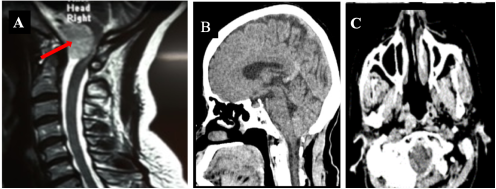
Figure 2. Preoperative MRI imaging of patient 3 reveals an anterior intradural extramedullary tumor (red arrow on A), and the control CT scan (B and C) confirms full excision.
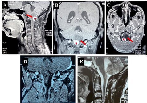
Figure 3. MRI of patient 4 showing a mixed intrabulbar cystic tumor (red arrows A, B, and C) with fleshy mural nodule (red star A, B, and C) and total excision leaving a residual cavity (D and E).
In all of our patients, this excision was carried out after a laminectomy; in two cases, it was combined with a hemi-laminectomy. We sectioned the C2 root in addition to performing coagulation in 2 patients. We had perioperative events included significant venous plexus bleeding in three cases and a dural breach without postoperative cerebrospinal fluid leak. In one case, there was a transient exacerbation of the neurological deficit. Another patient required mechanical breathing after surgery due to a lack of respiratory autonomy. He died on the seventh postoperative day. All these 2 patients had an anterior intradural extramedullary tumor. Neither radiation nor chemotherapy had been administered to any of our patients.
Four histological confirmations were found during the anatomopathological examination: 2 benign schwannomas, 1 neurofibroma, and 1 cavernous hemangioma. A specimen's histological analysis revealed a neurofibroma versus a low-grade MPNST (Malignant Peripheral Nerve Sheath Tumors). An immunohistochemical test was recommended, but not performed.
One surgical specimen's histological result was non-contributory (the imaging was in favor of pilocytic astrocytoma versus hemangioblastoma) while the other two patients' results were unreported (meningioma was indicated by imaging).
Seven of our patients had control imaging, which revealed tumor residue in two of them and total excision in five of the patients.
Patients had postoperative follow-up for an average of 21 months, with a range of 1 month to 5 years (60 months). We observed a satisfactory clinical progression, with three patients showing a complete regression of the motor deficit and four showing motor recovery with independent walking. During the follow-up period, we recorded no recurrences in our series.
Our patients' data summary is provided in Table 1 (manuscript end).
Table 1. Our patients' data summary.
Age Genre
Clinic
Preoperative imaging
Treatment
Clinical evolution
Postoperative imaging
histology
Patient1: 54 years (F)
Quadriparesis
MRI: intra- and extradural lesion in extra-intracanalaire in the left C1–C2 region
Partial excision / posteromedial approach on 08/26/2015
total recovery of motor function
MRI: residual tumors to the foraminal and anterolateral cord
Benign schwannoma
Patient2: 42 years (F)
Quadriparesis
MRI: extradural lesion C1–C2 posterolateral right extra-intracanal
Total excision / posteromedial approach on 09/19/2018
Motor recovery and Walking independently
MRI: spinal cord hypersignal at C2 level without tumor residue
Benign schwannoma
Patient3: 62 years (F)
1) Pyramidal syndrome
2) neck pain
MRI: intradural extramedullary lesion with a broad insertion base that runs from the clivus tip to the odontoid
Total excision / posterolateral approach on 11/14/2018
Transient quadriparesis followed by motor recovery with independent walking
CT scan: absence of tumor residue
Not reported
Patient4: 41 years (M)
Hemiparesia
MRI: Mixed intrabulbar lesion of the FM with a peripheral right posterior-paramedian mural nodule C0-C1 and a retrobulbar cyst
Total excision / posteromedial approach on 02/12/2020
Motor recovery with independent walking and weakness of right-hand grip
MRI: a bulbo-medullary junction excision cavity free of tumor residue
Uncertain
Patient5: 15 years (F)
1) Quadriparesis
2) VRN
IRM: extradural lesion C1–C2 posterolateral right extra-intracanal
Total excision / posteromedial approach on 06/05/2020
total recovery of motor function
CT scan: absence of tumor residue
Neurofibroma versus MPNST low grade.
Patient6: 30 years (M)
1) Quadriparesis
2) Cerebellar syndrome
3) cervical posterior scar
4) VHL
MRI: several mixed intraparenchymal lesions of the FCP, primarily cystic, with an extension towards the diencephalon and cervical cord, a mass effect on the fourth ventricle, and the brain stem
Partial excision / posteromedial approach on 06/09/2021
Recovery of independent walking and partial cerebellar regression syndrome
CT scan: decompression of the brain stem and V4
Cavernous hemangioma
Patient7: 8 years (M)
1) Quadriplegia
2) VRN
MRI: extra- intradural lesion C1–C2 posterolateral left extra-intracanal
Complete excision / posteromedial approach on 09/22/2021
total recovery of motor function
CT scan: left hemilaminectomy C1 and C2 without tumor residue
Neurofibroma
Patient8: 62 years (F)
Quadriplegia
MRI: anterior intradural lesion compressing the cervical spine and medulla oblongata, extending from FM to C2
Partial excision / posterolateral approach on 07/12/2022
Death on 7th post-operative day due to hypotension and lack of recovery of respiratory autonomy
According to the information in the literature, just 1.1% to 3.8% of brain and spinal cord tumors are foramen magnum tumors
[5]
Komotar RJ, Zacharia BE, McGovern RA, Sisti MB, Bruce JN, D'Ambrosio AL. Approaches to anterior and anterolateral foramen magnum lesions: A critical review. J Craniovert Junction Spine. 2010; 1(2): 86-99.
. The reason for our higher percentage could be attributed to the sample's weakness and potential bias in the selection process for brain tumors inside our structure.
In fact, Fann teacher university hospital’s neurosurgery department receives admissions for most brain tumors, and it's the first reference center for brain diseases.
There was one case of VHL illness with tumor recurrence in our study. Black Africans were not frequently reported to have VHL illness, according to two studies done in Togo by Belo et al.
[6]
Belo M, Guinhouya KM, Monkam Y. et al. La maladie de von-hippel lindau dans une famille togolaise. Afr J of Neurological Sciences. 2014; 33(1): 87-95.
[6]
and Kumako et al.
[7]
Kumako VK, Apetse K, Guinhouya KM, et al. La maladie de von Hippel-Lindau chez une jeune togolaise. Prat Neurol - FMC. Sept 2018; 9(3): 208-13.
The research reveals significant regional variations in the duration between the start of symptoms and the diagnosis
[8]
Yu Y, Hu F, Zhang X, Gu Y, Xie T, Ge J. Application of the Hemi-Semi-Laminectomy Approach in the Microsurgical Treatment of C2 Schwannomas. J Spinal Disord Tech. Août 2014; 27(6): E199-204. h
Bernard F, Lemee JM, Delion M, Fournier HD. Lower third clivus and foramen magnum intradural tumor removal: The plea for a simple posterolateral approach. Neurochirurgie. Avr 2016; 62(2): 86-93.
Hajhouji F. La prise en charge des tumeurs du foramen magnum. Marrakech: Université Cadi Ayyad; 2011, thèse N°92. 142p.
[11]
Das KK, Kumar R, Ashish K, et al. Extramedullary foramen magnum tumors and their surgical management: An experience with 29 cases. Asian J Neurosurg 2014; 9(04): 223-232.
. This could be explained by inequalities in the accessibility of healthcare. In fact, the majority of our patients in poor nations do not have health insurance; instead, they pay for their care with their own resources.
As a result, there is frequently a major delay in therapeutic and diagnostic care because of the populations' financial constraints as well as the prevalence of traditional medical practices, which may cause diagnostic inaccuracy. It should be emphasized, nonetheless, that in the case of craniocervical junction tumors, the principal reason for the delay in diagnosis could be attributed to the clinical signs gradually developing over a rather extended period of time and the diagnosis frequently occurring only after deficiency signs manifest.
An important consideration is the tumor's location. It provides the surgeon the opportunity to select the most efficient approach for accessing the lesion with a minimum of compromise to the vascular and nervous structures. We got a majority of the posterolateral tumors based on the implantation in the anteroposterior plane, as reported by Bruneau et al.
[13]
Bruneau M, George B. Foramen magnum meningiomas: approches chirurgicales détaillées et aspects techniques à l’hôpital Lariboisière et revue de la littérature. Neurochirurg Rév. 2008; 31: 19-32.
[13]
.
Moreover, the angiographic investigation offers a comprehensive examination of the arterial circulation, specifically focusing on the tumor's primary interactions with the vertebral artery. This enables the surgeon to evaluate the risks and challenges associated with the surgical treatment. Additionally, it makes it possible to evaluate the degree of vascularization present in specific tumors in order to potentially do a preoperative embolization
[10]
Hajhouji F. La prise en charge des tumeurs du foramen magnum. Marrakech: Université Cadi Ayyad; 2011, thèse N°92. 142p.
[10]
. Please take note that cerebral arteriography, both therapeutic and diagnostic, is still not available in our practice.
The patient's position during surgery depends on the tumor location and the approach used by the surgeon to enable easier access to the tumor. In our study, all patients were operated on in the ventral decubitus position, as in that of Hajhouji
[10]
Hajhouji F. La prise en charge des tumeurs du foramen magnum. Marrakech: Université Cadi Ayyad; 2011, thèse N°92. 142p.
[10]
and Sahraoui et al
[14]
Sahraoui M, Abzoubi B, Hassani L, Ioualalen N. Les tumeurs du foramen magnum. A propos de 10 cas et revue de la littérature. Dans: Benbouzid T. Journal de neurochirurgie. 2004; 27-30.
[14]
. This posture has the benefit of offering straightforward and direct access to the posterior and lateral contents of the craniocervical junction. Its main disadvantage is musculoaponeurotic detachment, which may be a source of weakening of the posterior cervical muscles.
It also presents a limitation in access to anterior tumors. Unlike our study, in the series of Doleagbenou et al
[15]
Doleagbenou AK, Ekouélé MHB, Diawara S, et al. Foramen magnum meningiomas: A report of 10 cases and review of litterature. Afr J Neurol Sci. 2013; 32(1): 1-9.
[15]
, most patients were operated on in a sitting position.
The sitting position has the advantage of offering a clean view of the surgical field by promoting satisfactory venous drainage. The Park Bench position has the advantage of minimizing the danger of venous gas embolism, however it increases the risk of brachial plexus injury and predisposes to muscular injuries
[16]
Mavarez-Martinez A, Israelyan LA, Soghomonyan S, et al. The Effects of Patient Positioning on the Outcome During Posterior Cranial Fossa and Pineal Region Surgery. Front Surg. 13 mars 2020; 7: 9.
Pai SB, Raghuram G, Keshav GC, Rodrigues E. Far-lateral Transcondylar Approach to Anterior Foramen Magnum Lesions. Our Experience. Asian J Neurosurg 2018; 13(03): 651-655.
. The transoral approach is likewise not practiced in our hospitals. We don't employ these very specialized operating facilities in our practice because we don't have adequate technical support. Note that intraoperative neurophysiological monitoring is not present. Consequently, the location, size, and habits of the surgeon all have a role in the surgical strategy that is selected. The best approach should allow for direct access to the mass while preserving the biomechanical stability of this extremely dynamic site in addition to maintaining neurological function
[11]
Das KK, Kumar R, Ashish K, et al. Extramedullary foramen magnum tumors and their surgical management: An experience with 29 cases. Asian J Neurosurg 2014; 9(04): 223-232.
. Therefore, hemi-laminectomy is a less-invasive technique that permits the C1 and C2 bone structures to be preserved. Hemi-semi-laminectomy, however, is superior to hemi-laminectomy. It permits the mechanically important bone structures to be preserved as much as feasible
[19]
Banczerowski P, Vajda J, Veres R. Removal of intraspinal spaceoccupying lesions through unilateral partial approach, the “hemisemi-laminectomy”. Ideggyogy Sz. 2008; 61(3-4): 114-22.
[20]
Banczerowski P, Veres R, Vajda J. Modified minimally invasive surgical approach to cervical neuromas with intraforaminal components: hemi-semi-laminectomy and supraforaminal burr hole (modified foraminotomy) technique. Minim Invasive Neurosurg. 2009; 52(1): 56-8.
Two patients with anterolateral intradural extramedullary tumors developed a surgical complication which resulted in a deterioration of their motor deficit and in one case, death after becoming dependent on mechanical ventilation.
A patient's intraoperative cardiac arrest was described by Hajhouji
[10]
Hajhouji F. La prise en charge des tumeurs du foramen magnum. Marrakech: Université Cadi Ayyad; 2011, thèse N°92. 142p.
[10]
.
In addition to spinal cord manipulations or ischemia, hyperflexion contusion during surgical installation may potentially be a factor of postoperative neurological complications.
The financial and technical limitations previously discussed make it difficult to perform immediate postoperative control imaging, especially magnetic resonance imaging (MRI), to highlight these complications. In our public hospitals, immunohistochemical is still practically absent in this the same context. Some tumors in our series have not histological confirmation, which affects our study's overall histological distribution.
5. Conclusion
For the treatment of tumors of the craniocervical junction, excision remains the treatment of choice. It offers recovery from symptoms, particularly from compression-related neurological issues. The lesion's location and the surgeon's preferences influence the surgical strategy that is chosen. The posterior approach is the most frequent and appears to have good results in our practice context, where improving access to care and enhancing the technological platform continue to be challenges.
Banerji D, Behari S, Jain VK, Pandey T, Chhabra DK. Extreme lateral transcondylar approach to the skull base. Neurol India. 1999; 47(1) 22-30.
[2]
Bertalanffy H, Gilsbach JM, Mayfrank L, Klein HM, Kawase T, Seeger W. Microsurgical Management of Ventral and Ventrolateral Foramen Magnum Meningiomas. In: Fahlbusch R, Bock WJ, Brock M, Buchfelder M, Klinger M, (eds) Modern Neurosurgery of Meningiomas and Pituitary Adenomas. Acta Neurochirurgica, vol 65. Springer, Vienna.
M. Berete I. Tumeurs de la fosse cérébrale postérieure [thèse]. Faculté de médecine et de pharmacie Fès; 2009, N° 028. 159p.
[4]
Rakotozanany P, Randriamizao HMR, Tsifiregna RL, Hasiniatsy R, Ratovondrainy W, Andriamamonjy C. Tumeur de la fosse postérieure de l’enfant vue au service de Neurochirurgie du CHU-JRA Antananarivo. Rev Anesth-Réanim Med Urg Toxicol. 2006; 1(8): 41-6.
[5]
Komotar RJ, Zacharia BE, McGovern RA, Sisti MB, Bruce JN, D'Ambrosio AL. Approaches to anterior and anterolateral foramen magnum lesions: A critical review. J Craniovert Junction Spine. 2010; 1(2): 86-99.
Yu Y, Hu F, Zhang X, Gu Y, Xie T, Ge J. Application of the Hemi-Semi-Laminectomy Approach in the Microsurgical Treatment of C2 Schwannomas. J Spinal Disord Tech. Août 2014; 27(6): E199-204. h
Bernard F, Lemee JM, Delion M, Fournier HD. Lower third clivus and foramen magnum intradural tumor removal: The plea for a simple posterolateral approach. Neurochirurgie. Avr 2016; 62(2): 86-93.
Hajhouji F. La prise en charge des tumeurs du foramen magnum. Marrakech: Université Cadi Ayyad; 2011, thèse N°92. 142p.
[11]
Das KK, Kumar R, Ashish K, et al. Extramedullary foramen magnum tumors and their surgical management: An experience with 29 cases. Asian J Neurosurg 2014; 9(04): 223-232.
Bruneau M, George B. Foramen magnum meningiomas: approches chirurgicales détaillées et aspects techniques à l’hôpital Lariboisière et revue de la littérature. Neurochirurg Rév. 2008; 31: 19-32.
[14]
Sahraoui M, Abzoubi B, Hassani L, Ioualalen N. Les tumeurs du foramen magnum. A propos de 10 cas et revue de la littérature. Dans: Benbouzid T. Journal de neurochirurgie. 2004; 27-30.
[15]
Doleagbenou AK, Ekouélé MHB, Diawara S, et al. Foramen magnum meningiomas: A report of 10 cases and review of litterature. Afr J Neurol Sci. 2013; 32(1): 1-9.
[16]
Mavarez-Martinez A, Israelyan LA, Soghomonyan S, et al. The Effects of Patient Positioning on the Outcome During Posterior Cranial Fossa and Pineal Region Surgery. Front Surg. 13 mars 2020; 7: 9.
Pai SB, Raghuram G, Keshav GC, Rodrigues E. Far-lateral Transcondylar Approach to Anterior Foramen Magnum Lesions. Our Experience. Asian J Neurosurg 2018; 13(03): 651-655.
Banczerowski P, Vajda J, Veres R. Removal of intraspinal spaceoccupying lesions through unilateral partial approach, the “hemisemi-laminectomy”. Ideggyogy Sz. 2008; 61(3-4): 114-22.
[20]
Banczerowski P, Veres R, Vajda J. Modified minimally invasive surgical approach to cervical neuromas with intraforaminal components: hemi-semi-laminectomy and supraforaminal burr hole (modified foraminotomy) technique. Minim Invasive Neurosurg. 2009; 52(1): 56-8.
Diop, S., Basse, A. S., Diallo, S., Tine, I., Thioub, M., et al. (2024). Surgical Treatment of Craniocervical Junction Tumors: Neurosurgery Department Experience of “Hôpital Principal De Dakar”. International Journal of Neurosurgery, 8(2), 28-34. https://doi.org/10.11648/j.ijn.20240802.12
Diop, S.; Basse, A. S.; Diallo, S.; Tine, I.; Thioub, M., et al. Surgical Treatment of Craniocervical Junction Tumors: Neurosurgery Department Experience of “Hôpital Principal De Dakar”. Int. J. Neurosurg.2024, 8(2), 28-34. doi: 10.11648/j.ijn.20240802.12
Diop S, Basse AS, Diallo S, Tine I, Thioub M, et al. Surgical Treatment of Craniocervical Junction Tumors: Neurosurgery Department Experience of “Hôpital Principal De Dakar”. Int J Neurosurg. 2024;8(2):28-34. doi: 10.11648/j.ijn.20240802.12
@article{10.11648/j.ijn.20240802.12,
author = {Sagar Diop and Ababacar Sall Basse and Souleymane Diallo and Ibrahima Tine and Mbaye Thioub and Abdou Azize Diop},
title = {Surgical Treatment of Craniocervical Junction Tumors: Neurosurgery Department Experience of “Hôpital Principal De Dakar”
},
journal = {International Journal of Neurosurgery},
volume = {8},
number = {2},
pages = {28-34},
doi = {10.11648/j.ijn.20240802.12},
url = {https://doi.org/10.11648/j.ijn.20240802.12},
eprint = {https://article.sciencepublishinggroup.com/pdf/10.11648.j.ijn.20240802.12},
abstract = {Introduction: Craniocervical tumors are uncommon but represent a medical emergency for diagnosis and treatment. Their approaches require of course surgical expertise in addition to adapted technical platform. In light of the limited resources available in our practice, we consequently made the decision to share our experience with the surgical treatment of this pathology. Patients and method: We performed a retrospective analysis on eight observations of patients treated in the neurosurgery department of “Hôpital Principal de Dakar” between July 2015 and December 2022 for progressive tumor spinal cord compression at the craniocervical junction. Results: We observed a 10% frequency, a mean age of 39.25 years, and extremes between 8 and 62 years. There was a 0.6 sex ratio. The most common risk factor was type I neurofibromatosis, and one case of Von Hippel-Lindau disease. All of our patients had pyramidal syndrome. Overall, there were 2 tumors with posterolateral extradural site and 4 tumors with extramedullary intradural location, including 2 posterolateral and 2 anterolateral. The location was intramedullary and cerebella-medullary in one case each. The posteromedial occipitocervical approach, which was employed on six patients, was the most utilized method. Five patients had total tumor resection, whereas three had partial resection. Two of our patients had postoperative complications: a worsening of motor deficit and a death following dependence on mechanical ventilation. We only received four histological confirmations for the anatomopathological samples. Our patients' evolution was positive over an average follow-up of 21 months, with neurological improvement and walking autonomy. Conclusion: Tumors affecting the craniocervical junction are still treated surgically. Despite the need for improvement in our working conditions, our results appear to be satisfactory.
},
year = {2024}
}
TY - JOUR
T1 - Surgical Treatment of Craniocervical Junction Tumors: Neurosurgery Department Experience of “Hôpital Principal De Dakar”
AU - Sagar Diop
AU - Ababacar Sall Basse
AU - Souleymane Diallo
AU - Ibrahima Tine
AU - Mbaye Thioub
AU - Abdou Azize Diop
Y1 - 2024/09/23
PY - 2024
N1 - https://doi.org/10.11648/j.ijn.20240802.12
DO - 10.11648/j.ijn.20240802.12
T2 - International Journal of Neurosurgery
JF - International Journal of Neurosurgery
JO - International Journal of Neurosurgery
SP - 28
EP - 34
PB - Science Publishing Group
SN - 2640-1959
UR - https://doi.org/10.11648/j.ijn.20240802.12
AB - Introduction: Craniocervical tumors are uncommon but represent a medical emergency for diagnosis and treatment. Their approaches require of course surgical expertise in addition to adapted technical platform. In light of the limited resources available in our practice, we consequently made the decision to share our experience with the surgical treatment of this pathology. Patients and method: We performed a retrospective analysis on eight observations of patients treated in the neurosurgery department of “Hôpital Principal de Dakar” between July 2015 and December 2022 for progressive tumor spinal cord compression at the craniocervical junction. Results: We observed a 10% frequency, a mean age of 39.25 years, and extremes between 8 and 62 years. There was a 0.6 sex ratio. The most common risk factor was type I neurofibromatosis, and one case of Von Hippel-Lindau disease. All of our patients had pyramidal syndrome. Overall, there were 2 tumors with posterolateral extradural site and 4 tumors with extramedullary intradural location, including 2 posterolateral and 2 anterolateral. The location was intramedullary and cerebella-medullary in one case each. The posteromedial occipitocervical approach, which was employed on six patients, was the most utilized method. Five patients had total tumor resection, whereas three had partial resection. Two of our patients had postoperative complications: a worsening of motor deficit and a death following dependence on mechanical ventilation. We only received four histological confirmations for the anatomopathological samples. Our patients' evolution was positive over an average follow-up of 21 months, with neurological improvement and walking autonomy. Conclusion: Tumors affecting the craniocervical junction are still treated surgically. Despite the need for improvement in our working conditions, our results appear to be satisfactory.
VL - 8
IS - 2
ER -