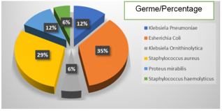
Introduction: Laser ureteroscopy is an effective method in the treatment of upper urinary tract stones. Our hospital is equipped with semi-rigid and flexible uretroscopes, equipped with a 35-watt Storz calculasse III holmium laser generator. The objective of this study is to evaluate the results of laser ureteroscopy. Materials and methods: The study was carried out in the urology department of the Pr Bocar Sidy Sall University Hospital in Kati. It was a prospective study, which took place from December 1, 2023 to March 31, 2024. Included in our study were patients diagnosed with renal and proximal or distal ureteral lithiasis and operated by flexible or semi-rigid laser ureteroscopy. The parameters studied were: sociodemographic data, reason for consultation, urological history, number, size and topography of stones, type of urethroscopy performed, postoperative follow-up, length of hospitalization. A survey form was established for data collection. Data analysis was done on Word 2016, Excel 2016 and SPSS version 23.0 software after data verification. The anonymity of patients was guaranteed with their consent for the use of personal data. Results: We performed 82 cases of laser ureteroscopy. The average age was 36.60 years with extremes from 7 to 79 years. The male sex was the most represented (57% of cases). Renal colic was the most frequent reason for consultation (82.9%). The insertion of a double J ureteral catheter was the most represented urological antecedent in 15.9% of cases. The left lumbar ureter was the most represented side (28% of cases). In the majority of cases, the size of the stone varied between 18-28 mm (37.8%). The urine culture was positive in 18% of cases and E. coli represented 35%. Flexible laser ureteroscopy was the most performed (75.6% of cases). The stone-free rate "without residual fragment" was successful in 69 patients, or 84.1% of cases, and revision surgery was performed in 13 patients, or 15.9% of cases. The hospital stay was 1 day in 90.2% of cases. Conclusion: Laser ureteroscopy is the treatment of choice for upper urinary tract stones. The challenge lies primarily in urologists mastering this technique and acquiring the necessary equipment.
| Published in | International Journal of Clinical Urology (Volume 9, Issue 1) |
| DOI | 10.11648/j.ijcu.20250901.20 |
| Page(s) | 62-67 |
| Creative Commons |
This is an Open Access article, distributed under the terms of the Creative Commons Attribution 4.0 International License (http://creativecommons.org/licenses/by/4.0/), which permits unrestricted use, distribution and reproduction in any medium or format, provided the original work is properly cited. |
| Copyright |
Copyright © The Author(s), 2025. Published by Science Publishing Group |
Stone, Upper Urinary Tract, Laser Ureteroscopy
Age group | Effective | Percentage (%) |
|---|---|---|
7-23 | 17 | 20.7 |
24-40 | 30 | 36.6 |
41-56 | 25 | 30.0 |
57-72 | 8 | 9.8 |
73-79 | 2 | 2.4 |
Total | 82 | 100 |
Reason for consultation | Effective | Percentage (%) |
|---|---|---|
Renal colic | 68 | 82.9 |
Low back pain and urinary symptom | 7 | 8.5 |
Low back pain + hydronephrosis without obstruction | 5 | 6.1 |
Low back pain + hydronephrosis with obstruction | 2 | 2.4 |
Total | 82 | 100 |
Urological history | Effective | Percentage (%) |
|---|---|---|
The rise of the JJ probe | 13 | 15.9 |
Flexible laser ureteroscopy | 7 | 8.5 |
Percutaneous nephrolithotomy | 4 | 4.9 |
Schistosomiasis urinairy | 2 | 2.4 |
Semi-rigid Laser ureteroscopy | 1 | 1.2 |
Extracorporeal shock wave lithotripsy | 1 | 1.2 |
Upper Prostatecotmy | 1 | 1.2 |
None | 53 | 64.6 |
Total | 82 | 100,0 |
Topography of the stones | Effective | Percentage (%) |
|---|---|---|
Left lumbar ureter | 23 | 28.0 |
Right renal | 21 | 25.6 |
Left renal | 14 | 17.1 |
Left pelvic ureter | 11 | 13.4 |
Left iliac ureter | 4 | 4.9 |
Left PU junction | 2 | 2.4 |
Bilateral renal | 2 | 2.4 |
Right PU junction | 1 | 1.2 |
Right lumbar ureter | 1 | 1.2 |
Right iliac ureter | 1 | 1.2 |
Total | 82 | 100 |
Size of the stone(s) (mm) | Effective | Percentage (%) |
|---|---|---|
7-17 | 30 | 36.6 |
18-28 | 31 | 37.8 |
29-39 | 12 | 14.6 |
40-50 | 8 | 9.8 |
Over 50 | 1 | 1.2 |
Total | 82 | 100 |
Type of ureteroscopy | Effectifs | Percentage (%) |
|---|---|---|
Flexible laser ureteroscopy | 62 | 75.6 |
Semi-Rigide laser ureteroscoy | 20 | 24.4 |
Total | 82 | 100 |
Duration of hospitalization | Effectifs | Pourcentage (%) |
|---|---|---|
1 day | 74 | 90.2 |
2 days | 7 | 8.5 |
5 days | 1 | 1.2 |
Total | 82 | 100 |
LASER | Light Amplification by Stimulated Emission of Radiation |
UUT | Upper Urinary Tract |
FLU | Flexible Laser Ureteroscopy |
SR-LU | Semi-Rigid Laser ureteroscopy |
YAG | Yttrium-Aluminum-Garnet |
PCNL | Percutaneous Nephrolithotomy |
ESWL | Extracorporeal Shock Wave Lithotripsy |
AVH | Upper Prostatecotmy |
| [1] | Padja E, Ibarra VA, Lmezguidi K, Janane A, Ghadouane M, Ameur A, Abbar M. Flexible laser ureteroscopy in the treatment of upper urinary tract stones: results about 166 interventions. Pan Afr Med J 2015; 8: 22: 13. |
| [2] | Lechevalier E, Saussine C, Traxer O. Ureteroscopy for upper urinary tract stones. Prog. Urol. 2008; 18(12): 912-916. |
| [3] | Traxer O, Lechevallier E, Saussine C. Flexible ureteroscopy with Holmium laser: The tools. Prog. Urol. 2008; 18(12): 917-928. |
| [4] | Berthe HJG, Cisse D, Diallo MS, Kassogue A, Diarra A, Coulibaly MT, Yattara I, Kouldjim A, Diakite ML, Tembely A. Our first experience of ureteroscopy with Holmium-YAG laser in the treatment of lithiasis of the upper urinary tract. Uro’ Andro 2020; 2(3): 108-111. |
| [5] | Kassogue A, Sissoko I, Sangare D, Diallo MS, Sylla H, Traore M, Coulibaly B, Coulibaly S, Diakite M, Togola M, Tall F, Guindo I, Kamel BN, Diakite ML. Ureteroscopy with Holmium: YAG Laser-A Initial Study in the Urology Department of the Pr Bocar Sidy Sall University Hospital of Kati. Open Journal of Urology 2024; 14: 207-216. |
| [6] | Niang L, Pare, AK, Ndoye, M, Samassékou, A, Avakoudjo, DJG, Agounkpé, MM, Jalloh, M, Ndiaye, A, Labou, I. and Gueye, S. M. Retrograde Ureteroscopy: Experience of the Grand Yoff General Hospital in Dakar. African Journal of Urology 2016; 22: 110-114. |
| [7] | Mbaeri TU, Onu OA, Odo C, Nwadi UV. Ureteroscopy and Holmium:YAG Laser Lithotripsy For Upper Tract Stones in a New Urology Centre: Our Initial Experience. Niger Med J 2023; 64(2): 259-2676. |
| [8] | Fall B., Mouracade, P., Bergerat, S. and Saussine, C. Flexible Ureteroscopy and Laser Lithotripsy for Kidney and Ureter Stone: Indications, Morbidity and Outcome. Prog. Urol. 2014; 24: 771-776. |
| [9] | Yameogo CAMKD, Ky B, Ouattara A, Kiraoya B, Kabore A. Holmium Laser Ureteroscopy in the Treatment of the Upper Urinary Tract Stones: Concerning 54 Cases in the Surgical Department of the Saint Camille Hospital of Ouagadougou (Burkina Faso). Open Journal of Urology 2020; 10(2): 17-24. |
| [10] | Chunlin Y, Wanlin D, Jinhua D. Analysis of the efficacy of holmium laser and pneumatic ballistic in the treatment of impacted ureteral calculi. Medicine 2020; 99(36): 1-5. |
| [11] | Carpentier X, Meria P, Bensalah K, Chabannes E, Estrade V, Denis E, Yonneau L, Mozer P, Hadjadj HA. Update for the Management of Kidney Stones in 2013. Stone Group Comity of the French Association of Urology. Prog. Urol. 2014; 24: 319-326. |
| [12] | Breda A, Ogunyemi O, Leppert JT, Schulam PG. Flexible ureteroscopy and laser lithotripsy for multiple unilateral intrarenal stones. Eur Urol 2009; 55(5): 1190-6. |
| [13] | Ben Saddik, MA, Al-Qahtani Sejiny S, Ndoye M, Gil-diez-de-Medina S, Merlet B, Thomas A, Haab F, Traxer O. Flexible Ureteroscopy in the Treat ment of Kidney Stone between 2 and 3 cm. Prog. Urol. 2011; 21: 327-332. |
| [14] | Faïs PO, Albert T, Gaillet, S. Flexible Ureteroscopy with Laser for Upper Urinary Tract Stone. Prog. Urol. 2011; 21: 811-815. |
| [15] | Kassogue A, Diarra A, Traore I, Djiguiba K, Diallo MS, Sissoko I, Traoré M, Guindo I, Coulibaly S. Preliminary Study on the Results of the Analysis of Urinary Stones by Infrared Spectrophotometry in the Urology Department of the Pr BSS University Hospital in Kati. Internationnal of clinical urology 2025; 9(1): 49-54. |
| [16] | Delbarre B, Baowaidan F, Culty T, Khelfat L, Brassier M, Ferragu M, Magnier A, Secourgeon A, Tariel F, Lebdai S, Bigo P. Prospective Comparison of Thulium and Holmium Laser Lithotripsy for the Treatment of Upper Urinary Tract Lithiasis. European urology open science 2023; 51: 7-12. |
APA Style
Kassogue, A., Sissoko, I., Diallo, M. S., Ouedrago, S., Sangaré, D., et al. (2025). Place of Holmium Laser Ureteroscopy in the Management of Upper Urinary Tract Stones: Results About 82 Cases. International Journal of Clinical Urology, 9(1), 62-67. https://doi.org/10.11648/j.ijcu.20250901.20
ACS Style
Kassogue, A.; Sissoko, I.; Diallo, M. S.; Ouedrago, S.; Sangaré, D., et al. Place of Holmium Laser Ureteroscopy in the Management of Upper Urinary Tract Stones: Results About 82 Cases. Int. J. Clin. Urol. 2025, 9(1), 62-67. doi: 10.11648/j.ijcu.20250901.20
@article{10.11648/j.ijcu.20250901.20,
  author = {Amadou Kassogue and Idrissa Sissoko and Moussa Salifou Diallo and Souleymane Ouedrago and Daouda Sangaré and Hamed Sylla and Mamadou Tidiani Coulibaly and Mahamadou Traoré and Honore Jean Gabriel Berthé},
  title = {Place of Holmium Laser Ureteroscopy in the Management of Upper Urinary Tract Stones: Results About 82 Cases
},
  journal = {International Journal of Clinical Urology},
  volume = {9},
  number = {1},
  pages = {62-67},
  doi = {10.11648/j.ijcu.20250901.20},
  url = {https://doi.org/10.11648/j.ijcu.20250901.20},
  eprint = {https://article.sciencepublishinggroup.com/pdf/10.11648.j.ijcu.20250901.20},
  abstract = {Introduction: Laser ureteroscopy is an effective method in the treatment of upper urinary tract stones. Our hospital is equipped with semi-rigid and flexible uretroscopes, equipped with a 35-watt Storz calculasse III holmium laser generator. The objective of this study is to evaluate the results of laser ureteroscopy. Materials and methods: The study was carried out in the urology department of the Pr Bocar Sidy Sall University Hospital in Kati. It was a prospective study, which took place from December 1, 2023 to March 31, 2024. Included in our study were patients diagnosed with renal and proximal or distal ureteral lithiasis and operated by flexible or semi-rigid laser ureteroscopy. The parameters studied were: sociodemographic data, reason for consultation, urological history, number, size and topography of stones, type of urethroscopy performed, postoperative follow-up, length of hospitalization. A survey form was established for data collection. Data analysis was done on Word 2016, Excel 2016 and SPSS version 23.0 software after data verification. The anonymity of patients was guaranteed with their consent for the use of personal data. Results: We performed 82 cases of laser ureteroscopy. The average age was 36.60 years with extremes from 7 to 79 years. The male sex was the most represented (57% of cases). Renal colic was the most frequent reason for consultation (82.9%). The insertion of a double J ureteral catheter was the most represented urological antecedent in 15.9% of cases. The left lumbar ureter was the most represented side (28% of cases). In the majority of cases, the size of the stone varied between 18-28 mm (37.8%). The urine culture was positive in 18% of cases and E. coli represented 35%. Flexible laser ureteroscopy was the most performed (75.6% of cases). The stone-free rate "without residual fragment" was successful in 69 patients, or 84.1% of cases, and revision surgery was performed in 13 patients, or 15.9% of cases. The hospital stay was 1 day in 90.2% of cases. Conclusion: Laser ureteroscopy is the treatment of choice for upper urinary tract stones. The challenge lies primarily in urologists mastering this technique and acquiring the necessary equipment.
},
 year = {2025}
}
TY  - JOUR
T1  - Place of Holmium Laser Ureteroscopy in the Management of Upper Urinary Tract Stones: Results About 82 Cases

AU  - Amadou Kassogue
AU  - Idrissa Sissoko
AU  - Moussa Salifou Diallo
AU  - Souleymane Ouedrago
AU  - Daouda Sangaré
AU  - Hamed Sylla
AU  - Mamadou Tidiani Coulibaly
AU  - Mahamadou Traoré
AU  - Honore Jean Gabriel Berthé
Y1  - 2025/04/19
PY  - 2025
N1  - https://doi.org/10.11648/j.ijcu.20250901.20
DO  - 10.11648/j.ijcu.20250901.20
T2  - International Journal of Clinical Urology
JF  - International Journal of Clinical Urology
JO  - International Journal of Clinical Urology
SP  - 62
EP  - 67
PB  - Science Publishing Group
SN  - 2640-1355
UR  - https://doi.org/10.11648/j.ijcu.20250901.20
AB  - Introduction: Laser ureteroscopy is an effective method in the treatment of upper urinary tract stones. Our hospital is equipped with semi-rigid and flexible uretroscopes, equipped with a 35-watt Storz calculasse III holmium laser generator. The objective of this study is to evaluate the results of laser ureteroscopy. Materials and methods: The study was carried out in the urology department of the Pr Bocar Sidy Sall University Hospital in Kati. It was a prospective study, which took place from December 1, 2023 to March 31, 2024. Included in our study were patients diagnosed with renal and proximal or distal ureteral lithiasis and operated by flexible or semi-rigid laser ureteroscopy. The parameters studied were: sociodemographic data, reason for consultation, urological history, number, size and topography of stones, type of urethroscopy performed, postoperative follow-up, length of hospitalization. A survey form was established for data collection. Data analysis was done on Word 2016, Excel 2016 and SPSS version 23.0 software after data verification. The anonymity of patients was guaranteed with their consent for the use of personal data. Results: We performed 82 cases of laser ureteroscopy. The average age was 36.60 years with extremes from 7 to 79 years. The male sex was the most represented (57% of cases). Renal colic was the most frequent reason for consultation (82.9%). The insertion of a double J ureteral catheter was the most represented urological antecedent in 15.9% of cases. The left lumbar ureter was the most represented side (28% of cases). In the majority of cases, the size of the stone varied between 18-28 mm (37.8%). The urine culture was positive in 18% of cases and E. coli represented 35%. Flexible laser ureteroscopy was the most performed (75.6% of cases). The stone-free rate "without residual fragment" was successful in 69 patients, or 84.1% of cases, and revision surgery was performed in 13 patients, or 15.9% of cases. The hospital stay was 1 day in 90.2% of cases. Conclusion: Laser ureteroscopy is the treatment of choice for upper urinary tract stones. The challenge lies primarily in urologists mastering this technique and acquiring the necessary equipment.

VL  - 9
IS  - 1
ER  -
Urology Department, Kati University Hospital Pr Bocar Sidy Sall, Kati, Mali
Urology Department, Kati University Hospital Pr Bocar Sidy Sall, Kati, Mali
Urology Department, Kati University Hospital Pr Bocar Sidy Sall, Kati, Mali
Urology Department, Point G University Hospital, Bamako, Mali
Information