Research Article | | Peer-Reviewed

Prevalence and Factors Associated with Toxoplasma gondii Infection in Dogs and Cats in the Boke Mining Area, Republic of Guinea

Received: 20 October 2026     Accepted: 4 November 2026     Published: 19 November 2026
Views:       Downloads:
Abstract

Toxoplasmosis is a major zoonosis in which domestic carnivores play a central role in transmission. This study determined the prevalence of Toxoplasma gondii infection and its associated factors in dogs and cats from the Boke mining area, Guinea. Between October 2021 and November 2024, blood samples were collected from 180 dogs and 106 cats in five localities of the prefecture. Anti-T. gondii antibodies were detected by indirect ELISA and parasite DNA by real-time PCR. Associated factors were analysed using Pearson's χ2 test and crude odds ratios (OR) in univariable analyses. Infection was defined as a positive result by ELISA and/or real-time PCR. All seropositive animals were also PCR-positive, so the overall prevalence of infection was equal to the molecular prevalence, namely 42.78% [35.78-50.08] in dogs and 51.89% [42.48-61.16] in cats. Real-time PCR detected T. gondii DNA in roughly twice as many animals as ELISA detected antibodies, with seroprevalences of only 17.78% in dogs and 27.36% in cats. Animals positive by PCR only showed late amplification (median Ct 37.82 in dogs and 36.65 in cats, against 27.70 and 28.50 in seropositive animals), and excluding Ct values above 35 reduced prevalence to 20.00% in dogs and 33.96% in cats. Agreement between the two techniques was moderate, with kappa values of 0.45 [0.31-0.59] in dogs and 0.52 [0.36-0.68] in cats. In dogs, infection was associated with age (OR = 2.37 [1.28-4.39]) and lack of deworming (OR = 3.22 [1.37-7.54]). In cats, it was associated with a free-roaming lifestyle (OR = 4.36 [1.87-10.15]), lack of deworming (OR = 4.82 [1.47-15.84]) and lack of vaccination (OR = 5.68 [1.16-27.70]). No significant difference was found between localities or between urban and rural settings. These findings indicate active circulation of T. gondii among domestic carnivores in the Boke mining area, in association with the way animals are kept, although the PCR-based prevalence should be regarded as an upper estimate. Their role in environmental contamination and human exposure was not assessed and should be investigated within a One Health framework, while better supervision of dogs and cats and control of stray populations could help limit transmission.

Published in International Journal of Animal Science and Technology (Volume 10, Issue 4)
DOI 10.11648/j.ijast.20261004.13
Page(s) 114-127
Creative Commons

This is an Open Access article, distributed under the terms of the Creative Commons Attribution 4.0 International License (http://creativecommons.org/licenses/by/4.0/), which permits unrestricted use, distribution and reproduction in any medium or format, provided the original work is properly cited.

Copyright

Copyright © The Author(s), 2026. Published by Science Publishing Group

Keywords

Domestic Carnivores, ELISA, Guinea, Mining Area, Real-time PCR, Toxoplasma gondii

1. Introduction
Toxoplasma gondii is an obligate intracellular apicomplexan protozoan with a worldwide distribution. It infects almost all warm-blooded species, including humans, livestock, birds and companion animals . About one third of the world's human population is thought to be infected, although prevalence varies widely between regions . Domestic and wild felids are the only definitive hosts, and the sexual phase of the parasite takes place in their small intestine. After a primary infection, a cat can shed several million oocysts over one to three weeks . These oocysts sporulate within a few days and remain infective for months in soil and water, which makes the cat a key contributor to environmental contamination . In other hosts, infection leads to the formation of bradyzoite-containing tissue cysts in various organs. Felids can also support this asexual cycle and act as intermediate hosts .
Infection is asymptomatic in most animal species, but toxoplasmosis can be fatal in some hosts, including domestic carnivores . In cats, clinical disease occurs mainly in immunocompromised animals, particularly those infected with feline immunodeficiency virus or feline leukaemia virus. It then presents as ocular, respiratory, hepatic or neurological involvement . Clinical toxoplasmosis is rare in dogs and usually follows immunosuppression. It combines non-specific signs such as fever, lymphadenopathy and digestive disorders with neurological, muscular or ocular disease . Noise hypersensitivity, myositis, ocular lesions and skin lesions in dogs under immunosuppressive treatment have also been described .
Seropositivity in domestic carnivores helps with differential diagnosis, but it also carries epidemiological weight because it reflects parasite circulation in the animals' surroundings . Free-roaming dogs and cats share the same environmental exposures as humans and wildlife and can therefore serve as sentinel species. Monitoring their seroprevalence provides an indirect measure of environmental contamination by oocysts . Dogs may also contribute to the spread of the parasite. After eating infected cat faeces, they can pass infective oocysts mechanically through the gut , and they can carry oocysts on their coat after rolling in feline faeces . Humans may then become infected through contact with the animal's coat, mouth or paws, especially in households where dogs and cats live together .
The seroprevalence of T. gondii in domestic carnivores depends on the region, the setting and the lifestyle of the animal. In dogs, the risk of infection rises with age because exposure accumulates over time, and seroprevalence is higher in rural than in urban areas . Dogs kept outdoors are more exposed than indoor dogs . Contact with cats, coprophagy and predation further increase the risk . In domestic cats, global seroprevalence is estimated at 35-37.5% and reaches 51% in Africa, compared with 59-64% in wild felids . Only 2.6% of domestic cats, however, are shedding oocysts at the time of examination . As in dogs, feline seroprevalence increases with age and outdoor access, and stray cats are more exposed than house cats . Predation on rodents and birds and feeding on raw meat are the main sources of infection .
In West Africa, toxoplasmosis in domestic carnivores remains poorly documented, but the available data indicate active circulation of the parasite. In Senegal, canine seroprevalence reached 43.9% in Dakar and 68% in Saint-Louis . A survey covering four regions of the country found anti-T. gondii antibodies in 67% of dogs, with higher seroprevalence in village dogs than in dogs from Dakar fed on commercial food . In Nigeria, 25% of dogs examined in Maiduguri were seropositive, and prevalence was higher in dogs aged three years or more . In stray cats from Oyo State, Nigeria, seroprevalence reached 86.7%, and 21.4% of the animals were shedding oocysts . Rodents, the main prey of cats, are also infected, as shown by the detection of parasite DNA in 15.2% of small mammals trapped in Cotonou, Benin . In Guinea, the only available study, conducted in Coyah, reported prevalences of 51.4% in dogs and 51.5% in cats . No data exist for mining areas, even though these areas combine several conditions that favour transmission. Boke Prefecture, the main bauxite mining hub of Guinea, has received a large influx of people seeking mining jobs, and its urban growth has been rapid and largely unplanned . This trend is expected to intensify under the Simandou 2040 programme, which places mining at the centre of national development. New households settling with their dogs and cats, precarious housing, waste accumulation and abundant rodents create favourable conditions for T. gondii transmission between animals and humans. The aim of this study was to determine the prevalence of T. gondii infection and to identify factors associated with infection in dogs and cats from the Boke mining area, Guinea.
2. Materials and Methods
2.1. Study Area
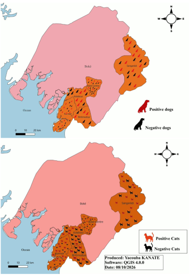
The study was carried out from October 2021 to November 2024 in Boke Prefecture, Maritime Guinea, about 300 km north-west of Conakry (Figure 1). The prefecture covers 11,053 km2 and had 552,704 inhabitants in 2021, a density of about 50 inhabitants/km2 . It comprises ten localities and is bordered by Guinea-Bissau to the north, Gaoual Prefecture to the north-east, Telimele Prefecture to the east, Boffa Prefecture to the south and the Atlantic Ocean to the west. Sampling relied on sera from 180 dogs and 106 cats collected for research or diagnostic purposes in five localities, namely the urban commune of Boke and four sub-prefectures. Three of these localities were urban (Boke Centre, Sangaredi and Kamsar) and two were rural (Bintimodia and Kolaboui) (Figure 1). The climate is tropical maritime Guinean, with a dry season from November to April and a rainy season from May to October. Mean annual temperature is 27.4 °C and annual rainfall is close to 2,496 mm . These hot and humid conditions favour the sporulation and survival of T. gondii oocysts in the environment .
Boke Prefecture is the leading bauxite centre of Guinea, with the Sangaredi mines and the Kamsar ore port . The mining boom has brought an influx of people and rapid, often unplanned, urban growth. In peri-urban neighbourhoods, dogs and cats usually roam freely and feed on kitchen scraps, slaughter waste and small prey. Free roaming, combined with waste accumulation and the presence of rodents, creates favourable conditions for the exposure of domestic carnivores to T. gondii.
Figure 1. Geographical distribution of the dogs and cats included in the study in Boke Prefecture. Sampled localities are highlighted in orange. Black icons represent negative animals and red icons represent animals positive for Toxoplasma gondii infection, placed in their respective areas of origin.
2.2. Ethical Approval and Consent to Participate
The study was approved by the Institutional Ethics Committee of ISSMV Dalaba (CEI-ISSMV) under reference No. 13/2021/ISSMV/D/DG. Prior authorisation was also obtained from the Prefectural Livestock Directorate of Boke before fieldwork began. In each locality, neighbourhood and district leaders were informed of the objectives and procedures of the study. Owners gave free and informed consent before any sampling, and refusal had no consequence for them. Blood sampling and chemical restraint were performed by qualified veterinarians using standard veterinary techniques and in compliance with animal welfare standards. Each animal was monitored until full recovery. Owner anonymity was preserved, as survey forms carried only a locality code and a serial number.
2.3. Study Design and Population
This study, conducted from October 2021 to November 2024, combined a questionnaire survey with serological (ELISA) and molecular (real-time PCR) analyses. The study population consisted of owned and ownerless dogs and cats present in five localities of Boke Prefecture.
2.4. Sampling of Dogs and Cats
The minimum sample size was estimated using the formula of Pourhoseingholi et al. :
n=Z2×P(1-P)d2
where n is the sample size for an infinite population, Z = 1.96 for a 95% confidence level, P the expected prevalence and d the absolute precision. The expected prevalence was taken from the Coyah study, namely 51.4% in dogs and 51.5% in cats . Because stray animals were difficult to capture, absolute precision was set at 7.3% for dogs and 9.5% for cats, giving a total of 180 dogs and 106 cats sampled across the five study localities. In the absence of a sampling frame for dogs and cats in the study area, non-probability convenience sampling was used. Animals were sampled in five localities of Boke Prefecture, namely the urban commune of Boke (Boke Centre) and the sub-prefectures of Bintimodia, Kamsar, Kolaboui and Sangaredi. In each locality, owned dogs and cats were included with their owners’ consent, while stray animals were included as they were captured in public areas, markets and around households. Sampling continued until the required sample size was reached for each species. Overall, 60 owned (33.3%) and 120 stray (66.7%) dogs, and 40 owned (37.7%) and 66 stray (62.3%) cats were sampled. Their distribution by locality is given in Table 3. Each animal was classified by sex, age class (young: < 3 years; adult: ≥ 3 years), setting (urban or rural), lifestyle (stray or non-stray), deworming status and vaccination status. Sampled animals were marked to avoid repeat sampling.
2.5. Owner Questionnaire Survey
A structured, pre-tested questionnaire was administered to the owners of the sampled animals. It covered the age, sex, lifestyle and diet of the animal, as well as its health care (deworming, vaccination and use of veterinary services). Cohabitation of dogs and cats within the household was also recorded. For owned animals, age was taken from the owner's declaration. For stray animals, age was estimated by oral examination of the dentition. A complete permanent dentition, which is usually in place by about seven months of age in both species, identified post-juvenile animals . Older animals were then distinguished by crown wear, tooth discoloration and the amount of dental calculus, these being markers whose accuracy decreases with age and varies between individuals . Because these signs only allow approximate ageing, stray animals were assigned to the broad age classes used in the analysis (< 3 years and ≥ 3 years) rather than given a precise age.
2.6. Restraint, Sampling and Sample Storage
Owned animals were restrained with the help of their owners. Stray cats were caught in bags baited with sardines, and stray dogs were held by assistants. Chemical restraint combined ketamine (Imalgene®, Boehringer Ingelheim, 10 mg/kg) and acepromazine (Calmivet®, Vetoquinol, 0.1 mg/kg) given intramuscularly. A 5 mL blood sample was drawn from the cephalic vein in dogs and a 3 mL sample from the femoral vein in cats, in line with recommendations for single sampling . Blood was collected into plain tubes labelled with a code indicating locality, species, age and sex. Tubes were transported on ice to the laboratory of the Boke health centre and centrifuged at 3,500 rpm for 20 min. Sera were aliquoted into cryotubes and stored at -20 °C, then shipped under cold chain to the Parasitology-Mycology Unit of the Central Veterinary Laboratory of Bingerville (Cote d'Ivoire).
2.7. Laboratory Analyses
2.7.1. Detection of Anti-Toxoplasma gondii Antibodies
Anti-T. gondii IgG antibodies were detected by indirect ELISA using the ID Screen® Toxoplasmosis Indirect Multi-species kit (Innovative Diagnostics, Grabels, France), whose plates are coated with the T. gondii P30 (SAG1) antigen. The assay was performed according to the manufacturer's instructions, as described by Turlewicz-Podbielska et al. . Each serum (10 µL) was diluted 1:10 in 90 µL of dilution buffer and added to the wells in single replicates. The kit positive and negative controls were run in duplicate on each plate. After 45 min of incubation at room temperature (21 ± 5 °C), plates were washed three times with 300 µL of wash solution per well. Peroxidase-labelled multi-species anti-IgG conjugate (100 µL) was then added, followed by a 30-min incubation under the same conditions and three further washes. Colour was developed with 100 µL of TMB (3,3′,5,5′-tetramethylbenzidine) substrate for 15 min in the dark. The reaction was stopped by adding 100 µL of 0.5 M sulfuric acid to each well, which turned the colour from blue to yellow. Optical density (OD) was then read at 450 nm on a microplate reader. A plate was validated when the mean OD of the positive control (ODPC) exceeded 0.350 and the ODPC/ODNC ratio exceeded 3. For each sample, the sample-to-positive ratio (S/P) was calculated as follows :
S/P %=ODsample-ODNCODPC-ODNC×100
where ODsample, ODNC and ODPC are the optical densities of the sample, the negative control and the positive control, respectively. Samples were classified as negative when S/P ≤ 40%, doubtful when S/P was between 40% and 50%, and positive when S/P ≥ 50% . Doubtful samples were classified as negative.
2.7.2. Detection of T. gondii DNA
(i). DNA Extraction
Genomic DNA was extracted from 200 µL of serum with the QIAamp® DNA Mini kit (Qiagen, Hilden, Germany), following the manufacturer's "Blood or body fluids" protocol . Each sample was lysed with 20 µL of proteinase K and 200 µL of buffer AL, then incubated at 56 °C for 10 min. After addition of 200 µL of absolute ethanol, the lysate was loaded onto a QIAamp silica column. The column was washed with 500 µL of buffer AW1 and then 500 µL of buffer AW2. DNA was eluted in 100 µL of buffer AE and stored at −20 °C until amplification. An extraction negative control (ultrapure water) was processed with each batch.
(ii). Real-time PCR Amplification
T. gondii DNA was detected by duplex real-time PCR using the ID Gene™ Toxoplasma gondii Duplex kit (Innovative Diagnostics, Grabels, France). The kit targets a T. gondii-specific repeated sequence of the 529-bp element type . Primers and probe were supplied by the manufacturer as a ready-to-use master mix, and their sequences are not disclosed. The mix detected, in the same well, T. gondii DNA (6-FAM-labelled probe) and an endogenous internal control corresponding to the host gapdh gene (VIC-labelled probe). Each reaction contained 15 µL of master mix and 5 µL of extracted DNA, for a final volume of 20 µL. Amplification was run on a 7500 Real-Time PCR System (Applied Biosystems, Foster City, USA) with the following programme: 10 min at 95 °C, then 45 cycles of 15 s at 95 °C and 60 s at 60 °C, with fluorescence read at the end of each cycle. The kit positive control and an amplification negative control (ultrapure water) were included in each run.
A run was validated when the positive control produced a FAM signal and neither the extraction nor the amplification negative control did. A sample was considered positive when the FAM signal showed a sigmoid amplification curve with a Ct ≤ 40 . Amplification was run for 45 cycles, but signals appearing after cycle 40 were not considered positive. The 529-bp target is a repetitive element present in 200 to 300 copies per parasite genome , which allows the detection of very low parasite loads but also increases the likelihood of late signals near the detection limit. A sample without FAM amplification at or before cycle 40 was considered negative only if the internal control (VIC) was amplified. When neither signal was present, the result was deemed uninterpretable and the sample was re-tested from a new extract.
2.8. Statistical Analysis
Data were analysed with R software, version 4.5.2, using the stats package . An animal was considered infected when it was positive by ELISA, by real-time PCR or by both tests, and the overall prevalence of infection was calculated on this basis. Prevalences were expressed as proportions with their 95% confidence intervals (95% CI), calculated by the Wilson method . ELISA and PCR results, obtained on the same animals, were compared with McNemar's test with continuity correction, and their agreement was estimated by Cohen's kappa coefficient, interpreted according to the scale of Landis and Koch (1977) . Ct values of PCR-positive animals were described by their median, interquartile range (IQR) and range, and compared between ELISA-positive and ELISA-negative animals with the Mann-Whitney U test. A sensitivity analysis was performed by recalculating prevalence after excluding PCR-positive results with a Ct above 35. Prevalences were compared by sub-prefecture, sex, age class, setting, lifestyle, deworming status and vaccination status using Pearson's χ2 test, or Fisher's exact test when an expected count was below 5. Crude odds ratios (OR) with 95% CI were calculated in univariable analyses to measure the association between each variable and infection status. No multivariable model was fitted, so the reported associations were not adjusted for potential confounders. A p-value < 0.05 was considered statistically significant.
3. Results
3.1. Prevalence of Toxoplasma Gondii Infection by Detection Method and Distribution of Ct Values
Of the 180 dogs examined, 17.78% [12.88-24.02] (n = 32) had anti-T. gondii antibodies by ELISA. Real-time PCR detected parasite DNA in 42.78% [35.78-50.08] (n = 77) of dogs, a significantly higher proportion than the seroprevalence (χ2 = 43.02; p < 0.001). The same pattern was observed in the 106 cats, with a seroprevalence of 27.36% [19.78-36.52] (n = 29) against a molecular prevalence of 51.89% [42.48-61.16] (n = 55) (χ2 = 24.04; p < 0.001) (Table 1). All seropositive animals were also PCR-positive, whereas 45 dogs and 26 cats were detected by PCR alone. Agreement between ELISA and PCR was moderate, with a kappa coefficient of 0.45 [0.31-0.59] in dogs and 0.52 [0.36-0.68] in cats (Table 1).
Table 1. Seroprevalence (ELISA) and molecular prevalence (real-time PCR) of Toxoplasma gondii in dogs and cats from the Boke mining area.

Species

ELISA % (n/No.)

PCR % (n/No.)

ELISA−/PCR+

McNemar χ2

p-value

Kappa (95% CI)

Dogs

17.78 (32/180)

42.78 (77/180)

45

43.02

< 0.001

0.45 [0.31-0.59]

Cats

27.36 (29/106)

51.89 (55/106)

26

24.04

< 0.001

0.52 [0.36-0.68]

No.: number tested; n: number positive; ELISA−/PCR+: animals positive by PCR only; McNemar: McNemar's test with continuity correction; kappa: Cohen's agreement coefficient.
Ct values of the 132 PCR-positive animals ranged from 20.40 to 39.82, with a median of 34.71 (IQR: 27.85-37.62) (Table 2). In dogs, the median Ct was 27.70 (IQR: 24.55-30.84) in ELISA-positive animals and 37.82 (IQR: 36.12-38.81) in animals positive by PCR only, a significant difference (U = 30.5; p < 0.001). The same pattern was observed in cats, with median Ct values of 28.50 (IQR: 24.22-30.43) and 36.65 (IQR: 35.15-37.68), respectively (U = 20.0; p < 0.001). A Ct above 35 was recorded in 83.10% (n = 59) of animals positive by PCR only, compared with 1.64% (n = 1) of seropositive animals. When PCR-positive results with a Ct above 35 were excluded, prevalence fell to 20.00% (n = 36) in dogs and 33.96% (n = 36) in cats.
Table 2. Distribution of real-time PCR cycle threshold (Ct) values in Toxoplasma gondii PCR-positive dogs and cats according to detection profile.

Species

Detection profile

n

Median Ct (IQR)

Range

Ct > 35, n (%)

Mann-Whitney U

p-value

Dogs

ELISA+/PCR+

32

27.70 (24.55-30.84)

20.40-38.40

1 (3.13)

30.5

< 0.001

ELISA−/PCR+

45

37.82 (36.12-38.81)

33.75-39.82

40 (88.89)

All PCR+

77

35.16 (28.40-37.90)

20.40-39.82

41 (53.25)

Cats

ELISA+/PCR+

29

28.50 (24.22-30.43)

20.73-34.62

0 (0.00)

20.0

< 0.001

ELISA−/PCR+

26

36.65 (35.15-37.68)

26.84-38.59

19 (73.08)

All PCR+

55

32.35 (27.29-36.39)

20.73-38.59

19 (34.55)

Overall

132

34.71 (27.85-37.62)

20.40-39.82

60 (45.45)

-

-

Ct: cycle threshold; IQR: interquartile range; n: number of animals; ELISA+/PCR+: animals positive by both ELISA and real-time PCR; ELISA−/PCR+: animals positive by real-time PCR only; All PCR+: all animals positive by real-time PCR; Mann-Whitney U and p-value: comparison of Ct values between ELISA+/PCR+ and ELISA−/PCR+ animals within each species. A Ct ≤ 40 was considered positive (45-cycle run).
3.2. Geographical Distribution of Infection in the Boke Mining Area
Because all ELISA-positive animals were also PCR-positive (Section 3.1), the overall prevalence of T. gondii infection in Boke Prefecture, defined as a positive result by ELISA and/or PCR, was identical to the molecular prevalence, namely 42.78% [35.78-50.08] (77/180) in dogs and 51.89% [42.48-61.16] (55/106) in cats. Infected animals of both species were found in all five localities studied (Figure 1). In dogs, prevalence ranged from 40.74% [30.70-51.62] (33/81) in Boke Centre to 52.38% [32.37-71.66] (11/21) in Sangaredi. In cats, it ranged from 36.36% [15.17-64.62] (4/11) in Bintimodia to 60.00% [40.74-76.60] (15/25) in Kamsar. None of these differences between localities was significant (p > 0.05) (Table 3; Figure 1).
Table 3. Prevalence of Toxoplasma gondii infection in dogs and cats by locality.

Species

Locality

No. tested

No. positive

Prevalence (%)

95% CI

p-value

OR [95% CI]

Dogs

Boke Centre

81

33

40.74

[30.70-51.62]

Ref.

1

Bintimodia

15

7

46.67

[24.81-69.88]

0.669

1.27 [0.42-3.85]

Kamsar

41

17

41.46

[27.76-56.63]

0.939

1.03 [0.48-2.21]

Kolaboui

22

9

40.91

[23.26-61.27]

0.989

1.01 [0.39-2.63]

Sangaredi

21

11

52.38

[32.37-71.66]

0.337

1.60 [0.61-4.20]

Total

180

77

42.78

[35.78-50.08]

-

-

Cats

Boke Centre

31

17

54.84

[37.77-70.84]

Ref.

1

Bintimodia

11

4

36.36

[15.17-64.62]

0.292

0.47 [0.11-1.94]

Kamsar

25

15

60.00

[40.74-76.60]

0.698

1.24 [0.42-3.59]

Kolaboui

18

8

44.44

[24.56-66.28]

0.483

0.66 [0.20-2.12]

Sangaredi

21

11

52.38

[32.37-71.66]

0.862

0.91 [0.30-2.75]

Total

106

55

51.89

[42.48-61.16]

-

-

No.: number; 95% CI: 95% confidence interval (Wilson method); p-value and OR: comparison with Boke Centre using Pearson's χ2 test.
3.3. Factors Associated with Toxoplasma gondii Infection in Dogs
Among the factors studied, only age and deworming were significantly associated with infection in dogs. Prevalence was 55.88% [44.08-67.05] (n = 38) in adults aged three years or more, compared with 34.82% [26.63-44.01] (n = 39) in dogs younger than three years (p = 0.006; OR = 2.37 [1.28-4.39]). Prevalence reached 47.92% [39.92-56.02] (n = 69) in dogs that had not been dewormed, against 22.22% [11.72-38.09] (8/36) in dewormed dogs (p = 0.005; OR = 3.22 [1.37-7.54]). Prevalence was 48.04% [38.59-57.63] (49/102) in males and 35.90% [26.15-46.97] (28/78) in females, 44.59% [36.82-52.64] (66/148) in unvaccinated dogs and 34.38% [20.41-51.69] (11/32) in vaccinated dogs, and 45.83% [37.19-54.74] (55/120) in stray dogs and 36.67% [25.62-49.32] (22/60) in owned dogs. None of these differences was significant (0.103 ≤ p ≤ 0.289). Prevalence was similar in urban (42.66% [34.85-50.85]) and rural areas (43.24% [28.67-59.09]) (p = 0.949) (Table 4).
Table 4. Prevalence of Toxoplasma gondii infection and associated factors in dogs from the Boke mining area.

Variable

Category

No. tested

No. positive

Prevalence (%)

95% CI

p-value

OR [95% CI]

Sex

Female

78

28

35.90

[26.15-46.97]

0.103

1

Male

102

49

48.04

[38.59-57.63]

1.65 [0.90-3.02]

Age class

Young (< 3 years)

112

39

34.82

[26.63-44.01]

0.006

1

Adult (≥ 3 years)

68

38

55.88

[44.08-67.05]

2.37 [1.28-4.39]

Vaccination

Vaccinated

32

11

34.38

[20.41-51.69]

0.289

1

Unvaccinated

148

66

44.59

[36.82-52.64]

1.54 [0.69-3.41]

Deworming

Dewormed

36

8

22.22

[11.72-38.09]

0.005

1

Not dewormed

144

69

47.92

[39.92-56.02]

3.22 [1.37-7.54]

Lifestyle

Owned

60

22

36.67

[25.62-49.32]

0.241

1

Stray

120

55

45.83

[37.19-54.74]

1.46 [0.77-2.76]

Setting

Urban

143

61

42.66

[34.85-50.85]

0.949

1

Rural

37

16

43.24

[28.67-59.09]

1.02 [0.49-2.13]

No.: number; 95% CI: 95% confidence interval (Wilson method); p-value: Pearson's χ2 test; OR: odds ratio, with the first category of each variable as reference (OR = 1).
3.4. Factors Associated with Toxoplasma gondii Infection in Cats
In cats, vaccination, deworming and lifestyle were significantly associated with infection. Prevalence was 65.15% [53.11-75.52] (n = 43) in stray cats and 30.00% [18.07-45.43] (n = 12) in owned cats (p < 0.001; OR = 4.36 [1.87-10.15]). It reached 57.95% [47.52-67.72] (n = 51) in cats that had not been dewormed, compared with 22.22% [9.00-45.21] (n = 4) in dewormed cats (p = 0.006; OR = 4.82 [1.47-15.84]). Unvaccinated cats also showed a higher prevalence, 55.79% [45.77-65.36] (53/95), than vaccinated cats, 18.18% [5.14-47.70] (p = 0.018; OR = 5.68 [1.16-27.70]). Prevalence was 54.84% [37.77-70.84] in males and 50.67% [39.60-61.67] in females, and 56.82% [42.22-70.32] in adults aged three years or more and 48.39% [36.41-60.55] in younger cats, with no significant difference (0.392 ≤ p ≤ 0.696). Cats from urban areas, 55.84% [44.74-66.39] (43/77), and rural areas, 41.38% [25.51-59.26], did not differ significantly (p = 0.184) (Table 5).
Table 5. Prevalence of Toxoplasma gondii infection and associated factors in cats from the Boke mining area.

Variable

Category

No. tested

No. positive

Prevalence (%)

95% CI

p-value

OR [95% CI]

Sex

Female

75

38

50.67

[39.60-61.67]

0.696

1

Male

31

17

54.84

[37.77-70.84]

1.18 [0.51-2.74]

Age class

Young (< 3 years)

62

30

48.39

[36.41-60.55]

0.392

1

Adult (≥ 3 years)

44

25

56.82

[42.22-70.32]

1.40 [0.65-3.05]

Vaccination

Vaccinated

11

2

18.18

[5.14-47.70]

0.018

1

Unvaccinated

95

53

55.79

[45.77-65.36]

5.68 [1.16-27.70]

Deworming

Dewormed

18

4

22.22

[9.00-45.21]

0.006

1

Not dewormed

88

51

57.95

[47.52-67.72]

4.82 [1.47-15.84]

Lifestyle

Owned

40

12

30.00

[18.07-45.43]

< 0.001

1

Stray

66

43

65.15

[53.11-75.52]

4.36 [1.87-10.15]

Setting

Urban

77

43

55.84

[44.74-66.39]

0.184

1

Rural

29

12

41.38

[25.51-59.26]

0.56 [0.23-1.33]

No.: number; 95% CI: 95% confidence interval (Wilson method); p-value: Pearson's χ2 test; OR: odds ratio, with the first category of each variable as reference (OR = 1).
4. Discussion
The present study provides the first data on T. gondii infection in domestic carnivores from the Boke mining area. By combining serology and molecular detection, infection was found in 42.78% of dogs and 51.89% of cats, which points to active circulation of the parasite in both species, although the PCR-based estimate is probably an upper bound. These rates are close to those reported by Konate et al. in Coyah, Maritime Guinea, where prevalence was 51.4% in dogs and 51.5% in cats. The feline rate is also in line with the African seroprevalence of about 51% estimated in the meta-analyses of Hatam-Nahavandi et al. and Montazeri et al. . In dogs, prevalence was higher than the 25% reported by Kamani et al. in Maiduguri, Nigeria, but lower than the 67% recorded by Davoust et al. across four regions of Senegal. These comparisons should be made with caution, since most of the cited studies relied on serology alone, whereas the overall prevalence reported here combines serology and DNA detection. The seroprevalences of 17.78% in dogs and 27.36% in cats are therefore the most directly comparable figures.
Climate may account for part of these differences. Oocyst sporulation and survival depend on temperature and humidity , and the semi-arid climate of Maiduguri is less favourable to oocysts than that of Boke, where annual rainfall is close to 2,500 mm . Diagnostic methods are a second source of variation, since sensitivity and positivity thresholds differ according to the test and antigen used . In the Maiduguri study, serology relied on a latex agglutination test with a cut-off of 1:64 . Such a high cut-off may have excluded weakly positive animals and led to an underestimate of prevalence. The Senegalese survey, in contrast, used a so-called "high-sensitivity" direct agglutination test , which is better suited to detecting low antibody titres. The multi-species ELISA kit used in Boke may also miss some infected dogs. Bellatreche et al. compared this kit with the modified agglutination test, used as the reference, in hunting dogs in Algeria and found that nearly a quarter of the animals positive by the reference test had not been detected. This lack of sensitivity may partly explain the low seroprevalence of 17.78% obtained by ELISA in the present study.
The composition of the sampled populations is a third factor. In Senegal, village dogs fed on scraps and living freely were more often seropositive than dogs from Dakar fed on commercial food . Husbandry conditions act as genuine exposure factors, as shown by Arruda et al. and Bazan et al. in Brazil, where home-made diets and offal increased the risk of infection. In the present study, the odds of infection were more than four times higher in stray cats than in owned cats, a finding widely reported in the literature . Rubbish dumps provide them with a constant supply of rodents and birds carrying tissue cysts, which are the main source of infection for cats . In dogs, the difference in prevalence between stray and owned animals was not significant, probably because owned dogs in Boke also roam and feed partly on waste. Deworming was associated with a lower prevalence in both species, as was vaccination in cats. Neither measure protects against T. gondii, since anthelmintics have no effect on protozoa and no toxoplasmosis vaccine is available for domestic carnivores . Both are therefore best regarded as indicators of the general level of care given to the animal, which is likely to go together with closer supervision, less roaming and reduced access to prey and contaminated food. As these associations come from univariable analyses and may be confounded by lifestyle, they should not be interpreted as independent risk factors.
In dogs, age was associated with infection, and the odds of infection were 2.37 times higher in adults aged three years or more than in young dogs. This result is consistent with the nature of T. gondii infection. Once acquired, the parasite persists for the lifetime of the host as tissue cysts, and antibodies remain detectable for a long time . Each additional year of life therefore increases the cumulative probability of having ingested oocysts or infected tissues, and prevalence rises with age. Kamani et al. made the same observation in Maiduguri, Nigeria, where dogs aged three years or more had a risk of infection nearly three times that of younger dogs. In cats, by contrast, age was not associated with infection, unlike the findings of Majid et al. in Pakistan, Schafer et al. in Europe and Yang et al. in China. Where infection pressure is high, as around the rubbish dumps of Boke, most cats become infected during their first months of hunting and prevalence quickly reaches a plateau. The small number of cats also limited the statistical power of this comparison. Sex was not associated with infection in either species, since risk depends above all on exposure . Rezaei et al. in Iran and Majid et al. in Pakistan reported the same absence of association.
Infection was found in all five localities, with no difference between urban and rural settings. This contrasts with the study of Yan et al. in China, where seroprevalence in stray dogs increased along an urban-rural gradient. Unplanned urban growth in the mining area, waste accumulation and abundant rodents, which are important reservoirs of the parasite in West Africa , probably recreate in towns exposure conditions close to those of rural areas. PCR also detected T. gondii DNA in more than twice as many animals as ELISA detected antibodies. Rezaei et al. described a similar profile in rural dogs from southern Iran, with 28.3% positive by PCR against 8.3% by serology. The Ct values help to interpret this discrepancy. Animals positive by PCR only showed late amplification, with a median Ct above 36 in both species and a Ct above 35 in 83.10% of cases, whereas seropositive animals had a median Ct below 29. Such late signals indicate very small amounts of target DNA. They may correspond to recent infections, since IgG antibodies appear only several weeks after infection , or to animals missed by the ELISA kit, which failed to detect nearly a quarter of infected dogs in the Algerian study of Bellatreche et al. . Because the parasite circulates in the blood only briefly before encysting , serum is, however, a matrix of limited sensitivity, and late signals close to the detection limit may also result from non-specific amplification of the multicopy 529-bp target . In the absence of confirmation by sequencing or a second molecular target, PCR-positive results are therefore interpreted as detection of T. gondii DNA rather than evidence of active infection, and the PCR-based prevalence should be regarded as an upper estimate. Excluding Ct values above 35 gave prevalences of 20.00% in dogs and 33.96% in cats, which are closer to the seroprevalences.
From a One Health perspective, the Boke mining area combines the three components that sustain T. gondii transmission: abundant free-roaming dogs and cats, a rapidly growing human population living in precarious housing, and a warm, humid environment where waste accumulation attracts rodents. A single infected cat can shed several million oocysts that remain infective in soil for months , and Awobode et al. documented such soil contamination around stray cats in Nigeria. Dogs can also carry oocysts on their coat . In Africa, cat ownership is associated with T. gondii infection in pregnant women , and Eyre et al. showed in precarious urban neighbourhoods of Brazil that environmental exposure alone can sustain transmission to humans. The present study, however, demonstrates only the presence of antibodies and parasite DNA in the sampled animals. Oocyst shedding, soil or water contamination and human infection were not investigated, so the contribution of these animals to human exposure remains a hypothesis. Other limitations should be considered: sampling was not random, some subgroups were small, the analysis was univariable and PCR-positive results were not confirmed by sequencing.
5. Conclusion
This study shows that Toxoplasma gondii circulates actively among dogs and cats in the Boke mining area, with prevalences of 42.78% and 51.89%, respectively, based on serology and DNA detection. The parasite is present in all the localities studied, in both urban and rural settings. In univariable analyses, infection was more frequent in animals that roam freely or receive little care, namely stray cats and animals that had not been dewormed, and, in dogs, in older individuals. PCR detected T. gondii DNA in more animals than ELISA detected antibodies, but most PCR-only results showed late amplification, so these prevalences should be regarded as upper estimates. The combined use of both techniques, with confirmation of PCR-positive results, is recommended for future surveys.
These results suggest that domestic carnivores, and stray cats in particular, could contribute to environmental contamination by T. gondii in the Boke mining area and represent a potential source of human exposure, although this was not directly assessed. A One Health approach linking veterinary services, public health services and municipal waste management would be appropriate, combining better control of waste and stray animals with awareness campaigns for pet owners and pregnant women. Further studies should confirm and genotype the circulating strains, investigate oocyst shedding in cats and environmental contamination, and assess human infection in the mining area.
Abbreviations

95% CI

95% Confidence Interval

bp

Base Pair

CEI-ISSMV

Institutional Ethics Committee of ISSMV Dalaba

Ct

Cycle Threshold

DNA

Deoxyribonucleic Acid

ELISA

Enzyme-Linked Immunosorbent Assay

FAM

6-Carboxyfluorescein

gapdh

Glyceraldehyde-3-Phosphate Dehydrogenase Gene

IgG

Immunoglobulin G

IQR

Interquartile Range

ISSMV

Higher Institute of Veterinary Science and Medicine of Dalaba

MAT

Modified Agglutination Test

NC

Negative Control

OD

Optical Density

OR

Odds Ratio

PC

Positive Control

PCR

Polymerase Chain Reaction

rpm

Revolutions Per Minute

S/P

Sample-to-Positive Ratio

SAG1

Surface Antigen 1

TMB

3,3′,5,5′-Tetramethylbenzidine

Acknowledgments
The authors thank the Prefectural Livestock Directorate of Boke for its support in the field, the local authorities and animal owners for their cooperation, and the Parasitology-Mycology Unit of the Central Veterinary Laboratory of Bingerville (Cote d'Ivoire) for performing the analyses.
Authors Contributions
Yacouba Konate: Conceptualization, Formal Analysis, Funding acquisition, Investigation, Writing – original draft
Albert Sourou Salako: Formal Analysis, Investigation, Writing – original draft, Writing – review & editing
Lancei Kaba: Formal Analysis, Investigation, Writing – original draft
Noumouke Kante: Writing – review & editing
Francis Saoulomou: Formal Analysis, Investigation, Writing – original draft
Germaine Millimono: Formal Analysis, Investigation, Writing – original draft
Mamadou Fode Camara: Writing – review & editing
Youssouf Sidime: Conceptualization, Funding acquisition
Data Availability Statement
The datasets that were analysed in this study are available from the corresponding author and the lead author.
Conflicts of Interest
The authors declare that they have no competing interests.
References
[1] Attias, M., Teixeira, D. E., Benchimol, M., Vommaro, R. C., Crepaldi, P. H. and De Souza, W. (2020). The life-cycle of Toxoplasma gondii reviewed using animations. Parasites & Vectors. 13: 588.
[2] Djurković-Djaković, O., Dupouy-Camet, J., Van der Giessen, J. and Dubey, J. P. (2019). Toxoplasmosis: overview from a One Health perspective. Food and Waterborne Parasitology. 15: e00054.
[3] Dubey, J. P., Murata, F. H. A., Cerqueira-Cézar, C. K., Kwok, O. C. H., Yang, Y. and Su, C. (2020). Toxoplasma gondii infections in dogs: 2009–2020. Veterinary Parasitology. 287: 109223.
[4] Milne, G., Webster, J. P. and Walker, M. (2020). Toxoplasma gondii: an underestimated threat? Trends in Parasitology. 36(12): 959–969.
[5] Smith, N. C., Goulart, C., Hayward, J. A., Kupz, A., Miller, C. M. and van Dooren, G. G. (2021). Control of human toxoplasmosis. International Journal for Parasitology. 51(2–3): 95–121.
[6] Dabritz, H. A. and Conrad, P. A. (2010). Cats and Toxoplasma: implications for public health. Zoonoses and Public Health. 57(1): 34–52.
[7] Dubey, J. P., Cerqueira-Cézar, C. K., Murata, F. H. A., Kwok, O. C. H., Yang, Y. R. and Su, C. (2020). All about toxoplasmosis in cats: the last decade. Veterinary Parasitology. 283: 109145.
[8] Shapiro, K., Bahia-Oliveira, L., Dixon, B., Dumètre, A., de Wit, L. A., VanWormer, E., et al. (2019). Environmental transmission of Toxoplasma gondii: oocysts in water, soil and food. Food and Waterborne Parasitology. 15: e00049.
[9] Dubey, J. P. (2022). Toxoplasmosis of Animals and Humans. 3rd ed. CRC Press, Boca Raton, USA.
[10] Calero-Bernal, R. and Gennari, S. M. (2019). Clinical toxoplasmosis in dogs and cats: an update. Frontiers in Veterinary Science. 6: 54.
[11] Migliore, S., La Marca, S., Stabile, C., Di Marco Lo Presti, V. and Vitale, M. (2017). A rare case of acute toxoplasmosis in a stray dog due to infection of T. gondii clonal type I: public health concern in urban settings with stray animals? BMC Veterinary Research. 13: 249.
[12] Papini, R., Mancianti, F. and Saccardi, E. (2009). Noise sensitivity in a dog with toxoplasmosis. Veterinary Record. 165(2): 62.
[13] Alvarado-Esquivel, C., Romero-Salas, D., Cruz-Romero, A., García-Vázquez, Z., Peniche-Cardeña, A., Ibarra-Priego, N., et al. (2014). High prevalence of Toxoplasma gondii antibodies in dogs in Veracruz, Mexico. BMC Veterinary Research. 10: 191.
[14] Meireles, L. R., Galisteo, A. J., Pompeu, E. and Andrade, H. F. (2004). Toxoplasma gondii spreading in an urban area evaluated by seroprevalence in free-living cats and dogs. Tropical Medicine & International Health. 9(8): 876–881.
[15] Salb, A. L., Barkema, H. W., Elkin, B. T., Thompson, R. C. A., Whiteside, D. P., Black, S. R., et al. (2008). Dogs as sources and sentinels of parasites in humans and wildlife, northern Canada. Emerging Infectious Diseases. 14(1): 60–63.
[16] Yan, C., Fu, L. L., Yue, C. L., Tang, R. X., Liu, Y. S., Lv, L., et al. (2012). Stray dogs as indicators of Toxoplasma gondii distributed in the environment: the first report across an urban-rural gradient in China. Parasites & Vectors. 5: 5.
[17] Schares, G., Pantchev, N., Barutzki, D., Heydorn, A. O., Bauer, C. and Conraths, F. J. (2005). Oocysts of Neospora caninum, Hammondia heydorni, Toxoplasma gondii and Hammondia hammondi in faeces collected from dogs in Germany. International Journal for Parasitology. 35(14): 1525–1537.
[18] Frenkel, J. K., Lindsay, D. S., Parker, B. B. and Dobesh, M. (2003). Dogs as possible mechanical carriers of Toxoplasma, and their fur as a source of infection of young children. International Journal of Infectious Diseases. 7(4): 292–293.
[19] Lindsay, D. S., Dubey, J. P., Butler, J. M. and Blagburn, B. L. (1997). Mechanical transmission of Toxoplasma gondii oocysts by dogs. Veterinary Parasitology. 73(1–2): 27–33.
[20] Cañón-Franco, W. A., Bergamaschi, D. P., Labruna, M. B., Camargo, L. M. A., Silva, J. C. R., Pinter, A., et al. (2004). Occurrence of anti-Toxoplasma gondii antibodies in dogs in the urban area of Monte Negro, Rondônia, Brazil. Veterinary Research Communications. 28(2): 113–118.
[21] Cano-Terriza, D., Puig-Ribas, M., Jiménez-Ruiz, S., Cabezón, Ó., Almería, S., Galán-Relaño, Á., et al. (2016). Risk factors of Toxoplasma gondii infection in hunting, pet and watchdogs from southern Spain and northern Africa. Parasitology International. 65(5): 363–366.
[22] Huertas-López, A., Sukhumavasi, W., Álvarez-García, G., Martínez-Subiela, S., Cano-Terriza, D., Almería, S., et al. (2021). Seroprevalence of Toxoplasma gondii in outdoor dogs and cats in Bangkok, Thailand. Parasitology. 148(7): 843–849.
[23] Wang, S., Zhou, Y. H., Xie, Q., Zheng, B., Zhang, H. Z., Yao, Z. J., et al. (2017). Prevalence of Toxoplasma gondii antibodies in dogs in central China. Israel Journal of Veterinary Medicine. 72(3): 11–15.
[24] Zarra-Nezhad, F., Borujeni, M. P., Mosallanejad, B. and Hamidinejat, H. (2017). A seroepidemiological survey of Toxoplasma gondii infection in referred dogs to Veterinary Hospital of Ahvaz, Iran. International Journal of Veterinary Science and Medicine. 5(2): 148–151.
[25] Dini, F. M., Stancampiano, L., Poglayen, G. and Galuppi, R. (2024). Risk factors for Toxoplasma gondii infection in dogs: a serological survey. Acta Veterinaria Scandinavica. 66: 14.
[26] Mendoza Roldan, J. A. and Otranto, D. (2023). Zoonotic parasites associated with predation by dogs and cats. Parasites & Vectors. 16: 55.
[27] Hatam-Nahavandi, K., Calero-Bernal, R., Rahimi, M. T., Pagheh, A. S., Zarean, M., Dezhkam, A., et al. (2021). Toxoplasma gondii infection in domestic and wild felids as public health concerns: a systematic review and meta-analysis. Scientific Reports. 11: 9509.
[28] Montazeri, M., Mikaeili Galeh, T., Moosazadeh, M., Sarvi, S., Dodangeh, S., Javidnia, J., et al. (2020). The global serological prevalence of Toxoplasma gondii in felids during the last five decades (1967–2017): a systematic review and meta-analysis. Parasites & Vectors. 13: 82.
[29] Ding, H., Gao, Y. M., Deng, Y., Lamberton, P. H. L. and Lu, D. B. (2017). A systematic review and meta-analysis of the seroprevalence of Toxoplasma gondii in cats in mainland China. Parasites & Vectors. 10: 27.
[30] Afonso, E., Thulliez, P., Pontier, D. and Gilot-Fromont, E. (2007). Toxoplasmosis in prey species and consequences for prevalence in feral cats: not all prey species are equal. Parasitology. 134(14): 1963–1971.
[31] Coulibaly, F., Ndour, A. P. N., Gbati, O. B., Efoua-Tomo, N., Allanonto, V., Adjé Koffi, J. F., et al. (2012). Séroprévalence et facteurs de risque associés de la toxoplasmose et de la néosporose chez la femme en consultation prénatale et chez les carnivores domestiques dans la région de Dakar, Sénégal [Seroprevalence and associated risk factors of toxoplasmosis and neosporosis in women attending antenatal care and in domestic carnivores in the Dakar region, Senegal]. In: Conférence de la Société Ouest Africaine de Parasitologie (SOAP) [Conference of the West African Society of Parasitology], Dakar, Sénégal.
[32] Kamga-Waladjo, A. R., Allanonto, V., Gbati, O. B., Kone, P. S., Adjé Koffi, J. F., Coulibaly, F., et al. (2013). Seroprevalence of Neospora caninum and Toxoplasma gondii in dogs and risk of infection of dogs and women in the city of Saint Louis, Senegal. Scientia Parasitologica. 14: 129–137.
[33] Davoust, B., Mediannikov, O., Roqueplo, C., Perret, C., Demoncheaux, J.-P., Sambou, M., et al. (2015). Enquête de séroprévalence de la toxoplasmose animale au Sénégal [Seroprevalence survey of animal toxoplasmosis in Senegal]. Bulletin de la Société de Pathologie Exotique. 108(1): 73–77.
[34] Kamani, J., Mani, A. U., Kumshe, H. A., Dogo, G. I., Yidawi, J. P., Pauline, D. K., et al. (2010). Serosurvey for Toxoplasma gondii in dogs in Maiduguri, Borno State, Nigeria. Journal of Infection in Developing Countries. 4(1): 15–18.
[35] Awobode, H. O., Ohiolei, J. A., Adekeye, T. A., Adeyi, A. O. and Anumudu, C. I. (2020). Shedding proportion of Toxoplasma gondii-like oocysts in feral cats and soil contamination in Oyo State, Nigeria. Parasite Epidemiology and Control. 11: e00181.
[36] Etougbétché, J. R., Hamidović, A., Dossou, H.-J., Coan-Grosso, M., Roques, R., Plault, N., et al. (2022). Molecular prevalence, genetic characterization and patterns of Toxoplasma gondii infection in domestic small mammals from Cotonou, Benin. Parasite. 29: 58.
[37] Konaté, Y., Keyra, M., Diallo, A. O. S., Gbati, O. B., Pangui, L. J. and Sidimé, Y. (2018). Seroprevalence and risk factors of toxoplasmosis in domestic carnivores, sheep and women in prenatal consultation in the city of Coyah (Republic of Guinea). Medicină Veterinară. 49: 122–130.
[38] Human Rights Watch (2018). "What Do We Get Out of It?" The Human Rights Impact of Bauxite Mining in Guinea. Human Rights Watch, New York, USA. Available from:
[39] Institut National de la Statistique [National Institute of Statistics] (2023). La région de Boké en chiffres 2022 [The Boke Region in Figures 2022]. INS, Conakry, Guinea. Available from:
[40] Ministère de l'Environnement et du Développement Durable de Guinée [Ministry of Environment and Sustainable Development of Guinea] (2026). Plan National d'Adaptation (PNA) au changement climatique [National Adaptation Plan for Climate Change]. Conakry, Guinea. Available from:
[41] Sidiki, S. (2019). Bauxite mining in the Boké region (Western Guinea): method used and impacts on physical environment. European Journal of Sustainable Development Research. 3: em0087.
[42] Pourhoseingholi, M. A., Vahedi, M. and Rahimzadeh, M. (2013). Sample size calculation in medical studies. Gastroenterology and Hepatology From Bed to Bench. 6(1): 14–17.
[43] Wright, A. (2026). Estimation of Age by Examination of the Teeth in Animals. In: MSD Veterinary Manual. Merck & Co., Inc., Rahway, USA. Available from:
[44] Park, K., Ahn, J., Kang, S., Lee, E., Kim, S., Park, S., et al. (2014). Determining the age of cats by pulp cavity/tooth width ratio using dental radiography. Journal of Veterinary Science. 15(4): 557–561.
[45] Diehl, K.-H., Hull, R., Morton, D., Pfister, R., Rabemampianina, Y., Smith, D., et al. (2001). A good practice guide to the administration of substances and removal of blood, including routes and volumes. Journal of Applied Toxicology. 21(1): 15–23.
[46] Turlewicz-Podbielska, H., Ruszkowski, J. J., Gogulski, M. and Pomorska-Mól, M. (2023). Seroprevalence of Toxoplasma gondii in domestic cats, dogs and rabbits from Poland. Veterinary Research Communications. 47(3): 1753–1758.
[47] Jacobson, R. H. (1998). Validation of serological assays for diagnosis of infectious diseases. Revue Scientifique et Technique (International Office of Epizootics). 17(2): 469–526.
[48] QIAGEN (2016). QIAamp® DNA Mini and Blood Mini Handbook. 5th ed. QIAGEN, Hilden, Germany.
[49] Edvinsson, B., Lappalainen, M. and Evengård, B. (2006). Real-time PCR targeting a 529-bp repeat element for diagnosis of toxoplasmosis. Clinical Microbiology and Infection. 12(2): 131–136.
[50] Homan, W. L., Vercammen, M., De Braekeleer, J. and Verschueren, H. (2000). Identification of a 200- to 300-fold repetitive 529 bp DNA fragment in Toxoplasma gondii, and its use for diagnostic and quantitative PCR. International Journal for Parasitology. 30(1): 69–75.
[51] R Core Team (2025). R: A Language and Environment for Statistical Computing. R Foundation for Statistical Computing, Vienna, Austria. Available from:
[52] Brown, L. D., Cai, T. T. and DasGupta, A. (2001). Interval estimation for a binomial proportion. Statistical Science. 16(2): 101–133.
[53] Landis, J. R. and Koch, G. G. (1977). The measurement of observer agreement for categorical data. Biometrics. 33(1): 159–174.
[54] Yan, C., Liang, L. J., Zheng, K. Y. and Zhu, X. Q. (2016). Impact of environmental factors on the emergence, transmission and distribution of Toxoplasma gondii. Parasites & Vectors. 9: 137.
[55] Liu, Q., Wang, Z. D., Huang, S. Y. and Zhu, X. Q. (2015). Diagnosis of toxoplasmosis and typing of Toxoplasma gondii. Parasites & Vectors. 8: 292.
[56] Liyanage, K. L. D. T. D., Wiethoelter, A., Hufschmid, J. and Jabbar, A. (2021). Descriptive comparison of ELISAs for the detection of Toxoplasma gondii antibodies in animals: a systematic review. Pathogens. 10(5): 605.
[57] Bellatreche, A. Y., Bouzid, R., Blaizot, A., Aubert, D., Blaga, R., Ait-Oudhia, K., et al. (2022). Comparison of a commercial enzyme-linked immunosorbent assay (ELISA) with the modified agglutination test (MAT) for the detection of antibodies against Toxoplasma gondii in a cohort of hunting dogs. Animals. 12(20): 2813.
[58] Arruda, I. F., Millar, P. R., Barbosa, A. S., Abboud, L. C. S., Reis, I. C., Moreira, A. S. C., et al. (2021). Toxoplasma gondii in domiciled dogs and cats in urban areas of Brazil: risk factors and spatial distribution. Parasite. 28: 56.
[59] Bazan, L., Argibay, H. D., Borges-Silva, W., Pita Gondim, L. F., dos Santos Mattos, T. A., Santana, J. O., et al. (2025). Seroprevalence and risk factors for Toxoplasma gondii infection in wild, domestic and companion animals in urban informal settlements from Salvador, Brazil. PLoS Neglected Tropical Diseases. 19: e0013303.
[60] Majid, A., Ahmad, N., Haleem, S., Akbar, N. U., Zareen, S., Taib, M., et al. (2021). Detection of toxoplasmosis in pets and stray cats through molecular and serological techniques in Khyber Pakhtunkhwa, Pakistan. BMC Veterinary Research. 17: 357.
[61] Hartmann, K., Addie, D., Belák, S., Boucraut-Baralon, C., Egberink, H., Frymus, T., et al. (2013). Toxoplasma gondii infection in cats: ABCD guidelines on prevention and management. Journal of Feline Medicine and Surgery. 15(7): 631–637.
[62] Schäfer, I., Binder, F., Kerner, K., Breu, D. and Müller, E. (2024). Serological and molecular detection of Toxoplasma gondii in cats in Europe with evaluation of associated risk factors for pathogen contact/infection. Journal of Feline Medicine and Surgery. 26: 1098612X241281223.
[63] Yang, C., Liu, S., Tao, C., Yu, J., Yang, M., Guo, L., et al. (2024). Serological and molecular survey of Toxoplasma gondii infection and associated risk factors in urban cats in Kunming, Southwest China. Frontiers in Veterinary Science. 11: 1393236.
[64] Rezaei, Z., Zeighami, A., Shahriarirad, R., Erfani, A., Rastegarian, M., Arefkhah, N., et al. (2021). Serosurvey and molecular detection of Toxoplasma gondii in dogs in rural areas of Kazeroun District, Fars Province, Southern Iran. Journal of Parasitology Research. 2021: 4499086.
[65] Brouat, C., Diagne, C. A., Ismaïl, K., Aroussi, A., Dalecky, A., Bâ, K., et al. (2018). Seroprevalence of Toxoplasma gondii in commensal rodents sampled across Senegal, West Africa. Parasite. 25: 32.
[66] Gelaw, Y. M., Worku Dagnew, G., Degu Alene, G., Gangneux, J.-P. and Robert-Gangneux, F. (2024). Toxoplasma gondii seroprevalence among pregnant women in Africa: a systematic review and meta-analysis. PLoS Neglected Tropical Diseases. 18: e0012198.
[67] Eyre, M. T., Wang, J. Y., Carneiro, I. O., Reis, R. B., Wunder, E. A., Júnior, N. N., et al. (2026). Social marginalisation, environmental degradation and Toxoplasma gondii exposure in urban informal settlements in Brazil. PLoS Neglected Tropical Diseases. 20: e0014453.
Cite This Article
  • APA Style

    Konate, Y., Salako, A. S., Kaba, L., Kante, N., Saoulomou, F., et al. (2026). Prevalence and Factors Associated with Toxoplasma gondii Infection in Dogs and Cats in the Boke Mining Area, Republic of Guinea. International Journal of Animal Science and Technology, 10(4), 114-127. https://doi.org/10.11648/j.ijast.20261004.13

    Copy | Download

    ACS Style

    Konate, Y.; Salako, A. S.; Kaba, L.; Kante, N.; Saoulomou, F., et al. Prevalence and Factors Associated with Toxoplasma gondii Infection in Dogs and Cats in the Boke Mining Area, Republic of Guinea. Int. J. Anim. Sci. Technol. 2026, 10(4), 114-127. doi: 10.11648/j.ijast.20261004.13

    Copy | Download

    AMA Style

    Konate Y, Salako AS, Kaba L, Kante N, Saoulomou F, et al. Prevalence and Factors Associated with Toxoplasma gondii Infection in Dogs and Cats in the Boke Mining Area, Republic of Guinea. Int J Anim Sci Technol. 2026;10(4):114-127. doi: 10.11648/j.ijast.20261004.13

    Copy | Download

  • @article{10.11648/j.ijast.20261004.13,
      author = {Yacouba Konate and Albert Sourou Salako and Lancei Kaba and Noumouke Kante and Francis Saoulomou and Germaine Millimono and Mamadou Fode Camara and Youssouf Sidime},
      title = {Prevalence and Factors Associated with Toxoplasma gondii Infection in Dogs and Cats in the Boke Mining Area, Republic of Guinea},
      journal = {International Journal of Animal Science and Technology},
      volume = {10},
      number = {4},
      pages = {114-127},
      doi = {10.11648/j.ijast.20261004.13},
      url = {https://doi.org/10.11648/j.ijast.20261004.13},
      eprint = {https://article.sciencepublishinggroup.com/pdf/10.11648.j.ijast.20261004.13},
      abstract = {Toxoplasmosis is a major zoonosis in which domestic carnivores play a central role in transmission. This study determined the prevalence of Toxoplasma gondii infection and its associated factors in dogs and cats from the Boke mining area, Guinea. Between October 2021 and November 2024, blood samples were collected from 180 dogs and 106 cats in five localities of the prefecture. Anti-T. gondii antibodies were detected by indirect ELISA and parasite DNA by real-time PCR. Associated factors were analysed using Pearson's χ2 test and crude odds ratios (OR) in univariable analyses. Infection was defined as a positive result by ELISA and/or real-time PCR. All seropositive animals were also PCR-positive, so the overall prevalence of infection was equal to the molecular prevalence, namely 42.78% [35.78-50.08] in dogs and 51.89% [42.48-61.16] in cats. Real-time PCR detected T. gondii DNA in roughly twice as many animals as ELISA detected antibodies, with seroprevalences of only 17.78% in dogs and 27.36% in cats. Animals positive by PCR only showed late amplification (median Ct 37.82 in dogs and 36.65 in cats, against 27.70 and 28.50 in seropositive animals), and excluding Ct values above 35 reduced prevalence to 20.00% in dogs and 33.96% in cats. Agreement between the two techniques was moderate, with kappa values of 0.45 [0.31-0.59] in dogs and 0.52 [0.36-0.68] in cats. In dogs, infection was associated with age (OR = 2.37 [1.28-4.39]) and lack of deworming (OR = 3.22 [1.37-7.54]). In cats, it was associated with a free-roaming lifestyle (OR = 4.36 [1.87-10.15]), lack of deworming (OR = 4.82 [1.47-15.84]) and lack of vaccination (OR = 5.68 [1.16-27.70]). No significant difference was found between localities or between urban and rural settings. These findings indicate active circulation of T. gondii among domestic carnivores in the Boke mining area, in association with the way animals are kept, although the PCR-based prevalence should be regarded as an upper estimate. Their role in environmental contamination and human exposure was not assessed and should be investigated within a One Health framework, while better supervision of dogs and cats and control of stray populations could help limit transmission.},
     year = {2026}
    }
    

    Copy | Download

  • TY  - JOUR
    T1  - Prevalence and Factors Associated with Toxoplasma gondii Infection in Dogs and Cats in the Boke Mining Area, Republic of Guinea
    AU  - Yacouba Konate
    AU  - Albert Sourou Salako
    AU  - Lancei Kaba
    AU  - Noumouke Kante
    AU  - Francis Saoulomou
    AU  - Germaine Millimono
    AU  - Mamadou Fode Camara
    AU  - Youssouf Sidime
    Y1  - 2026/11/19
    PY  - 2026
    N1  - https://doi.org/10.11648/j.ijast.20261004.13
    DO  - 10.11648/j.ijast.20261004.13
    T2  - International Journal of Animal Science and Technology
    JF  - International Journal of Animal Science and Technology
    JO  - International Journal of Animal Science and Technology
    SP  - 114
    EP  - 127
    PB  - Science Publishing Group
    SN  - 2640-1312
    UR  - https://doi.org/10.11648/j.ijast.20261004.13
    AB  - Toxoplasmosis is a major zoonosis in which domestic carnivores play a central role in transmission. This study determined the prevalence of Toxoplasma gondii infection and its associated factors in dogs and cats from the Boke mining area, Guinea. Between October 2021 and November 2024, blood samples were collected from 180 dogs and 106 cats in five localities of the prefecture. Anti-T. gondii antibodies were detected by indirect ELISA and parasite DNA by real-time PCR. Associated factors were analysed using Pearson's χ2 test and crude odds ratios (OR) in univariable analyses. Infection was defined as a positive result by ELISA and/or real-time PCR. All seropositive animals were also PCR-positive, so the overall prevalence of infection was equal to the molecular prevalence, namely 42.78% [35.78-50.08] in dogs and 51.89% [42.48-61.16] in cats. Real-time PCR detected T. gondii DNA in roughly twice as many animals as ELISA detected antibodies, with seroprevalences of only 17.78% in dogs and 27.36% in cats. Animals positive by PCR only showed late amplification (median Ct 37.82 in dogs and 36.65 in cats, against 27.70 and 28.50 in seropositive animals), and excluding Ct values above 35 reduced prevalence to 20.00% in dogs and 33.96% in cats. Agreement between the two techniques was moderate, with kappa values of 0.45 [0.31-0.59] in dogs and 0.52 [0.36-0.68] in cats. In dogs, infection was associated with age (OR = 2.37 [1.28-4.39]) and lack of deworming (OR = 3.22 [1.37-7.54]). In cats, it was associated with a free-roaming lifestyle (OR = 4.36 [1.87-10.15]), lack of deworming (OR = 4.82 [1.47-15.84]) and lack of vaccination (OR = 5.68 [1.16-27.70]). No significant difference was found between localities or between urban and rural settings. These findings indicate active circulation of T. gondii among domestic carnivores in the Boke mining area, in association with the way animals are kept, although the PCR-based prevalence should be regarded as an upper estimate. Their role in environmental contamination and human exposure was not assessed and should be investigated within a One Health framework, while better supervision of dogs and cats and control of stray populations could help limit transmission.
    VL  - 10
    IS  - 4
    ER  - 

    Copy | Download

Author Information
  • Abstract
  • Keywords
  • Document Sections

    1. 1. Introduction
    2. 2. Materials and Methods
    3. 3. Results
    4. 4. Discussion
    5. 5. Conclusion
    Show Full Outline
  • Abbreviations
  • Acknowledgments
  • Authors Contributions
  • Data Availability Statement
  • Conflicts of Interest
  • References
  • Cite This Article
  • Author Information