Abstract
Background: We assessed the differential sole and Doxorubicin-(Doxo)-combined chemotherapy of the phytomedicines Crocin and Flavocoxid-(flvcox), against the-mouse-Ehrlich-Ascites-Carcinoma-solid-tumor-model-(EAC). We further identified the underlying-molecular mechanisms of actions, interrelations of probed signals, as well as the relative-potency among all used drug modalities. Methods: Functional studies evaluated tumor-burden, animal-survival, serum/tumor redox-status, and levels of key-effectors coherent with tumorigenesis, inflammation, and host-immunity, namely (serum IL-10 and TNF-α) and with tumor-apoptosis (Caspase-3-expression). Furthermore, histopathological examinations were performed to envisage the associated structural changes. Results: EAC-bearing mice had significantly raised serum-TNF-α and tumor lipid-peroxide (MDA) levels, but lower serum IL-10-levels and total serum antioxidant-capacity-(TAC), thereby showing animal-fatalities after-3-weeks. Crocin administration significantly-shrank tumor-mass by (50%), -reduced tumor lipid-peroxide-(MDA) and serum-TNF-α levels; but raised serum-IL-10, TAC and tumor-caspase-3 levels; ultimately augmenting animal survival by (79%). Flvcox had weaker survival-effects (44%) than that of crocin. Correlation studies showed IL-10, contrary to TNF-α to boost animal-survival, and suppress tumor-size. Tumor caspase-3 levels augmented both animal-survival and the TAC-level, while opposed tumor-weight and tumor-MDA levels. Besides, tumor oxidative-stress boosted tumor growth, and reduced caspase-3 levels, thereby worsening animal survival. Histopathology analyses confirmed functional studies. Conclusions: 1)- The study reveals that Doxo confers superior cytotoxicity but inferior cytokine-balance, redox-status and animal-rescuing profiles; 2)- Crocin and Flvcox elicit prominent sole- and combined-cytotoxicity, and animal-rescuing potentials, by restoring the disrupted-balance of the cytokines (IL-10/TNF-α), optimizing serum/tumor redox-potentials and accelerating tumor-cell apoptosis; 3)- Cross-talk was evidently documented among (key-cytokines), (tumor-burden), (redox-status), and (tumor-apoptosis), in a manner that dictates the efficacy of sole (or) mutual-therapy, and their influence on animal-survival in response to cancer.
1. Introduction
Cancer is a malignant hysterical cellular proliferation that usually invades and disrupts remote body organs, thereby devastating their functions and the overall health status. Thus, subsequent to cardiovascular diseases (CVD), cancer remains as a leading cause of death in the Western world, thereby accounting for 24% of all death tolls. In European countries each year over three quarters of a million people die from cancer
| [1] | El-Mesery M, Al-Gayyar M, Salem H, Darweish M & El-Mowafy A. Chemopreventive and renal protective effects for docosahexaenoic acid (DHA): implications of CRP and lipid peroxides. Cell Div4, 6. https://doi.org/10.1186/1747/1028/4/6(2009) |
| [2] | Ferlay J, Soerjomataram I, Dikshit R, Eser S, Mathers C, Rebelo M, Parkin D M, Forma D & Bray F. editors Cancer incidence and mortality worldwide: sources, methods and major patterns in GLOBOCAN 2012. International journal of cancer, 136(5), E359–E386 (2015). https://doi.org/10.1002/ijc.29210 |
[1, 2]
. In developing countries, similar rank of incidence was observed, however; by the year 2050, a 3-fold higher fatality to cancer is anticipated due to progressive rises in the rates of obesity and smoking, two major cancer risk factors
| [3] | Forman D, Bray F, Brewster DH, Gombe Mbalawa C, Kohler B, Piñeros M, Steliarova-Foucher E, Swaminathan R & Ferlay J, editors. Cancer Incidence in Five Continents, 2014, Vol. X. IARC Scientific Publication No. 164. Lyon: International Agency for Research on Cancer. https://doi.org/10.1097/00004347-199001000-00001 |
[3]
.
Although chemotherapy confers effective cytotoxicity against cancer cells, it also suppresses other rapidly growing cells like epithelial- and bone marrow-cells, thereby causing considerable complications. Doxorubicin (Doxo, Adriamycin) has been a frontline chemotherapeutic drug for decades now. However, Doxo was documented to induce several serious adverse reactions, thereby forcing its dose-reduction and limiting its use
| [4] | Tacar O, Sriamornsak P & Dass CR. Doxorubicin: an update on anticancer molecular action, toxicity and novel drug delivery systems. J Pharm Pharmacol/ 652, 157-70 (2013). https://doi.org/10.1111/j.2042-7158 |
[4]
. Furthermore, cancer cell resistance to actions of chemotherapeutic drugs (chemoresistance) poses an appreciable challenge and a persisting hurdle in fighting cancer
| [5] | Kachalaki S, Ebrahimi M, Mohamed Khosroshahi L, Mohammadinejad S, Baradaran B. Cancer chemoresistance, biochemical and molecular aspects: a brief overview. Eur J Pharm Sci 89, 20-30 (2016). https://doi.org/10.1016/j.ejps.2016.03.025 |
[5]
. Together, these challenges with chemotherapy have mandated new therapeutic strategies to be explored, developed and implemented. In this cause, for several decades now, identification of new, safer and more effective cytotoxic compounds has led the development of anticancer therapeutics. Thus, more than 60% of the marketed anticancer drugs (like taxol) are of natural origin (phytochemicals). When jointly administered with traditional chemotherapeutic agents, phytochemicals can be intended not only to accentuate their cytotoxicity (chemosensitization), but also to ameliorate their organ toxicity, thereby improving overall therapeutic outcomes
| [6] | El-Mowafy AM. Herbal Therapy: Can Omics Technology Create Order from Chaos? Biochem Anal Biochem (2012). https://doi.org/10.4172/2161- 1009.1000e130 |
| [7] | El-Mowafy AM, Al-Gayyar MM, El-Mesery ME, Salem HA, Darweish M. Novel chemotherapeutic and renal protective effects for the green-tea EGCG: Role of oxidative stress and inflammatory-cytokine signaling. Phytomedicine, 1714, 1067-75 (2010). https://doi.org/10.1093/advances/nmy077 |
| [8] | Apaya MK, Chang MT & Shyur LF. Phytomedicine polypharmacology: Cancer therapy through modulating the tumor microenvironment and oxylipin dynamics. Pharmacol Ther 62, 58-68 (2016). https://doi.org/10.1016/j.pharmthera |
[6-8]
.
Crocin is a natural carotenoid of saffron, with reportedly potential anti-inflammatory, antioxidant, and anti-tumor effects that were demonstrated both
in vitro and
in vivo. These profiles may not relate to its conversion to vitamin-A, but alternatively involve a broad-range of mechanisms and cellular targets. The latter encompass the cell-cycle effectors, ERKs, IP3 and the cell-death machinery, which seemingly vary among systems and species with details that are still far from complete, and are getting explored/unfolding
| [9] | Gautam Y, Srivastav M, Mishra P, Singh M, Kausar H, Rao D. P, Srivastav A, Kumari L, Chauhan R. P. S, Verma N. Saffron: A Comprehensive Review of its Cancer-Preventive and Curative Properties in Different Types of Cancer. Orient J Chem, 40(3) (2024). http://dx.doi.org/10.13005/ojc/400303 |
| [10] | Vago R, Trevisani F, Vignolini P, Vita C, Fiorio F, Campo M, Leri F, Di Marco F, Andrea S, Romani A & Bettiga A: Evaluation of anti-cancer potential of saffron extracts against kidney and bladder cancer cells, Food Bioscience 57, 103501 (2024). https://doi.org/10.1016/j.fbio.2023.103501 |
[9, 10]
. On the other hand, flavocoxid (Flvcox) is a currently marketed anti-inflammatory drug that comprises a unique mixture of baicalin and catechin. At the molecular level, flvcox is a dual prostanoid-inhibitor, which ablates both of the peroxidase (PO) activities of COX-1 and COX-2, and the 5-lipoxygenase (LOX) enzyme
| [11] | Burnett BP, Bitto A, Altavilla D, Squadrito F, Levy RM & Pillai L. Flavocoxid inhibits phospholipase A2, peroxidase moieties of the cyclooxygenases COX, and 5-lipoxygenase, modifies COX-2 gene expression, and acts as an antioxidant. Mediators Inflamm 385780 (2011). https://doi.org/10.1155/2011/385780 |
[11]
. In that vein, numerous studies have suggested the promise of dual Cox/Lox inhibition in fighting cancer, although the utility of this approach has been largely inconclusive. Moreover, flvcox possesses a strong redox-preserving profile as well, consonant with a potentially functional tumor-fighting capacity
| [12] | Bitto A, Squadrito F, Irrera N, Pizzino G, Pallio G, Mecchio A et al.. Flavocoxid, a nutraceutical approach to blunt inflammatory conditions. Mediators Inflamm 2014, 790851. https://doi.org/10.1155/2014/790851 |
[12]
.
Thus, motivated by their prominent profiles and achieved reports, we first sought the individual cytotoxic effects of crocin and flvcox, as well as their chemosensitizing (synergic) effects with Doxo, in EAC-tumor bearing mice. Next, of a more clinical impact, we probed the molecular and cellular mechanisms involved in the process of EAC-tumorigenesis, and the effect of such sole and combined drug-regimens thereon. In this regard, we monitored serum and tumor redox potential status, and estimated levels of key cytokine- and apoptotic-effectors (IL-10, TNF-α, caspase-3) that are widely implicated in settings of proliferative-, inflammatory-, immune-disrupted-, and drug-induced-health anomalies, such as cancer. Thus, IL-10 (contrary to TNF-α) elicits favorable anti-inflammatory and anti-metastatic effects, on top of its prominent immune-stimulant characters
| [13] | Landskron G, De la Fuente M, Thuwajit P, Thuwajit C & Hermoso MA. Chronic inflammation and cytokines in the tumor microenvironment. J Immunol Res 149185 (2014). https://doi.org/10.1155/2014/149185 |
| [14] | Dennis KL, Blatner NR, Gounari F & Khazaie K. Current status of interleukin-10 and regulatory T-cells in cancer. Curr Opin Oncol 256, 637-645 (2013). https://doi.org/10.1097/CCO.0000000000000006 |
| [15] | Abdel-Dayem MA, Elmarakby A, Abdel-Aziz A, Pye C, Said SA & El-Mowafy A. Valproate-Induced Liver Injury: Modulation by the Omega-3 Fatty Acid DHA Proposes a Novel Anticonvulsant Regimen. Drugs in R&D 142, (2014). https://doi.org/10.1007/s40268-014-0042-z |
[13-15]
. Moreover, we verified the incidence and extent of correlations (interplay) among levels of such machineries, rate of tumor-growth (tumor burden), redox-potential, and animal survival. Lastly, tumor histopathological analyses were undertaken to concurrently visualize the cellular structural changes coherent with tumorigenesis and their possible modifications by various drug treatments. Thus, this study avails whether, and how then; the phytomedicines crocin and flvcox would stand as sole (independent) anticancer agents and/or joint chemosensitizers that substantiate the overall therapeutic, prognostic and safety profiles of Doxo. Besides, the study defines (statistically) the exact cross-talk (interplay) relationship among critical aspects of tumorigenesis, namely; cytokine levels, tumor metrics, its microenvironment, apoptosis, and redox sensors.
2. Materials and Methods
2.1. Animals, Drug and Treatments
Female Swiss albino mice weighing 20–25 g were used. The mice were purchased from Urology and Nephrology Center, Mansoura University, Egypt and were housed in standard animal facility under controlled environmental conditions at room temperature 22±2 °C and 12-hour light-dark cycle with standard laboratory diet and water ad libitum. The study was conducted in accordance with the ethical guidelines for investigations in laboratory animals and the experimental design was approved by the Ethical Committee of Faculty of Pharmacy, Mansoura University, Egypt. Crocin (> 99.0 % purity) is now marketed (same trade name) as an analgesic/antipyretic drug. It was purchased from Sigma-Aldrich (St. Louis, MO, USA) and stored at 4 ºC at a stock concentration of 50 mM in phosphate buffered saline (PBS).
Flavocoxid (Limbrel®) was purchased from MedVantx (Sioux Falls, SD, USA), dissolved in 100% DMSO at a stock concentration of 100

mg/ml, and then diluted in PBS prior to injection. Final DMSO concentration was <0.1%. Dosages of drugs used were based on current preliminary experiments, as initially guided by relevant literature reports on these drugs.
2.2. Ehrlich Ascites Tumor (EAT) Model
Mouse EAC cells are routinely standardized and maintained by intraperitoneal inoculation in female albino mice, as per the earlier protocols
| [16] | Mishra S, Tamta A, Sarikhani M., Desingu PA, Kizkekra SM, Pandit AS, Kumar S, Khan D, Raghavan SC & Sundaresan NR. Subcutaneous Ehrlich Ascites Carcinoma mice model for studying cancer-induced c. Sci Rep 8, 5599 (2018). https://doi.org/10.1038/s41598-018-23669-9 |
| [17] | Schirner M, Hoffmann J, Menrad A & Schneider MR. Antiangiogenic chemotherapeutic agents: characterization in comparison to their tumor growth inhibition in human renal cell carcinoma models. Clin Cancer Res, 4, 1331–1336 (1998). http://clincancerres.aacrjournals.org/content/4/5/1331 |
[16, 17]
. Cells were kindly provided by the National Cancer Center, Cairo, Egypt. The viability of the cells was always above 99% as judged by Trypan blue exclusion assay. Mice (n=72) were inoculated with 0.1 ml containing 5 x 10
5 cells/mice in the right hind limb subcutaneously. Tumor growth was assessed by measuring the volume of solid tumor using digital caliper and was calculated using the formula A x B
2 x 0.5, where (A) is the largest diameter and (B) is its perpendicular width (Schirner
et al., 1998)
| [17] | Schirner M, Hoffmann J, Menrad A & Schneider MR. Antiangiogenic chemotherapeutic agents: characterization in comparison to their tumor growth inhibition in human renal cell carcinoma models. Clin Cancer Res, 4, 1331–1336 (1998). http://clincancerres.aacrjournals.org/content/4/5/1331 |
[17]
. Animal tumor size was always below 1 gm (5% of weight), as recommended per the university protocol.
The day of tumor implantation was assigned as day ‘0’. On day 1, the animals were randomized and divided into six groups (n=12 per group). The EAT-bearing mice received normal saline by oral gavage needle (control, group 1), Doxo (2 mg/kg/day i. p., group 2), Crocin (20 mg/kg by gavage, group 3), flavocoxid (30 mg/kg by gavage, group 4), Doxo and Crocin (group 5), Doxo and flavocoxid (group 6). In addition, 12 negative control mice received only normal saline (5 ml/kg/day, i. p.). Treatment continued for 21 days. Tumor volume was measured from the 5th day of tumor induction and every five days thereafter. At the end of the study, mice were sacrificed. Tumor mass was removed and preserved in buffered formalin and processed for histopathological and immunohistochemical investigations. Serum samples were collected for cytokine analyses and oxidative stress assays.
2.3. Mean Animal Survival Time (MST) and Percentage Increase in Life Span
The mean survival time, and percentage increase in life span (% ILS) were calculated according to the following reported formulas
| [17] | Schirner M, Hoffmann J, Menrad A & Schneider MR. Antiangiogenic chemotherapeutic agents: characterization in comparison to their tumor growth inhibition in human renal cell carcinoma models. Clin Cancer Res, 4, 1331–1336 (1998). http://clincancerres.aacrjournals.org/content/4/5/1331 |
[17]
.
2.4. Redox Potential
Redox potential markers (Antioxidant capacity and Oxidative stress measurements):
2.4.1. Serum-Total
Serum-Total antioxidant capacity (TAC): Serum TAC was estimated in freshly collected sera, using a commercially available colorimetric-assay kit from Biodiagnostic Company (Giza, Egypt), according to manufacturer’s instructions.
2.4.2. Tissue Lipid Peroxides
Tissue lipid peroxides, measured as thiobarbituric acid reactive species (TBARs):
Tumor samples were excised, quickly weighed, then placed and homogenized on ice, in 10 V w/v PBS (pH 7.0). The homogenate was centrifuged at 4000 rpm for 10 minutes at 4°C, and the supernatant was used for colorimetric determination of TBARS in terms of malondialdehyde (MDA), as we detailed before
| [18] | El-Mowafy AM, Qatary M, Pye C, Ibrahim AS & Elmarakby A. Novel molecular triggers underlie valproate-induced liver injury and its alleviation by the omega-3 fatty acid DHA: role of Inflammation and apoptosis. Heliyon 2016, https://doi.org/10.1016/j.heliyon. 2016. e00130 |
[18]
, using a MDA-kit from Biodiagnostic Company (Giza, Egypt).
2.5. Enzyme-linked Immunosorbent Assay (ELISA) Measurement of Serum Cytokines
ELISA was used to assess the serum levels of tumor necrosis factor-α (TNF-α) and interleukin-10 (IL-10) (eBioscience Inc., San Diego, CA, USA), according to manufacturer’s instructions.
2.6. Immunohistochemistry Analysis of Mature Caspase-3
Tumor tissue sections (5-μm-thick) were first deparaffinized with xylene for subsequent immuno-localization of the cleaved (mature) caspase-3. Briefly, following incubation with blocking solution, the tissue sections were incubated overnight with mouse monoclonal anti-cleaved caspase-3 antibody (1: 100 dilution, Millipore, AB3623, MA, USA) at 4ºC in a humidified chamber. After washing with PBS, the slides were further incubated with a secondary biotin-labeled rabbit anti-mouse IgG antibody (Dako, E035401, Denmark). This was followed by incubation with avidin- peroxidase enzyme, and developed with the substrate 3, 3'-diaminobenzidine (DAB). All sections were then viewed using with a Nikon-microscope connected to a digital camera-aided computer system (Nikon digital camera, Japan).
2.7. Statistical Analyses
Statistical and data-correlation analyses were performed using Prism-5 software package (Graph Pad, San Diego, CA). Data are reported as means SEM. Statistical significance among groups was inferred by using one-way analysis of variance (ANOVA) followed by Tukey’s post-hoc test. A probability value (P<0.05) was considered significant.
3. Results
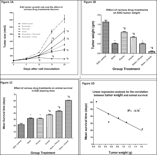
EAC, a breast-cancer- related solid tumor-model, was utilized to assess the differential cytotoxic/chemoenhancing capacity of Crocin and Flavocoxid (Flvcox) and consequent impact on animal life expectancy (survival). As compared to normal control animals, EAC-tumor grew progressively to reach a near maximal size and weight after 20 days (
Figures 1A, B). Treatment with Doxo significantly shrank tumor-size by (69%), and raised the mean animal survival time (MST) by 46% (
Figure 1C). Individual Flvcox treatment elicited also significant, but less prominent cytotoxicity, thereby shrinking tumor size by (33%); yet however, scored similar boosting of animal survival (MST) to that of Doxo (45%). Individual Crocin elicited 50% reduction in tumor weight, while extended the MST more significantly than Doxo (72%). Interestingly, the combined administration of Flvcox/Doxo showed equipotent cytotoxicity to that of Doxo (70%), but elicited a more pronounced elongation of animal survival (99%). In comparison, the joint Crocin/Doxo regimen afforded the most striking-cytotoxic response (reduction of tumor weight by 93%) and -promotion of animal-survival (180%), which significantly surpassed those of Doxo, Flvcox or their combination.
Figure 1D shows a linear-regression analysis that correlates tumor weight with animal survival. A significantly negative correlation coefficient (R
2= -0.76*) was obtained, indicating an inverse association (deterioration) of animal survival with bigger tumor weight/size. However, the magnitude of R
2, as supported also by individual responses of Doxo versus Flvocox, may further imply that tumor size is not the sole determinant or cellular trigger of animal life expectation. These notions prompted us to probe for possible variations among groups with respect to status of redox potential and expressional levels (or activity) of key cytokines or pro-apoptotic effectors.
Figure 1. (A-D): Time course for EAC tumor growth (A) and the effect of various drug regimens on tumor-weight (B) and on mean-animal survival time (C). (D) Linear regression analysis for the correlation between tumor-weight and animal survival.
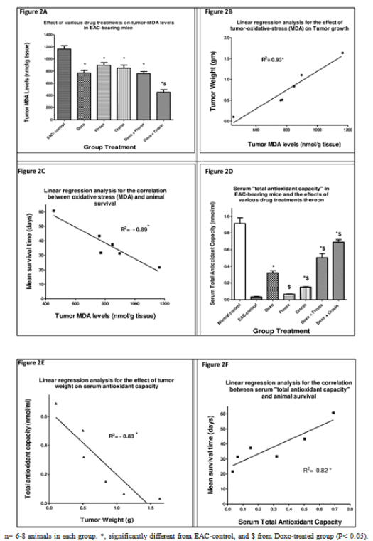
Figure 2A displays tumor-tissue levels of the lipid-peroxide-marker (MDA) in EAC-bearing mice and the effects of diverse drug regimens thereon. In absence of drug treatments, EAC-tumor tissues generated the highest (peak) MDA levels among all groups. Treatment with Doxo, Crocin, Flvcox or the (Doxo/Flvcox) combination afforded comparably significant reductions in tumor-MDA levels (24-34%). Uniquely, and consistent with its obtained cytotoxic response, the (Crocin/Doxo) combination regimen produced the most profound/significant reduction in tumor-MDA levels (61%). To further assess its prognostic value, tumor-MDA levels were correlated with data for tumor weight, and for animal survival.
Figures 2B, C elucidate that tumor MDA levels (microenvionment oxidative-stress) positively correlate with (enhance) tumor growth (R
2= 0.93), but, consequently, adversely affect animal survival (R
2= - 0.89).
Another global redox estimate that measures body defense against oxidative-stress is “total antioxidant capacity, TAC”.
Figure 2D delineates the levels of mouse-serum TAC before and after induction of EAC-tumor growth, and the impact of diverse drug regimens thereon. Thus, while peak TAC levels evidently associated with normal (tumor-free) mice, induction of EAC growth dramatically ablated such levels of “antioxidant capacity” to 4% of its original value. In EAC-bearing animals, Doxo or Crocin treatments (but not that of Flvcox) partly, but significantly restored TAC to 35% and 20% of normal peak levels; respectively. Responsiveness to concurrently-administered regimens of Doxo/Crocin or Doxo/Flvcox was far superior to individual drug components in restoring TAC, thereby scoring 55% and 77 % of normal control values; respectively. To gain insights into the basis of obtained diversity in TAC responses and its prognostic utility, we constructed two correlation curves for “TAC” values against data obtained for “tumor weight” and “animal survival” in diverse groups. Thus,
Figures 2E, F clearly indicate that TAC negatively correlates with tumor weight (growth) (R
2= - 0.83*), but positively associates with animal survival (R
2= 0.82*).
Figure 2. (A-F): (A) EAC-Tumor lipid-peroxide (MDA) levels and the effect of various drug regimens thereon. Correlation of MDA levels with tumor weight (B), and with animal survival (C). (D) Serum total antioxidant capacity (TAC) in EAC-bearing mice and the effect of various drugs thereon. (E) Correlation of (TAC) with tumor weight, and with animal survival (F).
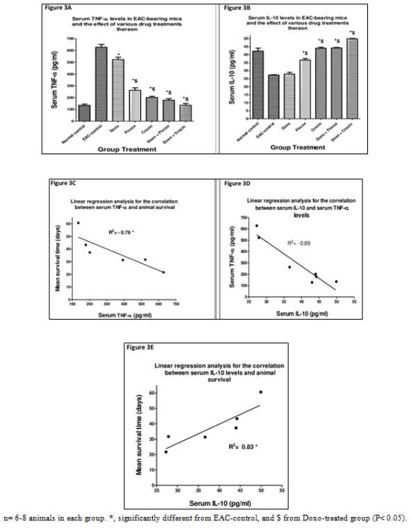
Besides, next we monitored serum cytokine levels of TNF-α and IL-10 in response to tumor induction, in presence and absence of various drug treatments. TNF-α has mostly pro-carcinogenic/ inflammatory implications, while IL-10 is now more accepted as a promoter of host immunity with positive impact on prognosis of cancer
| [13] | Landskron G, De la Fuente M, Thuwajit P, Thuwajit C & Hermoso MA. Chronic inflammation and cytokines in the tumor microenvironment. J Immunol Res 149185 (2014). https://doi.org/10.1155/2014/149185 |
| [14] | Dennis KL, Blatner NR, Gounari F & Khazaie K. Current status of interleukin-10 and regulatory T-cells in cancer. Curr Opin Oncol 256, 637-645 (2013). https://doi.org/10.1097/CCO.0000000000000006 |
[13, 14]
. Tumor growth (EAC-group) significantly raised serum TNF-α levels (4.7 folds), while reduced serum IL-10 levels by (36%) of control (tumor-free) levels, (
Figure 3A, B). Doxo treatment significantly attenuated the rises in TNF-α to 390% and decreased IL-10 levels by 35% of EAC-control. Flvcox more significantly lowered TNF-α levels to 197%, and reduced IL-10 by only 14% of EAC-control values. Either of Crocin, Doxo /Flvcox, or Doxo/Crocin regimens evoked more significant/sharper declines in TNF-α levels to (103-155%), but greater rises in IL-10 levels (105-119%) implying that TNF-α deteriorates, while IL-10 boosts, the overall drug “cytotoxic profiles”. Therefore, correlations were constructed to evaluate the potential and extent of cross-talk among levels of these cytokines and their possible influence on animal survival (
Figure 3C-E). TNF-α levels had significantly negative impact on animal survival (R
2= -0.76*). Moreover, interestingly, IL-10 correlated significantly negative with TNF-α (R
2= - 0.89*), but positively with animal survival (R
2= 0.83*).
Figure 3. (A-E): Serum TNF-α (A) and IL-10- (B) levels in control- and EAC-bearing-mice, and the effect of various drug regimens thereon. Correlation of TNF-α levels with animal survival (C) and with serum IL-10 levels (D). (E) Correlation of IL-10 levels with extent of animal survival.
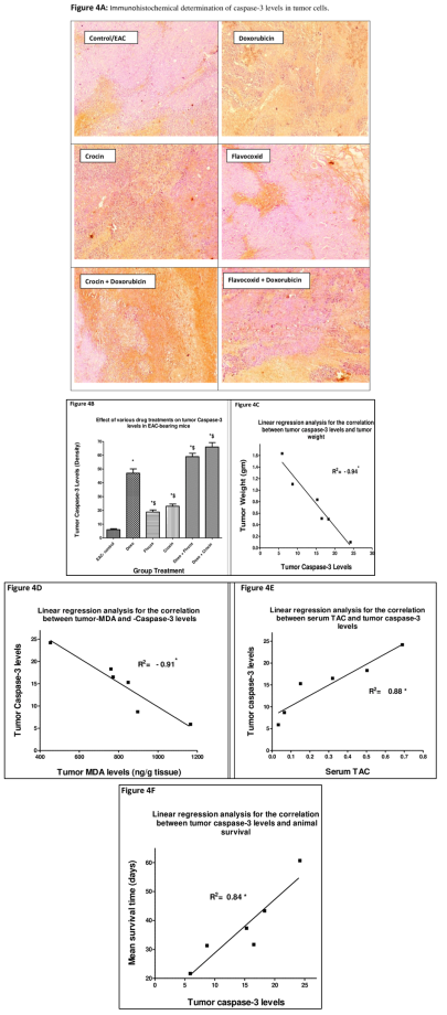
Apoptosis of tumor cells has now become a hallmark of cancer pharmacotherapy. Therefore, we measured its incidence and extent in diverse groups and further correlated these data with redox markers, tumor size, and mean animal survival.
Figure 4A, B indicates that Doxo induced a substantial (7.5 fold) rise in EAC-caspase-3 expression, a response that was mimicked by either or Flvcox or Crocin, albeit to a lower extent (3-4 folds). Remarkably, the combination regimens of the two latter drugs with Doxo significantly enhanced caspase-3 expression far beyond their obtained individual responses (9-10 folds). Linear-regression correlations (
Figure 4C-F) indicate that such caspase-3 expression inversely correlates with tumor weight, and with tumor MDA levels (-0.94*, -0.71*; respectively), but positively associates with TAC and with animal survival (0.88*, 0.84*; respectively).
Figure 4. (A-F): Microphotographs (A) and densitometric analysis (B) for immunohistochemical determination of mature caspase-3 levels in tumor sections from EAC-bearing mice (control/EAC) and the effect of various drug combinations thereon. Caspase-3 positive areas appear as brown granules. Correlation of caspase-3 levels with tumor weight (C), with MDA levels (D), with TAC (E), and with animal survival (F).
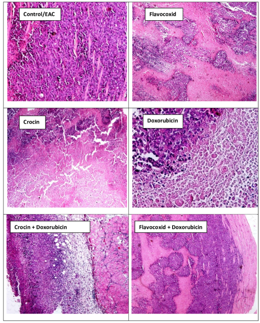
Figure 5 depicts the histopathological profiles of EAC-tumor sections from different animal groups. Thus, in EAC-sections, examination revealed malignant tumoral proliferation that is composed of highly atypical large cells with large pleomorphic nuclei, prominent large nucleoli and frequent mitotic figures, including atypical ones. Besides, wide areas of coagulative necrosis and hemorrhage are evident, while tumoral proliferation deeply invades the surrounding skeletal muscle fibers and fatty tissues. Likewise, marked inflammatory infiltrates are encountered at the tumor edge.
Figure 5. Histopathological analysis of Ehrlich Ascites Carcinoma (EAC) tumor and the effect of various drug treatments thereon.
Microphotographs through tumor stained with H&E showing sheets of large atypical cells with invasion of the muscle fibers in untreated muscles (control/EAC). Treatment of EAC bearing mice with Flavocoxid, Crocin, Doxorubicin or (Flavocoxid + Doxorubicin) showed less tumor invasion and cellular infiltrates. Treatment of EAC bearing mice with (Crocin + Doxorubicin) showed the highest protective effects against tumor invasion and cellular infiltrates. The migration of malignant cells stopped leaving a line of demarcation.
Doxo group examination revealed similar malignant tumors with the same pathological features, but with relatively more necrotic areas and less viable tumor tissue. A prominent characteristic is the deep EAC-cellular extension and infiltration into the surrounding skeletal muscle fibers, but with relatively less inflammatory cell infiltrates at the tumor edge, thereby indicating lower potential for metastasis.
Crocin treatment afforded high grade carcinoma with wide areas of coagulative necrosis that nearly involved half of the examined tumor tissue fields. Moreover, infiltrations into the surrounding skeletal muscle fibers were also observed, with less inflammatory infiltrates at the tumor edge, implying reduced metastasis.
Flvcox-treated animals displayed more viable tumor tissue with highly atypical cytological and nuclear features, along with less frequent areas of necrosis. Some prominent invasion of tumor cells to vascular and perineural tissues are seen as well. Furthermore, deep infiltrations into the surrounding skeletal muscle fibers and fatty tissue with relatively dense inflammatory infiltrates are observed at the tumor edge.
Examination of H&E stained slides from EAC-bearing mice treated with Flvcox and Doxo shows synergetic and protective effects in terms of tumor invasion and cellular infiltrates. The migration of malignant cells appears to have stopped, leaving a clear line of demarcation (less metastasis). Moreover, wide areas of coagulative necrosis, involving most of the tumor tissue with minor viable tumor cells, are noticed. Besides, mild inflammatory infiltrates can be seen at the tumor edge. On the other hand, EAC-bearing mice treated jointly with Crocin and Doxo showed synergetic and protective effects in terms of tumor invasion and cellular infiltrates, with fewer invasions into the surrounding muscle fibers and less inflammatory infiltrates at the tumor edge.
Table 1 summarizes major differential pathological findings among the treated groups with respect to mitotic and apoptotic indices as well as tumor and inflammatory infiltrations.
Table 1. Major cancer-related pathologic findings in EAC and the effect of different drug treatments thereon.
Group | Tumor infiltration at margin | Inflammatory infiltrates at edge | Mitotic index (no. of MFs in 12 HPFs) | Apoptotic index (no. of Abs in 12 HPFs) |
Control/EAC | ++++++ | ++++++ | 11 | 4 |
Doxorubicin | +++ | +++ | 4 | 10 |
Flavocoxid | +++++ | +++++ | 10 | 4 |
Crocin | ++++ | ++++ | 9 | 6 |
Flavocoxid + Doxorubicin | + | ++ | 3 | 11 |
Crocin + Doxorubicin | + | + | 3 | 13 |
- MF= Mitotic figures, HPF= HighPower Field.
4. Discussion
Our understanding of the cellular and molecular underpinnings of tumorigenesis and its drug-based cytotoxic manipulations are still far from complete. This dogma has been further complicated by two coherent aspects of chemotherapy, namely; the eruption of cancer-resistance to cytotoxicity and the observed prominent adverse reactions that can mount to life-threatening extents
| [19] | Holohan C, Van Schaeybroeck S, Longley DB & Johnston PG. Cancer drug resistance: an evolving paradigm. Nat Rev Cancer 10, 714-26 (2013). https://doi.org/10.1038/nrc3599 |
| [20] | El-Mowafy AM, Salem HA, Al-Gayyar MM, El-Mesery ME & El-Azab MF. Evaluation of the renal protective effects of the green-tea EGCG and red-grape's resveratrol: role of oxidative stress and inflammatory cytokines. Nat Prod Res 258, 850-6 (2011). https://doi.org/10.1080/14786419.2010.533669 |
| [21] | Bonita R &Pradhan R. Cardiovascular toxicities of cancer chemotherapy. Semin Oncol 402, 156-67, 2013 (2013). https://doi.org/10.1053/j.seminoncol..01.004 |
[19-21]
.
We currently employed a mouse EAC-solid tumor model to first probe the therapeutic and chemosensitizing capacity of two well tolerated natural drugs, crocin and flvcox. Next, we investigated the associated changes in expressional-levels of the cytokines (TNF-α and IL-10), in redox potential within tumor cells and the blood, and in tumor-cell susceptibility to undergo apoptosis. Besides, the extent of correlation and/or significance of inter-dependence among these factors, and their ultimate impact on animal-survival, next to malignancy, have all been delineated.
The study revealed that both drugs elicited significant individual cytotoxicity that was comparable to, but also further exceeded that by Doxo, thereby appreciably blunting tumor growth/metastasis and extending animal survival far beyond their individual drug responses. Overall, quantitatively, individual and synergy responses to crocin significantly surpassed those of flvcox. Correlation curves drawn among cytokine levels, oxidative-stress markers, and rate of tumor apoptosis, unequivocally indicated that favorable cytotoxicity and animal survival rates directly associate with serum-total antioxidant capacity, IL-10 levels and tumor caspase-3 levels, but inversely correlate with serum TNF-α- and tumor lipid-peroxide (MDA) levels. Intriguingly as well, significant negative association was observed between TNF-α and IL-10 levels, thereby suggesting a biomarker utility in evaluating tumor prognosis, and efficacy of drugs as cytotoxic and chemosensitizing agents. These results agree (in part) with ours performed on a hepatic-carcinogenesis model in rats with focus on the effectors of Nrf-2, and some apoptotic mediators
| [22] | Elsherbiny NM, Eisa NH, El-Sherbiny M & Said E. Chemo-preventive effect of crocin against experimentally-induced hepatocarcinogenesis via regulation of apoptotic and Nrf2 signaling pathways. Environ Toxicol Pharmacol 80, 103494 (2020). https://10.1016/j.etap.2020.103494 |
[22]
.
Oxidative stress is reportedly implicated in the etiology of cancer. It results from an imbalance in the production of reactive oxygen species (ROS) and cell’s own antioxidant defenses
. ROS disrupt the redox homeostasis and favor tumor development by promoting definitive signaling cascades and cellular machineries. Cancer stem cells, thanks to its anomalous redox system and subsequent manipulation of stress-transcription factors, continue to flourish and proliferate hysterically. Accordingly, several lines of evidence have indicated that oxidative stress and relevant-gene-environment can orchestrate serious pathophysiological cascades that culminate into the development of breast, prostate, pancreatic and colon cancers. For instance, oxidative stress and estrogen receptor-induced DNA disruption trigger proliferative changes to ultimately dictate estrogen-responsive breast cancers
| [24] | Acharya A, Das I, Chandhok D & Saha T. Redox regulation in cancer: a double-edged sword with therapeutic potential. Oxid Med Cell Longev 1: 23-34 (2010). https://doi.org/10.4161/oxim.3.1.10095 |
| [25] | Lee JH, Khor TO, Shu L & Su ZY, Fuentes F & Kong AN. Dietary phytochemicals and cancer prevention: Nrf2 signaling, epigenetics, and cell death mechanisms in blocking cancer initiation and progression. Pharmacol Ther 1372, 153-71 (2013). https://10.1016/j.pharmthera.2012.09.008 |
[24, 25]
. Because EAC-cells belong to breast cancer, these aspects of “tumor microenvironment” and the role of redox potential therein have been likewise currently investigated in a tumor model of these cells.
Thus, we showed that EAT proliferation is highly positively regulated by rises in tumor lipid-peroxides and falls in serum antioxidant-capacity (TAC). Accordingly, overall, oxidative stress evidently associated with tumor weight, but negatively associated with expression of tumor-caspase-3 (apoptosis), serum-IL-10 and animal survival extent. Such latter orchestrated cross-talks appear to ultimately favor inflammation, tumor-growth and metastasis, while attenuating host immune-responses and tumor cell apoptosis. Therefore, one pivotal way crocin and flvcox elicited cytotoxic responses, augmented chemotherapeutic actions, and enhanced safety profiles of Doxo, is by abrogating oxidative stress and inflammation. Our findings showed that albeit Doxo was currently the most potent cytotoxic drug, it elicited significantly inferior animal-survival profiles than crocin. These notions imply that the life-span outcomes are dictated not only by the extent of cytotoxicity but also by associated tumor-microenvironmental events and chemotherapy safety profiles, likely reasons as to why Doxo has clinically shown inconsistent therapeutic outcomes. Not surprisingly, therefore, jointly given crocin or flvcox alleviated Doxo-associated adversity on redox potential both at tumor microenvironment (MDA) and in the blood (TAC), to eventually boost its safety, efficacy and animal survival outcomes. These latter current outcomes were further reinforced and validated
via correlation studies performed with redox-markers against survival or tumor-size data. In support, BRCA1, a tumor suppressor against hormone-responsive cancers such as breast and prostate cancer, jointly plays a significant role in inhibiting ROS and estrogen-mediated DNA damage; thereby normalizing the redox homeostasis of the cells
| [24] | Acharya A, Das I, Chandhok D & Saha T. Redox regulation in cancer: a double-edged sword with therapeutic potential. Oxid Med Cell Longev 1: 23-34 (2010). https://doi.org/10.4161/oxim.3.1.10095 |
[24]
.
Inflammation has been tightly implicated in cancer development and spread. Moreover, over several decades, clinical and experimental evidence has accumulated to indicate a cross-talk interdependence (causal) relationship between cancer and inflammation. Thus, as early as 1863, the observation by Rudolf Virchow that inflammatory cells may well infiltrate tumors hypothesized also that cancer may erupt from inflammatory reactions
| [26] | David H & Rudolf Virchow. Modern aspects of tumor pathology. Pathology, research and practice, 183(3), 356–364 (1988). https://doi.org/10.1016/S0344-0338(88)80138-9 |
| [27] | Balkwill F &Mantovani A. Inflammation and cancer: back to Virchow? The Lancet 357, 9255. 539–545 (2001). |
[26, 27]
. Now, this postulation has been recently proven rational with the emergence of several reports implicating infection and chronic inflammatory disease in the development of cancer
. Besides, chronic inflammation can also foster malignant cell transformation in healthy tissues. Interestingly, inflammation deploys many common molecular effectors and signaling pathways with carcinogenesis, including accelerated cellular proliferation and angiogenesis. On the other hand, consonant therapeutic evidence came also from the use of nonsteroidal anti-inflammatory drugs, which lessened the incidence, severity and mortality of numerous cancer types
| [29] | Yan L, Anderson GM, DeWitte M & Nakada, MT. “Therapeutic potential of cytokine and chemokine antagonists in cancer therapy. European Journal of Cancer 42, 6: 793-802 (2006). https://doi.org/10.1016/j.ejca.2006.01.013 |
[29]
.
Tumor necrosis factor (TNF-α) is a paramount inflammatory mediator with wide implications in the early stages of carcinogenesis, including angiogenesis and invasion, rather than in the late progression of carcinogenesis
| [30] | Popa C, Netea MG, Van Riel PLC, Van Der Meer JWM & Stalenhoef AFH. The role of TNF-α in chronic inflammatory conditions, intermediary metabolism, and cardiovascular risk. Journal of Lipid Research 484, 751–762 (2007). https://api.semanticscholar.org/CorpusID:8382081 |
| [31] | Moore RJ, Owens DM, Stamp G, Arnott C, Burke F & East N et al. Mice deficient in tumor necrosis factor-alpha are resistant to skin carcinogenesis. Nat Med 5, 828-831 (1999). |
| [32] | Szlosarek P, Charles KA & Balkwill FR. Tumour necrosis factor-α as a tumour promoter. European Journal of Cancer 426, 745–750 (2006). https://doi.org/10.1016/j.ejca.2006.01.012 |
[30-32]
. As well, Moore et al. provided experimental evidence that TNF-α-deficient mice were markedly resistant to phorbol-ester-induced skin cancer
. Furthermore, in oral squamous cell carcinoma, intense TNF-α exposure can raise the proportion of cancer stem cell phenotypes and enhance over-expression of their growth-promoting transcription factors, thereby exacerbating tumorigenicity
.
A myriad of mechanisms have been proposed to define the exact role of TNF-α in tumorigenesis and metastasis. One such proposals has addressed the contribution of oxidative-stress, thus showing TNF-α to enhance generation of reactive oxygen and nitrogen species (RONS) to induce DNA damage, hence facilitating tumorigenesis. By contrast, a few reports have suggested that high concentrations of this cytokine can evoke an antitumoral response in a murine model of sarcoma
| [34] | Havell EA, Fiers W & North RJ. The antitumor function of tumor necrosis factor TNF-I. Therapeutic action of TNF against an established murine sarcoma is indirect, immunologically dependent, and limited by severe toxicity. Journal of Experimental Medicine 167, 1067–1085 (1988). https://doi.org/10.1084/jem.167.3.1067 |
[34]
. According to these findings, reconciliation considers that, within the tumor microenvironment, the ultimate pro- or anti-tumoral TNF-α response depends on its local concentration and expressional site in the tumor cell. Thus, in this vein, patients with raised levels of TNF-α in tumor islets from non-small cell lung cancer, that were mostly confined to macrophages and/or mast cells, displayed superior survival rates than patients with rising stromal TNF-α levels
| [35] | Ohri CM, Shikotra, Green RH, Waller DA & Bradding P. Tumour necrosis factor-alpha expression in tumour islets confers a survival advantage in non-small cell lung cancer. BMC Cancer 10, article 323 (2010). https://doi.org/10.1186/1471-2407-10-323 |
| [36] | Lee SH, Hong HS, Liu ZX, Kim RH, Kang MK & Park NH et al. TNFα enhances cancer stem cell-like phenotype via Notch-Hes1 activation in oral squamous cell carcinoma cells. Biochemical and Biophysical Research Communications 424, 58–64 (2012). https://doi.org/10.1016/j.bbrc.2012.06.065 |
[35, 36]
.
In contrast to TNF-α, IL-10 elicits mostly anti-inflammatory effects, enhances host immunity against cancer, and confers anti-angiogenic effects
. Almost all immune cells, including T-cells, B-cells, monocytes, macrophages and mast cells produce IL-10
. IL-10 inhibits NF-κB signaling; thereby blunting the expression of proinflammatory cytokine and favoring an overall antitumor environment
| [38] | Schottelius A J, Mayo MW, Balfour Sartor R & Baldwin Jr. AS. Interleukin-10 signaling blocks inhibitor of κB kinase activity and nuclear factor κB DNA binding. J Biol Chem 274, 31868–31874 (1999). https://doi.org/10.1074/jbc.274.45.31868 |
[38]
. Consistent with this finding, Berg et al. demonstrated that IL-10-deficient murine models are more prone to bacteria-induced carcinogenesis
| [39] | Berg D., Davidson N, Kühn R. Müller, Menon S & Holland G. et al. Enterocolitis and colon cancer in interleukin-10-deficient mice are associated with aberrant cytokine production and CD4+ Th1-like responses. Journal of Clinical Investigation 98, 1010–1020 (1996). https://doi.org/10.1172/JCI118861 |
[39]
.
Therefore, in contrast to the above reports, in EAC a paucity of information on cytokine functions appears to dominate. Thus, we differentially measured the levels of TNF-α and IL-10 in EAC, presented also on a conference for us
| [40] | El-Mowafy A., Said, E., Elsherbiny N. M., Abdelaziz R. R., and Zaki M. A. Novel chemosensitizing effects for crocin and flavocoxid in a mouse eac-tumor model: cellular and molecular triggers. A 4th European Biopharma Congress & 6th International Conference and Exhibition on Pharmacology and Ethnopharmacology. 2017, Viena, Austria. https://doi.org/10.4172/2167-065X-C1-026 |
[40]
, and further thereafter investigated how the levels of these cytokines may relate to each other (cross-talk), and to animal-survival or tumor-growth, in the absence and presence of single and combined drug regimens of Doxo, crocin and flvcox.
Ultimately, the measured levels of these cytokines revealed the impact of inflammation (TNF-α) and host immune response (IL-10) on tumor growth; and how far these responses are modulated by chemotherapy regimens under considerations. The translation value of current cytokine levels is also unequivocally inferred from correlation curves, which overtly indicate that TNF-α significantly associated with worsening, while IL-10 with promotion, of animal survival.
Doxo displayed inferior cytokine profiles in which it scored the highest serum levels of TNF-α, while failed to enhance that of IL-10, leaving its levels similar to basal EAC ones. These notions reveal that Doxo fosters a high level of inflammation, while blunts host ability to restrain tumor proliferation and metastasis, a likely compelling reason as to why it currently showed a limited animal rescuing caliber. Such envisions are further reinforced by the obtained individual and synergy results for crocin/flvcox, and Doxo. Thus, sole crocin or flvcox, albeit had lower cytotoxic effects than Doxo, they were far superior to it in reducing TNF-α- and raising IL-10-levels, thereby showing, per se, similar (Flvcox) or higher (Crocin) promotion of animal survival. Besides, crocin and flvcox elicited a much higher survival effect when they were combined with Doxo. Therefore, optimal modification of these cytokine by crocin and flvcox constitutes a molecular basis and a crucial trigger whereby they chemosensitize the cytotoxic actions of Doxo.
Chemosensitization is a strategy to counteract chemoresistance that usually erupts to traditional cytotoxic agents, by using an additional drug, thereby augmenting the overall responsiveness to chemotherapy. Among the potential chemosensitizers are natural agents such as catechins, flavonoids, carotenoids, omega-3-fatty-acids and resveratrol, which were proven capable of modulating key biological pathways and cellular triggers, consonant with antineoplastic effects
| [7] | El-Mowafy AM, Al-Gayyar MM, El-Mesery ME, Salem HA, Darweish M. Novel chemotherapeutic and renal protective effects for the green-tea EGCG: Role of oxidative stress and inflammatory-cytokine signaling. Phytomedicine, 1714, 1067-75 (2010). https://doi.org/10.1093/advances/nmy077 |
| [20] | El-Mowafy AM, Salem HA, Al-Gayyar MM, El-Mesery ME & El-Azab MF. Evaluation of the renal protective effects of the green-tea EGCG and red-grape's resveratrol: role of oxidative stress and inflammatory cytokines. Nat Prod Res 258, 850-6 (2011). https://doi.org/10.1080/14786419.2010.533669 |
| [41] | Abou-Zeid LA & El-Mowafy AM. Molecular dynamic characteristics of resveratrol interaction with human estrogen receptor-α: distinct recognition from diethylstilbestrol. Journal of Molecular Structure: Theochem 593, 1, 39-48 (2002). https://doi.org/10.1159/000058013 |
[7, 20, 41]
. Over the years, numerous natural products have been identified as potentially efficacious anti-cancer agents for their versatile/multitargeting properties, low cost and toxicity profiles, and instant availability. Not surprisingly, over 60% of the anticancer drugs available to date in the market are of natural origin
| [42] | Gupta SC, Kim JH, Prasad S & Aggarwal BB. Regulation of survival, proliferation, invasion, angiogenesis, and metastasis of tumor cells through modulation of inflammatory pathways by nutraceuticals. Cancer Metastasis Rev 29, 405–434 (2010). https://doi.org/10.1007/s10555-010-9235-2 |
| [43] | Newman DJ, Cragg GM & Snader KM. Natural products as sources of new drugs over the period 1981–2002. J Nat Prod 66, 1022–1037 (2003). https://doi.org/10.1021/np030096l |
[42, 43]
.
Another aspect in favor of crocin and flvcox antitumor/chemoenhancing utility is their well-tolerance and safety profiles. For instance, they possess additional anti-inflammatory, antioxidant, cell-protective and immunostimulatory effects, which are of superb value and utmost need to mitigate a myriad of fingerprinted health anomalies and complications entailed to cancer.
Moreover, besides to its anti-inflammatory effects, flvcox possesses a strong antioxidant activity which further helps downregulate inducible inflammatory gene expression and neutralizes ROS, thereby preventing the conversion of arachidonic acid to oxidized lipids
| [11] | Burnett BP, Bitto A, Altavilla D, Squadrito F, Levy RM & Pillai L. Flavocoxid inhibits phospholipase A2, peroxidase moieties of the cyclooxygenases COX, and 5-lipoxygenase, modifies COX-2 gene expression, and acts as an antioxidant. Mediators Inflamm 385780 (2011). https://doi.org/10.1155/2011/385780 |
[11]
. Accordingly, Flvcox has shown cellular protective and anti-inflammatory potential in ischemic stroke, pancreatitis and nephritis. Intriguingly as well, crocin has shown cellular protective effects against Doxo-induced renal- cardiac- and liver-toxicity
| [12] | Bitto A, Squadrito F, Irrera N, Pizzino G, Pallio G, Mecchio A et al.. Flavocoxid, a nutraceutical approach to blunt inflammatory conditions. Mediators Inflamm 2014, 790851. https://doi.org/10.1155/2014/790851 |
| [44] | El-Kashef DH, El-Kenawi AE, Suddek GM & Salem HA. Flavocoxid attenuates gentamicin-induced nephrotoxicity in rats. Naunyn Schmiedebergs Arch Pharmacol 38812, 1305-15 (2015). https://doi.org/10.1007/s00210-015-1164-8 |
| [45] | Elsherbiny NM, Salama MF, Said E, El-Sherbiny M & Al-Gayyar MM. Crocin protects against doxorubicin-induced myocardial toxicity in rats through down-regulation of inflammatory and apoptic pathways. Chem Biol Interact 247, 39-48 (2016). https://10.1016/j.cbi.2016.01.014 |
[12, 44, 45]
. Moreover, we currently show that both drugs elicit a marked tumor apoptotic effect on their own, while also significantly boost that of Doxo. Such profiles for these two candidates under investigation, suggest additional means whereby they may lessen the untoward reactions, while sensitize the cytotoxic effects of Doxo, thereby augmenting its overall therapeutic index. The capacity of current drugs to limit tumor growth and metastasis, while also expediting its apoptotic termination, was documented with outcomes of histopathological analyses. Intriguingly, currently, a high level of positive association existed between tumor rate of apoptosis and oxidative-stress within tumor microenvironment and circulation as inferred from negative correlation with tumor-MDA levels, and positive association with TAC. Together, these findings project the value of antioxidants (radical scavengers) as also superb accelerators of tumor demise. In support, reactive oxygen species (ROS) are engaged in regulating diverse biological systems, especially those with rapidly proliferating cells, as with tumor cells. Indeed, oxygen metabolism entails many hazardous byproducts that can induce carcinogenesis. ROS play a role in
ras-induced cellular transformation. Defective signaling through ROS pathways can contribute to concerted curbing of apoptosis, and development of cancer. Therefore, new approaches have been deployed to design and apply novel delivery methods to circumvent and/or obliterate cancer. Thus, the use of a recombinant adenoviral vector that expresses the radical-depleting enzyme Mn-SOD (Ad-Mn-SOD) showed antitumor effect. The latter was also more overt in the presence of conventional anticancer drugs, thereby appreciating the concept of synergy and sensitization
, as per the present study. Besides, a phase-I study has been conducted on human cancer with injection of Mn-SOD plasmid-liposomes
| [47] | Tarhini AA, Belani CP, Luketich JD, Argiris A, Ramalingam SS & Gooding W et al. A phase I study of concurrent chemotherapy paclitaxel and carboplatin and thoracic radiotherapy with swallowed manganese superoxide dismutase plasmid liposome protection in patients with locally advanced stage III-non-small-cell lung cancer. Hum Gene Ther 223, 336-342 (2011). https://doi.org/10.1089/hum.2010.078 |
[47]
.
Thus, conclusively, this study elucidates that EAC tumor growth and animal survival are controlled through a reciprocal balance in levels of two cytokines: TNF-α (negative prognosis) and IL-10 (positive prognosis). Other principal players include “tumor & environmental” redox-potential, and tumor expression of apoptotic effectors (caspase-3), which inversely correlate to each other to differentially regulate tumor growth and animal survival. Thus, interestingly, prominent cross-talk exists among these three pathways, thereby attesting to complexity of the cellular and molecular machinery whereby EAC tumor growth and prognosis are regulated. Doxo, albeit produced the top cytotoxicity, it displayed inferior cytokine and animal rescuing profiles. Crocin and flvcox, in a rank order of potency, significantly optimized the aforementioned pathways to confer significant anti-inflammatory, antioxidant, tumor apoptotic- and cytotoxic-effects on their own, and further substantially sensitize the therapeutic and animal rescuing capacity of Doxo. Per se, flvcox is a promising chemosensitizing agent that is worth of further experimental/clinical chemotherapeutic evaluations. Finally, the overall redox potential is a crucial regulator of tumor growth, modulator of responsiveness to chemotherapy, and a likely predictor of animal-survival in these settings.