1. Introduction
Environmental contamination with synthetic pyrethroid insecticides has become a growing public health concern due to their increasing use in agriculture, veterinary medicine, and domestic pest control. Although these compounds were initially considered relatively safe for mammals, accumulating evidence indicates that chronic or repeated exposure may adversely affect non-target organisms, particularly the male reproductive system
. Among these compounds, Bifenthrin, a widely used type I pyrethroid insecticide, has attracted attention because of its extensive application and environmental persistence. Several experimental studies have demonstrated that exposure to bifenthrin may impair male reproductive function by inducing structural and biochemical alterations in reproductive organs
. The epididymis plays a crucial role in sperm maturation, storage, and transport; therefore, toxicant-induced damage to this organ can significantly compromise sperm quality and male fertility
| [3] | Mboso, O. E., Ukam, I. O., Iwara, I. A., Ojonshua, I. A., Eteng, K. A., Ibor, M., & Eteng, M. U. (2025). Molecular and In-Silico Insights into Eremomastax speciosa Protection Against Cadmium Toxicity. Scientific African, e03102.
https://doi.org/10.1016/j.sciaf.2025.e03102 |
[3]
.
The mechanisms underlying pyrethroid-induced reproductive toxicity involve multiple interconnected molecular pathways. One of the primary mechanisms is the excessive generation of reactive oxygen species (ROS), which disrupts cellular redox homeostasis and leads to oxidative damage in reproductive tissues
. Elevated ROS levels promote lipid peroxidation, protein oxidation, and DNA fragmentation, thereby impairing epididymal epithelial integrity and sperm maturation processes
| [5] | Asadi, N., Bahmani, M., Kheradmand, A., & Rafieian-Kopaei, M. (2017). The impact of oxidative stress on testicular function and the role of antioxidants in improving it: a review. Journal of clinical and diagnostic research: JCDR, 11(5), IE01. https://doi.org/10.7860/jcdr/2017/23927.9886 |
[5]
. In addition to oxidative stress, pesticide-induced reproductive injury is often associated with activation of inflammatory signaling cascades, particularly the nuclear factor-kappa B (NF-κB) pathway, which stimulates the release of pro-inflammatory mediators such as tumor necrosis factor-alpha (TNF-α) and interleukin-1 beta (IL-1β)
| [6] | El Safadi, M., Shah, T. A., Zahara, S. S., Jardan, Y. A. B., & Bourhia, M. (2025). Regulation of TGF-β1, PI3K/PIP3/Akt, Nrf-2/Keap-1 and NF-κB signaling pathways to avert bifenthrin induced hepatic injury: A palliative role of daidzein. Tissue and Cell, 93, 102733.
https://doi.org/10.1016/j.tice.2025.102733 |
[6]
. Persistent inflammatory responses can further amplify oxidative damage and contribute to cellular dysfunction in reproductive tissues.
Another important signaling pathway involved in toxicant-induced tissue injury is the mitogen-activated protein kinase (MAPK) cascade, particularly the p38 MAPK pathway. Activation of p38 MAPK plays a central role in mediating cellular stress responses, inflammation, and apoptosis
| [7] | Dutta, S., Sengupta, P., Slama, P., & Roychoudhury, S. (2021). Oxidative stress, testicular inflammatory pathways, and male reproduction. International journal of molecular sciences, 22(18), 10043. https://doi.org/10.3390/ijms221810043 |
[7]
. Excessive activation of this pathway can trigger downstream apoptotic signaling, including the activation of caspase-3, a key executioner enzyme responsible for programmed cell death
| [8] | Ujong, U. P., Ibor, M. E., Iwara, I. A., & Eteng, M. U. (2025). Synergistic effect and biochemical evaluation of kolaviron and quercetin on rat-model benign prostate hyperplasia. Journal of Asian Natural Products Research, 1-16.
https://doi.org/10.1080/10286020.2025.2501022 |
[8]
. Although oxidative stress, inflammation, and apoptosis are recognized contributors to pesticide-induced reproductive toxicity, the integrated involvement of inflammatory mediators, p38 MAPK signaling, and caspase-3-mediated apoptosis in bifenthrin-induced epididymal injury remains insufficiently characterized. Understanding these mechanistic interactions is essential for identifying potential therapeutic interventions capable of mitigating reproductive damage.
Natural bioactive compounds with antioxidant and anti-inflammatory properties have attracted increasing attention as potential protective agents against environmental toxicants. One such compound is kolaviron, a biflavonoid complex isolated from the seeds of
Garcinia kola. Kolaviron has been extensively studied for its potent antioxidant, anti-inflammatory, and cytoprotective activities in several experimental models
. It has been shown to enhance endogenous antioxidant defense systems by increasing the activities of superoxide dismutase (SOD), catalase (CAT), and glutathione peroxidase (GPx), as well as by maintaining intracellular glutathione levels, thereby reducing oxidative stress-mediated cellular damage
| [10] | Abarikwu, S. O. (2015). Anti-inflammatory effects of kolaviron modulate the expressions of inflammatory marker genes, inhibit transcription factors ERK1/2, p-JNK, NF-κB, and activate Akt expressions in the 93RS2 Sertoli cell lines. Molecular and cellular biochemistry, 401(1), 197-208.
https://doi.org/10.1007/s11010-014-2307-9 |
[10]
. In addition, kolaviron has been reported to modulate inflammatory signaling pathways through suppression of NF-κB activation and inhibition of pro-inflammatory mediators such as inducible nitric oxide synthase (iNOS) and cyclooxygenase-2 (COX-2)
. These pharmacological properties suggest that kolaviron may be a promising candidate for protecting reproductive tissues against toxicant-induced injury.
Despite these well-documented biological activities, the potential role of kolaviron in protecting the epididymis against pesticide-induced reproductive toxicity has not been fully investigated. In particular, limited information exists regarding its ability to regulate stress-activated signaling pathways such as p38 MAPK and apoptotic processes mediated by caspase-3 during toxicant exposure. Consequently, the therapeutic relevance of kolaviron in mitigating bifenthrin-induced epididymal damage remains largely unexplored.
Therefore, the present study aimed to investigate the protective effects of kolaviron against bifenthrin-induced epididymal toxicity in male Wistar rats. Specifically, this study examined alterations in inflammatory mediators, stress-activated signaling pathways involving p38 MAPK, and apoptosis-related proteins such as caspase-3, in addition to oxidative stress parameters and histopathological changes. By elucidating these mechanistic pathways, this investigation provides new insight into the molecular basis of bifenthrin-induced reproductive injury and highlights the potential therapeutic value of kolaviron as a natural agent for protecting the male reproductive system against environmental toxicants.
2. Materials and Methods
2.1. Chemicals
Bifenthrin (97% TC) (with batch number 20211012) was procured from Nanjing Essence Fine-Chemical Co., Ltd. China. All other reagents were of analytical grade and obtained from Sigma-Aldrich Co. (St. Louis, MO, USA) through ND-Harris & associates Onitsha, Nigeria.
2.2. Isolation of Kolaviron
Seeds of Garcinia kola were obtained from a local vendor in Boki, Cross River State, Nigeria (GPS: 6.2500° N, 9.0333° E). The seeds were authenticated by Dr. Emmanuel Ekpiken, a qualified plant taxonomist at the Herbarium of the University of Cross River State, Nigeria, and a voucher specimen (Voucher No. UCS/HB/2025/012) was deposited for future reference. Prior to extraction, the seeds were screened for common mycotoxins (aflatoxins B1, B2, G1, G2) and pesticide residues, and were confirmed to be free of detectable contaminants.
The seeds were air-dried, pulverized, and kolaviron was isolated following a previously validated protocol with minor modifications
. Briefly, 1.5 kg of the pulverized seeds was defatted using light petroleum ether (60–80°C) in a Soxhlet extractor for 24 h. The defatted residue was air-dried and subsequently extracted with acetone using a Soxhlet apparatus for an additional 24 h. The acetone extract was concentrated under reduced pressure at 40°C using a rotary evaporator.
The concentrated extract was diluted with distilled water in a 1:2 (v/v) ratio and successively partitioned with ethyl acetate (6 × 250 mL). The combined ethyl acetate fractions were concentrated under reduced pressure to obtain a golden-yellow solid identified as kolaviron. The extraction yield of kolaviron was 9.4% (w/w) relative to the initial dry weight of the seeds.
Purity and structural validation of the isolated kolaviron were carried out using ¹H-NMR, ¹³C-NMR, and electron ionization mass spectrometry (EI–MS). The obtained spectral data were in agreement with previously reported reference spectra for kolaviron
| [12] | Farombi, E. O., Ajayi, B. O., Opata, E. K., Fafioye, A. O., & Akinade, A. T. (2023). Kolaviron modulates angiogenesis, apoptosis and inflammatory signaling in rat model of testosterone propionate-induced benign prostate hyperplasia. Inflammopharmacology, 31(4), 2121-2131.
https://doi.org/10.1007/s10787-023-01171-7 |
[12]
, confirming compound identity. Further purity assessment was performed using high-performance liquid chromatography (HPLC), which demonstrated a purity of ≥95%, and thin-layer chromatography (TLC), which revealed a single major spot, indicating minimal impurities. The purified kolaviron was stored in airtight amber-colored vials at −20°C until use to prevent photodegradation and oxidative degradation.
2.3. Animal Protocol
All experimental procedures involving animals were conducted in accordance with the ARRIVE 2.0 guidelines and complied with institutional and national regulations for the care and use of laboratory animals. Ethical approval was obtained from the Faculty of Basic Medical Sciences Animal Ethics Committee, University of Cross River State, Okuku Campus (Approval No.: FBMS/UNICROSS/25/10; approved on 12 March 2025).
A total of forty-eight (48) young adult male Wistar rats (Rattus norvegicus), aged 10–12 weeks and weighing 110–180 g at the start of the experiment, were obtained from the Animal House, Faculty of Basic Medical Sciences, University of Cross River State. All animals were sourced from the same breeding colony to ensure uniformity in age, genetic background, and health status. Following acclimatization for 7 days, rats with comparable body weights were randomly assigned to experimental groups to minimize inter-group variability. Only male rats were used because the study specifically focused on epididymal structure, function, and sperm parameters, which are exclusive to the male reproductive system.
Rats were housed in standard polypropylene cages (60 cm × 40 cm) fitted with stainless steel mesh covers, with eight animals per cage. Environmental conditions were maintained at 26 ± 2°C, relative humidity of approximately 45%, and a 12-hour light/12-hour dark cycle. Animals were provided with standard commercial pelletized rat chow (Mazuri®) and clean drinking water ad libitum. Cages were cleaned regularly to maintain hygienic conditions throughout the study.
Following a 7-day acclimatization period, animals were randomly assigned to six experimental groups (n = 8 per group):
1) Group I (Control): Canola oil vehicle only for 28 days.
2) Group II (Bifenthrin): Bifenthrin (1 mg/kg body weight) for 28 days.
3) Group III (Kolaviron): Kolaviron (200 mg/kg body weight) for 28 days.
4) Group IV (Kolaviron Pre-treatment): Kolaviron (200 mg/kg) for 28 days, followed immediately by bifenthrin (1 mg/kg) for an additional 28 days (total 56 days).
5) Group V (Kolaviron Post-treatment): Bifenthrin (1 mg/kg) for 28 days, followed immediately by kolaviron (200 mg/kg) for 28 days (total 56 days).
6) Group VI (Co-treatment): Bifenthrin (1 mg/kg) and kolaviron (200 mg/kg) administered concurrently for 28 days.
All compounds were administered orally by gavage once daily. Bifenthrin, being highly lipophilic, was dissolved in canola oil to ensure complete solubilization and accurate dosing. Kolaviron, the isolated biflavonoid fraction, was dissolved in a minimal volume of 0.5% dimethyl sulfoxide (DMSO) and then diluted with canola oil to achieve the desired concentration. The standardized dosing volume for all groups was 0.2 mL/100 g body weight (2 mL/kg). Vehicle control animals received an equivalent volume of canola oil to account for any potential vehicle effects.
The doses of bifenthrin (1 mg/kg) and kolaviron (200 mg/kg) were selected based on pilot investigations and previous reports demonstrating sub-chronic reproductive toxicity and protective efficacy, respectively, without inducing significant mortality or systemic toxicity
| [8] | Ujong, U. P., Ibor, M. E., Iwara, I. A., & Eteng, M. U. (2025). Synergistic effect and biochemical evaluation of kolaviron and quercetin on rat-model benign prostate hyperplasia. Journal of Asian Natural Products Research, 1-16.
https://doi.org/10.1080/10286020.2025.2501022 |
| [12] | Farombi, E. O., Ajayi, B. O., Opata, E. K., Fafioye, A. O., & Akinade, A. T. (2023). Kolaviron modulates angiogenesis, apoptosis and inflammatory signaling in rat model of testosterone propionate-induced benign prostate hyperplasia. Inflammopharmacology, 31(4), 2121-2131.
https://doi.org/10.1007/s10787-023-01171-7 |
| [13] | Ujong, U. P., & Ibor, M. E. Bifenthrin Causes Kidney Damage via Induction of Oxidative Stress, Activation of Pro-Inflammatory Cytokines, and Up Regulation of Apoptosis in Wistar Rats. African Journal of Biochemistry and Molecular Biology Research. 2024; 510-525.
https://doi.org/10.58578/ajbmbr.v1i1.3654 |
[8, 12, 13]
. This experimental design allowed a comprehensive assessment of structural, biochemical, molecular, and functional indices of reproductive toxicity, including epididymal histopathology, sperm number and abnormalities, oxidative stress markers, inflammatory mediators, and apoptosis-related proteins.
At the end of the experimental period, rats were euthanized under deep anesthesia induced by intraperitoneal administration of ketamine in accordance with institutional ethical guidelines. Adequate anesthesia was confirmed by the absence of pedal withdrawal and corneal reflexes. Death was further verified by cessation of respiration and cardiac activity before tissue collection. The epididymides were then rapidly excised, carefully trimmed of adhering fat and connective tissue, and rinsed in ice-cold normal saline to remove blood contaminants prior to further processing.
2.4. Determination of Epididymal Sperm Abnormality, Sperm Number and Glucose 6-Phosphate Dehydrogenase (G6PD) Activity
The epididymal tissues were minced and mechanically dispersed in a buffered medium (phosphate-buffered saline). A small aliquot of the sperm suspension was placed on a glass slide and a coverslip applied, and the sample was observed under a light microscope at 200x magnification. Abnormality grading was done according to WHO criteria as reported by Patel et al. (2018)
| [14] | Patel, A. S., Leong, J. Y., & Ramasamy, R. (2018). Prediction of male infertility by the World Health Organization laboratory manual for assessment of semen analysis: a systematic review. Arab journal of urology, 16(1), 96-102.
https://doi.org/10.1016/j.aju.2017.10.005 |
[14]
.
Hemocytometer was used to determined sperm number. Briefly, homogenize epididymal tissue were diluted and mixed in a trypan blue to immobilize sperm. 10 µL volume of the cell suspension was placed on the hemocytometer and the sperm heads in the central grid were counted under a microscope.
Calculation:
Sperm concentration =
Epididymal tissues were homogenized in ice-cold 0.1 M phosphate buffer (pH 7.4) using a Teflon homogenizer and centrifuged at 10,000g for 15 min at 4°C. The supernatant was collected for biochemical estimations. Glucose 6-phosphate dehydrogenase (G6PD) activity was assessed according to the method of Temel et al.
| [15] | Temel, Y., Bengü, A. Ş., Akkoyun, H. T., Akkoyun, M., & Ciftci, M. (2017). Effect of astaxanthin and aluminum chloride on erythrocyte G6PD and 6PGD enzyme activities in vivo and on erythrocyte G6PD in vitro in rats. Journal of biochemical and molecular toxicology, 31(10), e21954.
https://doi.org/10.1002/jbt.21954 |
[15]
.
2.5. Determination of Serum Luteinizing Hormone, Follicle Stimulating Hormone and Testosterone
Serum levels of luteinizing hormone (LH), follicle-stimulating hormone (FSH), and testosterone were quantified using commercially available ELISA kits (CUSABIO Life Science Inc., Wuhan, China). The catalog numbers, species validation (rat), assay detection ranges, and intra- and inter-assay coefficients of variation (%CV) were as follows: LH: Cat. No. CSB-E05122r; validated for rat; detection range 0.156–10 ng/mL; intra-assay CV < 8%, inter-assay CV < 10%. FSH: Cat. No. CSB-E05124r; validated for rat; detection range 0.156–10 ng/mL; intra-assay CV < 8%, inter-assay CV < 10%. Testosterone: Cat. No. CSB-E05140r; validated for rat; detection range 0.1–50 ng/mL; intra-assay CV < 7%, inter-assay CV < 9%.
All assays were performed according to the manufacturer’s instructions and following the procedure described by Ujong et al. (2025)
| [8] | Ujong, U. P., Ibor, M. E., Iwara, I. A., & Eteng, M. U. (2025). Synergistic effect and biochemical evaluation of kolaviron and quercetin on rat-model benign prostate hyperplasia. Journal of Asian Natural Products Research, 1-16.
https://doi.org/10.1080/10286020.2025.2501022 |
[8]
. Serum samples and standards were run in duplicate to ensure accuracy, and absorbance was measured using a microplate reader at the specified wavelengths recommended by the kit protocols.
2.6. Determination of Biomarkers of Epididymal Oxidative Stress
Epididymal tissues were homogenized in ice-cold 0.1 M phosphate buffer (pH 7.4) using a Teflon homogenizer and centrifuged at 10,000g for 15 min at 4°C. The supernatant was collected for biochemical estimations. Superoxide dismutase (SOD) activity was assessed according to the method of Zhang et al. (2017)
| [16] | Zhang, J., Chen, R., Yu, Z., & Xue, L. (2017). Superoxide dismutase (SOD) and catalase (CAT) activity assay protocols for Caenorhabditis elegans. Bio-protocol, 7(16), e2505-e2505. https://doi.org/10.21769/bioprotoc.2505 |
[16]
. Reduced glutathione (GSH) was determined at 412 nm according to the method of Hamad et al. (2019)
| [17] | Hamad, A., Elshahawy, M., Negm, A., & Mansour, F. R. (2019). Analytical methods for determination of glutathione and glutathione disulfide in pharmaceuticals and biological fluids. Reviews in analytical chemistry, 38(4), 20190019. https://doi.org/10.1515/revac-2019-0019 |
[17]
. Catalase (CAT) activity was determined using hydrogen peroxide as a substrate according to the method of Knikman et al. (2020)
| [18] | Knikman, J. E., Rosing, H., Guchelaar, H. J., Cats, A., & Beijnen, J. H. (2020). A review of the bioanalytical methods for the quantitative determination of capecitabine and its metabolites in biological matrices. Biomedical Chromatography, 34(1), e4732. https://doi.org/10.1002/bmc.4732 |
[18]
. Glutathione peroxidase (GPx) activity was determined according to the method of Ahmed et al. (2021)
. Lipid peroxidation was quantified as MDA according to the method described by Fauziah, (2018)
| [20] | Fauziah, P. N., Maskoen, A. M., Yuliati, T., & Widiarsih, E. (2018). Optimized steps in determination of malondialdehyde (MDA) standards on diagnostic of lipid peroxidation. Padjadjaran Journal of Dentistry, 30(2), 136-139.
https://doi.org/10.24198/pjd.vol30no2.18329 |
[20]
. Glutathione s-transferase (GST) activity was determined according to the method of Hajdinák et al. (2018)
| [21] | Hajdinák, P., Czobor, Á., Lőrincz, T., & Szarka, A. (2019). The problem of glutathione determination: A comparative study on the measurement of glutathione from plant cells. Periodica Polytechnica Chemical Engineering, 63(1), 1-10.
https://doi.org/10.3311/ppch.11785 |
[21]
.
2.7. Determination of Epididymal Inflammatory Proteins
Epididymal tissues were first rinsed with ice-cold 1× phosphate-buffered saline (PBS, pH 7.4) and then homogenized in four volumes of ice-cold 0.1 M phosphate buffer (pH 7.4) using a Teflon homogenizer. The homogenates were centrifuged at 10,000 × g for 15 minutes at 4 °C, and the supernatants were stored at −20 °C until further analysis. To ensure complete disruption of cell membranes, the samples underwent two freeze–thaw cycles, followed by a second centrifugation at 5,000 × g for 5 minutes at 4 °C.
Protein concentrations of tumor necrosis factor-alpha (TNF-α), inducible nitric oxide synthase (iNOS), cyclooxygenase-2 (COX-2), interleukin-1 beta (IL-1β), interleukin-4 (IL-4), caspase-3, and p38 MAPK were determined using commercially available ELISA kits (CUSABIO Life Science Inc., Wuhan, China). All assays were conducted strictly according to the manufacturer’s instructions, with samples and standards run in duplicate to ensure accuracy and reproducibility.
2.8. Histological Analysis
Excised epididymal tissues were immediately fixed in 10% neutral buffered formalin for 48 hours to preserve tissue architecture. Following fixation, tissues were dehydrated through graded ethanol series, cleared in xylene, and embedded in paraffin wax using standard histological procedures
| [8] | Ujong, U. P., Ibor, M. E., Iwara, I. A., & Eteng, M. U. (2025). Synergistic effect and biochemical evaluation of kolaviron and quercetin on rat-model benign prostate hyperplasia. Journal of Asian Natural Products Research, 1-16.
https://doi.org/10.1080/10286020.2025.2501022 |
[8]
.
From each animal, multiple serial sections were obtained, and at least three non-consecutive sections per animal were selected for analysis to ensure adequate tissue representation. Paraffin-embedded tissues were sectioned at 5 µm thickness using a rotary microtome. The sections were mounted on glass slides, deparaffinized, rehydrated, and stained with hematoxylin and eosin (H&E) following standard staining protocols
| [12] | Farombi, E. O., Ajayi, B. O., Opata, E. K., Fafioye, A. O., & Akinade, A. T. (2023). Kolaviron modulates angiogenesis, apoptosis and inflammatory signaling in rat model of testosterone propionate-induced benign prostate hyperplasia. Inflammopharmacology, 31(4), 2121-2131.
https://doi.org/10.1007/s10787-023-01171-7 |
[12]
.
Histological evaluation was performed using a light microscope by an investigator blinded to the treatment groups to minimize observational bias. For each section, a minimum of five randomly selected, non-overlapping microscopic fields were examined at ×400 magnification. Morphological features assessed included epithelial integrity, luminal sperm density, interstitial architecture, and evidence of cellular degeneration or inflammatory infiltration. Photomicrographs were captured using a digital camera attached to the microscope.
2.9. Statistical Analysis
Data were analyzed using one-way analysis of variance (ANOVA) to compare the various experimental groups followed by Duncan post hoc to identify significantly different groups (GraphPad Prism statistics, version 8.0). Values of P < 0.05 were considered significant.
3. Results
3.1. Kolaviron Increases Sperm Number, Sperm Morphology and Ameliorates Glucose 6-Phosphate Dehydrogenase (G6-PD) Activity in Bifenthrin-induced Epididymal Toxicity
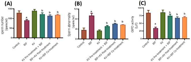
Figure 1. Effect of kolaviron (KV) on spermatic indices in bifenthrin-induced epididymal toxicity. (A) sperm count, (B) sperm abnormality and (C) glucose 6-phosphate dehydrogenase (G6PD) activity. Values are expressed as mean ± SD. aP < 0.05 versus the control group; bP < 0.05 versus the bifenthrin group. abP < 0.05 versus control and bifenthrin groups.
As shown in
Figure 1a, epididymal sperm number was significantly reduced (P < 0.05) in the bifenthrin-induced group compared to the normal control. However, kolaviron treatment alone, as well as kolaviron pretreatment before bifenthrin exposure, post-treatment following bifenthrin exposure, and co-treatment with kolaviron and bifenthrin, all led to a significant (P < 0.05) increase in sperm number relative to the bifenthrin-induced group.
As shown in
Figure 1b, epididymal sperm abnormality was significantly increased (P < 0.05) in the bifenthrin-induced group compared to the normal control. However, kolaviron treatment alone, as well as kolaviron pretreatment before bifenthrin exposure, post-treatment following bifenthrin exposure, and co-treatment with kolaviron and bifenthrin, all led to a significant (P < 0.05) reduction in sperm abnormality relative to the bifenthrin-induced group.
As shown in
Figure 1c, epididymal Glucose 6-phosphate dehydrogenase (G6PD) activity was significantly reduced (P < 0.05) in the bifenthrin-induced group compared to the normal control. However, treatment with kolaviron alone, kolaviron pretreatment followed by bifenthrin exposure, bifenthrin exposure followed by kolaviron post-treatment, and co-treatment with kolaviron and bifenthrin all led to a significant (P < 0.05) increase in G6PD activity relative to the bifenthrin-induced group.
3.2. Kolaviron Ameliorate the Level of Luteinizing Hormone, Follicle Stimulating Hormone and Testosterone in Bifenthrin induced Epididymal Toxicity
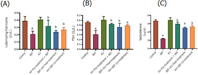
Figure 2. Effect of kolaviron (KV) on luteinizing hormone (A), follicle stimulating hormone (B) and testosterone (C) in bifenthrin-induced epididymal toxicity. Values are expressed as mean ± SD. aP < 0.05 versus the control group; bP < 0.05 versus the bifenthrin group.
As shown in
Figure 2a, serum luteinizing hormone level was significantly reduced (P < 0.05) in the bifenthrin-induced group compared to the normal control. However, kolaviron treatment alone, as well as kolaviron pretreatment before bifenthrin exposure, post-treatment following bifenthrin exposure, and co-treatment with kolaviron and bifenthrin, all led to a significant (P < 0.05) increase in luteinizing hormone level relative to the bifenthrin-induced group.
As shown in
Figure 2b, serum follicle stimulating hormone level was significantly reduced (P < 0.05) in the bifenthrin-induced group compared to the normal control. However, kolaviron treatment alone, as well as kolaviron pretreatment before bifenthrin exposure, post-treatment following bifenthrin exposure, and co-treatment with kolaviron and bifenthrin, all led to a significant (P < 0.05) increase in follicle stimulating hormone level relative to the bifenthrin-induced group.
As shown in
Figure 2c, serum testosterone level was significantly reduced (P < 0.05) in the bifenthrin-induced group compared to the normal control. However, kolaviron treatment alone, as well as kolaviron pretreatment before bifenthrin exposure, post-treatment following bifenthrin exposure, and co-treatment with kolaviron and bifenthrin, all led to a significant (P < 0.05) increase in testosterone level relative to the bifenthrin-induced group.
3.3. Kolaviron Improves Antioxidant Defenses in Bifenthrin Induced Epididymal Toxicity
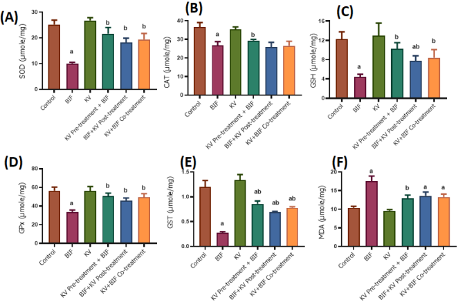
As shown in
Figure 3a, epididymal superoxide dismutase (SOD) activity was significantly reduced (P < 0.05) in the bifenthrin-induced group compared to the normal control. However, kolaviron treatment alone, as well as kolaviron pretreatment before bifenthrin exposure, post-treatment following bifenthrin exposure, and co-treatment with kolaviron and bifenthrin, all led to a significant (P < 0.05) increase in SOD activity relative to the bifenthrin-induced group.
As illustrated in
Figure 3b, catalase (CAT) activity in the epididymis was significantly reduced (P < 0.05) in the bifenthrin-treated group compared to the normal control. Treatment with kolaviron alone, as well as pretreatment with kolaviron prior to bifenthrin exposure, significantly increased CAT activity (P < 0.05) relative to the bifenthrin-only group. In contrast, post-treatment with kolaviron after bifenthrin exposure and co-administration of kolaviron with bifenthrin did not produce a statistically significant change in CAT activity compared to the bifenthrin group.
As shown in
Figure 3c, epididymal glutathione (GSH) activity was significantly reduced (P < 0.05) in the bifenthrin-induced group compared to the normal control. However, kolaviron treatment alone, as well as kolaviron pretreatment before bifenthrin exposure, post-treatment following bifenthrin exposure, and co-treatment with kolaviron and bifenthrin, all led to a significant (P < 0.05) increase in GSH activity relative to the bifenthrin-induced group.
As shown in
Figure 3d, epididymal glutathione peroxidase (GPx) activity was significantly reduced (P < 0.05) in the bifenthrin-induced group compared to the normal control. However, kolaviron treatment alone, as well as kolaviron pretreatment before bifenthrin exposure, post-treatment following bifenthrin exposure, and co-treatment with kolaviron and bifenthrin, all led to a significant (P < 0.05) increase in GPx activity relative to the bifenthrin-induced group.
As shown in
Figure 3e, epididymal glutathione s-transferase (GST) activity was significantly reduced (P < 0.05) in the bifenthrin-induced group compared to the normal control. However, kolaviron treatment alone, as well as kolaviron pretreatment before bifenthrin exposure, post-treatment following bifenthrin exposure, and co-treatment with kolaviron and bifenthrin, all led to a significant (P < 0.05) increase in GST activity relative to the bifenthrin-induced group.
As shown in
Figure 3f, epididymal malonaldehyde (MDA) activity was significantly increased (P < 0.05) in the bifenthrin-induced group compared to the normal control. However, kolaviron treatment alone, as well as kolaviron pretreatment before bifenthrin exposure, post-treatment following bifenthrin exposure, and co-treatment with kolaviron and bifenthrin, all led to a significant (P < 0.05) decrease in MDA activity relative to the bifenthrin-induced group.
Figure 3. Effect of kolaviron (KV) on oxidative stress biomarkers in bifenthrin-induced epididymal toxicity. (A) superoxide dismutase (SOD), (C) catalase (CAT), (C) glutathione (GSH), (D) glutathione peroxidase (GPx), (E) glutathione s-transferase (GST), (F) malonaldehyde (MDA). Values are expressed as mean ± SD. aP < 0.05 versus the control group; bP < 0.05 versus the bifenthrin group. abP < 0.05 versus control and bifenthrin groups.
3.4. Kolaviron Attenuates Inflammatory Markers in Bifenthrin induced Epididymal Toxicity
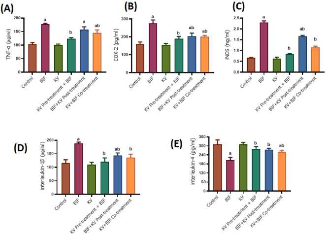
As shown in
Figure 4a, epididymal tumor necrosis factor-alpha (TNF-α) activity was significantly increased (P < 0.05) in the bifenthrin-induced group compared to the normal control. However, kolaviron treatment alone, as well as kolaviron pretreatment before bifenthrin exposure, post-treatment following bifenthrin exposure, and co-treatment with kolaviron and bifenthrin, all led to a significant (P < 0.05) decrease in TNF-α activity relative to the bifenthrin-induced group.
As shown in
Figure 4b, epididymal cyclooxygenase-2 (COX-2) activity was significantly increased (P < 0.05) in the bifenthrin-induced group compared to the normal control. However, kolaviron treatment alone, as well as kolaviron pretreatment before bifenthrin exposure, post-treatment following bifenthrin exposure, and co-treatment with kolaviron and bifenthrin, all led to a significant (P < 0.05) decrease in COX-2 activity relative to the bifenthrin-induced group.
As shown in
Figure 4c, epididymal inducible nitric oxide synthase (iNOS) activity was significantly increased (P < 0.05) in the bifenthrin-induced group compared to the normal control. However, kolaviron treatment alone, as well as kolaviron pretreatment before bifenthrin exposure, post-treatment following bifenthrin exposure, and co-treatment with kolaviron and bifenthrin, all led to a significant (P < 0.05) decrease in iNOS activity relative to the bifenthrin-induced group.
As shown in
Figure 4d, epididymal Interleukin-1 beta (IL-1β) activity was significantly increased (P < 0.05) in the bifenthrin-induced group compared to the normal control. However, kolaviron treatment alone, as well as kolaviron pretreatment before bifenthrin exposure, post-treatment following bifenthrin exposure, and co-treatment with kolaviron and bifenthrin, all led to a significant (P < 0.05) decrease in IL-1β activity relative to the bifenthrin-induced group.
As shown in
Figure 4e, epididymal Interleukin-4 (IL-4) activity was significantly reduced (P < 0.05) in the bifenthrin-induced group compared to the normal control. However, kolaviron treatment alone, as well as kolaviron pretreatment before bifenthrin exposure, post-treatment following bifenthrin exposure, and co-treatment with kolaviron and bifenthrin, all led to a significant (P < 0.05) increase in IL-4 activity relative to the bifenthrin-induced group.
Figure 4. Effect of kolaviron (KV) on inflammatory mediators in bifenthrin-induced epididymal toxicity. (A) tumor necrosis factor alpha (TNF-α), (B) cyclooxygenases-2 (COX-2), (C) inducible nitric oxide synthase (iNOS), (D) interleukin 1 beta (IL-1-β), (D) interleukin 4 (IL-4). Values are expressed as mean ± SD. aP < 0.05 versus the control group; bP < 0.05 versus the bifenthrin group. abP < 0.05 versus control and bifenthrin groups.
3.5. Kolaviron Promotes Apoptotic Balance in Bifenthrin induced Epididymal Toxicity
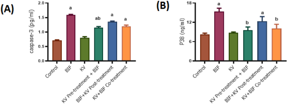
As shown in
Figure 5a, epididymal caspase-3 activity was significantly increased (P < 0.05) in the bifenthrin-induced group compared to the normal control. However, kolaviron treatment alone, as well as kolaviron pretreatment before bifenthrin exposure, post-treatment following bifenthrin exposure, and co-treatment with kolaviron and bifenthrin, all led to a significant (P < 0.05) decrease in caspase-3 activity relative to the bifenthrin-induced group.
As shown in
Figure 5b, epididymal p38 MAPK activity was significantly increased (P < 0.05) in the bifenthrin-induced group compared to the normal control. However, kolaviron treatment alone, as well as kolaviron pretreatment before bifenthrin exposure, post-treatment following bifenthrin exposure, and co-treatment with kolaviron and bifenthrin, all led to a significant (P < 0.05) decrease in p38 MAPK activity relative to the bifenthrin-induced group.
Figure 5. Effect of kolaviron (KV) on apoptotic-related proteins in bifenthrin-induced epididymal toxicity. (A) caspase-3, (B) p38 mitogen-activated protein kinase. Values are expressed as mean ± SD. aP < 0.05 versus the control group; bP < 0.05 versus the bifenthrin group. abP < 0.05 versus control and bifenthrin groups.
3.6. Kolaviron Improves Epididymal Histomorphology in Bifenthrin-exposed Rats
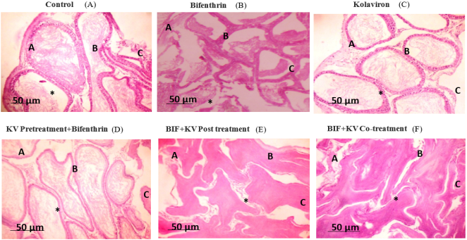
Figure 6. Representative photomicrographs of epididymal tissues from experimental groups stained with hematoxylin and eosin (H&E) and examined at ×400 magnification. (A) Control; (B) bifenthrin-induced; (C) kolaviron-only; (D) kolaviron pretreatment followed by bifenthrin exposure; (E) bifenthrin followed by kolaviron post-treatment; and (F) bifenthrin and kolaviron co-treatment. Internal annotations provisionally indicate the epididymal duct lumen and/or luminal spermatozoa (A), ductal epithelium (B), surrounding interstitial connective tissue (C), and the indicated luminal space or tissue alteration (*). Scale bar = 50 µm.
As shown in
Figure 6, histological examination revealed differences in epididymal architecture across the experimental groups. The control and kolaviron (KV)-only groups exhibited relatively preserved epididymal histoarchitecture, characterized by well-defined ducts containing abundant spermatozoa, intact pseudostratified columnar epithelium with prominent stereocilia, and organized interstitial connective tissue (
Figure 6A and C). In contrast, bifenthrin (BIF) exposure was associated with pronounced histopathological alterations, including distorted ductal lumina, reduced luminal spermatozoal content, cellular debris, epithelial degeneration, diminished stereocilia, and disorganization of the interstitial tissue (
Figure 6B). KV pretreatment appeared to attenuate these alterations, with improved luminal integrity, moderate spermatozoal content, better-preserved epithelial organization, and relatively organized interstitial tissue (
Figure 6D). Similarly, KV post-treatment following BIF exposure was associated with apparent improvement in luminal patency, reduced cellular debris, partial preservation of the epithelial lining and stereocilia, and improved organization of the interstitial connective tissue, although some alterations remained evident (
Figure 6E). Notably, the BIF + KV co-treatment group exhibited relatively preserved epididymal architecture, with defined lumina containing spermatozoa, less apparent cellular debris, a comparatively continuous pseudostratified epithelium, visible stereocilia, and organized interstitial connective tissue (
Figure 6F).
4. Discussion
This study employed a subchronic (28-day) oral exposure model to investigate bifenthrin (BIF)-induced epididymal toxicity and to evaluate the modulatory effects of kolaviron (KV), a biflavonoid complex isolated from Garcinia kola. The selected dose of BIF (1 mg/kg) reflects a repeated low-dose exposure paradigm suitable for assessing cumulative reproductive toxicity rather than acute toxicity, while the KV dose (200 mg/kg) was based on prior experimental validation demonstrating biological activity in rodent models. Using standard body surface area normalization, these doses correspond to biologically plausible human-equivalent doses, thereby enhancing the translational context of the findings. Given the lipophilic nature of BIF, which favors accumulation in lipid-rich tissues including reproductive organs, and the moderate lipophilicity of KV that supports tissue distribution and cellular interaction, the observed biochemical and histological changes are mechanistically plausible. Across hormonal, oxidative stress, inflammatory, apoptotic, and histoarchitectural parameters, the results demonstrate that subchronic BIF exposure disrupts epididymal integrity and function, whereas KV exerts significant protective and restorative effects, likely mediated through its antioxidant, anti-inflammatory, and anti-apoptotic properties.
Bifenthrin-induced reduction in sperm number (
Figure 1a) may be attributed to its pro-oxidant properties, which compromise the structural and functional integrity of the epididymal epithelium
| [22] | Ibor, M. E., Iwara, I. A., Ujong, U. P., Eteng, K. A., Ujong, V. P., Igile, G. O.,... & Eyong, E. U. (2026). A Novel Mixture Extract for Benign Prostatic Hyperplasia: Multi-Targeted Inhibition of 5α-Reductase and NF-κB Signaling with Biochemical and In Silico Insights in Rat-model Benign Prostate hyperplasia. Pharmacological Research-Natural Products, 100580.
https://doi.org/10.1016/j.prenap.2026.100580 |
[22]
. Oxidative damage to the epididymal microenvironment can impair sperm storage and maturation, leading to increased sperm loss and reduced fertility
. Additionally, bifenthrin is known to interfere with hormonal regulation by disrupting the hypothalamic–pituitary–gonadal (HPG) axis, reducing testosterone levels essential for epididymal function and sperm production
| [24] | Zhang, X., Zhang, T., Ren, X., Chen, X., Wang, S., & Qin, C. (2021). Pyrethroids toxicity to male reproductive system and offspring as a function of oxidative stress induction: rodent studies. Frontiers in Endocrinology, 12, 656106.
https://doi.org/10.3389/fendo.2021.656106 |
[24]
. The observed increase in sperm abnormalities (
Figure 1b), such as head or tail deformities, likely reflects oxidative damage to the sperm membrane and cytoskeletal proteins, further impairing fertility potential.
The significant decrease in epididymal G6PD activity (
Figure 1c) in the bifenthrin group further supports the role of oxidative stress in mediating its toxic effects. G6PD plays a pivotal role in maintaining redox homeostasis by generating NADPH through the pentose phosphate pathway
| [25] | Chen, P. H., Tjong, W. Y., Yang, H. C., Liu, H. Y., Stern, A., & Chiu, D. T. Y. (2022). Glucose-6-phosphate dehydrogenase, redox homeostasis and embryogenesis. International Journal of Molecular Sciences, 23(4), 2017.
https://doi.org/10.3390/ijms23042017 |
[25]
. NADPH is essential for the regeneration of reduced glutathione (GSH), a major intracellular antioxidant
| [26] | Wu, C., Xu, D., Ge, M., Luo, J., Chen, L., Chen, Z., & Shi, J. (2022). Blocking glutathione regeneration: Inorganic NADPH oxidase nanozyme catalyst potentiates tumoral ferroptosis. Nano Today, 46, 101574.
https://doi.org/10.1016/j.nantod.2022.101574 |
[26]
. Reduced G6PD activity thus compromises the cellular antioxidant defense system, rendering epididymal cells more susceptible to oxidative insult and leading to lipid peroxidation, protein oxidation, and cell death
| [27] | Olaniyi, K. S., Amusa, O. A., Oniyide, A. A., Ajadi, I. O., Akinnagbe, N. T., & Babatunde, S. S. (2020). Protective role of glutamine against cadmium-induced testicular dysfunction in Wistar rats: involvement of G6PD activity. Life sciences, 242, 117250. https://doi.org/10.1016/j.lfs.2019.117250 |
[27]
.
Importantly, kolaviron administration whether alone, as a pretreatment, post-treatment, or in co-treatment with bifenthrin significantly ameliorated these adverse effects. Kolaviron restored sperm count and morphology to near-normal levels and significantly increased G6PD activity compared to the bifenthrin-only group. These effects can be attributed to the well-established antioxidant and anti-inflammatory properties of kolaviron, a biflavonoid complex rich in garcinoic acid and related polyphenols.
The study further reveals that bifenthrin exposure significantly disrupts endocrine function in male Wistar rats, as indicated by reductions in serum luteinizing hormone (LH), follicle-stimulating hormone (FSH), and testosterone levels (
Figure 2a-c). These findings are consistent with previous reports indicating that pyrethroid insecticides act as endocrine-disrupting chemicals (EDCs), interfering with the hypothalamic–pituitary–gonadal (HPG) axis and ultimately impairing male reproductive health
| [28] | Ye, X., & Liu, J. (2019). Effects of pyrethroid insecticides on hypothalamic-pituitary-gonadal axis: a reproductive health perspective. Environmental pollution, 245, 590-599.
https://doi.org/10.1016/j.envpol.2018.11.031 |
[28]
.
The observed reduction in LH and FSH levels suggests that bifenthrin interferes with the normal regulatory function of the anterior pituitary. LH and FSH are gonadotropins critical for testicular function
| [29] | Oduwole, O. O., Huhtaniemi, I. T., & Misrahi, M. (2021). The roles of luteinizing hormone, follicle-stimulating hormone and testosterone in spermatogenesis and folliculogenesis revisited. International journal of molecular sciences, 22(23), 12735. https://doi.org/10.3390/ijms222312735 |
[29]
. LH stimulates Leydig cells to produce testosterone, while FSH acts on Sertoli cells to support spermatogenesis
| [29] | Oduwole, O. O., Huhtaniemi, I. T., & Misrahi, M. (2021). The roles of luteinizing hormone, follicle-stimulating hormone and testosterone in spermatogenesis and folliculogenesis revisited. International journal of molecular sciences, 22(23), 12735. https://doi.org/10.3390/ijms222312735 |
[29]
. Disruption in their levels reflects impaired signaling from the hypothalamus and pituitary, potentially due to bifenthrin’s neurotoxic effects or its interaction with hormone receptors. In particular, pyrethroids have been shown to alter GnRH pulsatility, impairing gonadotropin release, and to interfere with the expression of key genes involved in steroidogenesis, such as steroidogenic acute regulatory protein (StAR) and 17β-hydroxysteroid dehydrogenase
| [30] | Moreira, S., Silva, R., Carrageta, D. F., Alves, M. G., Seco-Rovira, V., Oliveira, P. F., & de Lourdes Pereira, M. (2022). Carbamate pesticides: shedding light on their impact on the male reproductive system. International Journal of Molecular Sciences, 23(15), 8206. https://doi.org/10.3390/ijms23158206 |
[30]
.
The significant decline in serum testosterone (
Figure 2c) observed in the bifenthrin-exposed group may be a direct consequence of reduced LH stimulation of Leydig cells. Additionally, bifenthrin-induced oxidative stress likely contributes to Leydig cell dysfunction and decreased testosterone synthesis. Oxidative damage to testicular mitochondria impairs cholesterol transport and conversion to pregnenolone, a critical step in steroidogenesis
| [31] | Bassi, G., Sidhu, S. K., & Mishra, S. (2021). The expanding role of mitochondria, autophagy and lipophagy in steroidogenesis. Cells, 10(8), 1851. https://doi.org/10.3390/cells10081851 |
[31]
.
Notably, kolaviron administration in all treatment regimens significantly restored serum LH, FSH, and testosterone levels compared to the bifenthrin group. These findings highlight kolaviron’s capacity to modulate the HPG axis and protect endocrine tissues from toxicant-induced disruption. By restoring LH and FSH levels, kolaviron ensures appropriate stimulation of testicular steroidogenesis and spermatogenesis, as evidenced by the corresponding improvements in testosterone concentration and sperm parameters.
Oxidative stress plays a central role in the pathogenesis of bifenthrin-induced reproductive toxicity, as evidenced by the marked alterations in key epididymal antioxidant biomarkers in this study (
Figure 3a-f). Bifenthrin exposure significantly impaired enzymatic and non-enzymatic antioxidant defenses reducing superoxide dismutase (SOD), catalase (CAT), reduced glutathione (GSH), glutathione peroxidase (GPx), and glutathione S-transferase (GST) activities while concomitantly increasing malondialdehyde (MDA) levels, a lipid peroxidation marker.
SOD and CAT serve as the primary line of defense against oxidative insult
| [32] | Ighodaro, O. M., & Akinloye, O. A. (2018). First line defence antioxidants-superoxide dismutase (SOD), catalase (CAT) and glutathione peroxidase (GPX): Their fundamental role in the entire antioxidant defence grid. Alexandria journal of medicine, 54(4), 287-293. https://doi.org/10.1016/j.ajme.2017.09.001 |
[32]
. SOD catalyzes the dismutation of superoxide radicals (O₂⁻) into hydrogen peroxide (H₂O₂), which is subsequently detoxified by CAT into water and oxygen
| [32] | Ighodaro, O. M., & Akinloye, O. A. (2018). First line defence antioxidants-superoxide dismutase (SOD), catalase (CAT) and glutathione peroxidase (GPX): Their fundamental role in the entire antioxidant defence grid. Alexandria journal of medicine, 54(4), 287-293. https://doi.org/10.1016/j.ajme.2017.09.001 |
[32]
. The observed reductions in SOD and CAT activities in bifenthrin-treated rats indicate compromised capacity to neutralize ROS, allowing accumulation of peroxides and exacerbating oxidative injury to epididymal tissue. Interestingly, kolaviron administration in all treatment protocols significantly restored SOD activity (
Figure 3a), suggesting its ability to enhance the first tier of antioxidant defense. However, the increase in CAT activity (
Figure 3b) was significant only in the kolaviron-alone and pretreatment groups, indicating that early intervention may be more effective in preserving catalase function compared to post-exposure or co-treatment strategies.
GSH, GPx, and GST are critical components of the glutathione-based antioxidant system. GSH serves as a direct ROS scavenger and a cofactor for GPx and GST. GPx reduces hydrogen peroxide and lipid peroxides to water and alcohols, while GST facilitates the conjugation of GSH to electrophilic xenobiotics and oxidative stress byproducts
| [33] | Hatem, E., El Banna, N., & Huang, M. E. (2017). Multifaceted roles of glutathione and glutathione-based systems in carcinogenesis and anticancer drug resistance. Antioxidants & redox signaling, 27(15), 1217-1234.
https://doi.org/10.1089/ars.2017.7134 |
[33]
. The significant decline in GSH (
Figure 3c), GPx (
Figure 3d), and GST (
Figure 3e) activities in the bifenthrin group reflects depletion of cellular redox reserves and impaired detoxification capacity. Kolaviron significantly restored these parameters across all treatment regimens, underscoring its ability to replenish GSH levels and activate GSH-dependent enzymes. This is likely due to kolaviron’s polyphenolic structure, which allows it to directly scavenge free radicals and upregulate nuclear factor erythroid 2–related factor 2 (Nrf2), a transcription factor that governs the expression of antioxidant response elements (ARE) including genes encoding GPx and GST.
MDA, a byproduct of lipid peroxidation
| [34] | Wang, Z., He, Z., Emara, A. M., Gan, X., & Li, H. (2019). Effects of malondialdehyde as a byproduct of lipid oxidation on protein oxidation in rabbit meat. Food Chemistry, 288, 405-412. https://doi.org/10.1016/j.foodchem.2019.02.126 |
[34]
, was significantly elevated in the bifenthrin group (
Figure 3f), reflecting extensive oxidative degradation of membrane lipids and cellular damage. This increase corresponds with the observed impairment of antioxidant defenses. Notably, kolaviron significantly reduced MDA levels across all treatment conditions, indicating effective protection against lipid peroxidation and preservation of membrane integrity.
This study provides compelling evidence that bifenthrin induces a pronounced proinflammatory response in the epididymis, as reflected by significant elevations in tumor necrosis factor-alpha (TNF-α), cyclooxygenase-2 (COX-2), inducible nitric oxide synthase (iNOS), and interleukin-1 beta (IL-1β), alongside a marked reduction in the anti-inflammatory cytokine interleukin-4 (IL-4) (
Figure 4a–e). These changes are consistent with bifenthrin’s known ability to activate redox-sensitive and inflammatory signaling pathways, including nuclear factor-kappa B (NF-κB) and mitogen-activated protein kinases (MAPKs), which play pivotal roles in mediating inflammation and tissue injury
| [35] | Sung, J., Ju, S. Y., Park, S., Jung, W. K., Je, J. Y., & Lee, S. J. (2025). Lysine-Proline-Valine peptide mitigates fine dust-induced keratinocyte apoptosis and inflammation by regulating oxidative stress and modulating the MAPK/NF-κB pathway. Tissue and Cell, 95, 102837.
https://doi.org/10.46793/lle25.048jc |
[35]
.
TNF-α is a master regulator of inflammation that initiates the inflammatory cascade by promoting leukocyte recruitment, enhancing vascular permeability, and stimulating the production of other proinflammatory mediators
| [36] | Zhao, H., Wu, L., Yan, G., Chen, Y., Zhou, M., Wu, Y., & Li, Y. (2021). Inflammation and tumor progression: signaling pathways and targeted intervention. Signal transduction and targeted therapy, 6(1), 263.
https://doi.org/10.1038/s41392-021-00658-5 |
[36]
. The significant elevation of TNF-α in the bifenthrin-treated group (
Figure 4a) suggests an acute inflammatory response within the epididymis, likely contributing to the observed histological disruptions and impaired sperm quality. TNF-α also induces apoptosis of germ cells, further compromising epididymal function and male fertility
.
COX-2, an inducible isoform of cyclooxygenase, catalyzes the conversion of arachidonic acid to proinflammatory prostaglandins
| [38] | Rojas, A., Chen, D., Ganesh, T., Varvel, N. H., & Dingledine, R. (2019). The COX-2/prostanoid signaling cascades in seizure disorders. Expert opinion on therapeutic targets, 23(1), 1-13. https://doi.org/10.1080/14728222.2019.1554056 |
[38]
. Its upregulation (
Figure 4b) in response to bifenthrin exposure is indicative of an active inflammatory state and oxidative stress. Prostaglandins produced by COX-2 can disrupt epithelial barrier function and promote tissue remodeling, both of which are detrimental to the tightly regulated environment of the epididymis
| [39] | Tran-Guzman, A., & Culty, M. (2022). Eicosanoid biosynthesis in male reproductive development: effects of perinatal exposure to NSAIDs and analgesic drugs. Frontiers in toxicology, 4, 842565. https://doi.org/10.3389/ftox.2022.842565 |
[39]
.
Similarly, the elevation of iNOS (
Figure 4c) reflects increased nitric oxide (NO) production, which, while serving immune defense roles, can become cytotoxic at high concentrations. Excess NO reacts with superoxide to form peroxynitrite, a potent oxidizing and nitrating agent capable of damaging lipids, proteins, and DNA
| [40] | L Piacenza, L., Zeida, A., Trujillo, M., & Radi, R. (2022). The superoxide radical switch in the biology of nitric oxide and peroxynitrite. Physiological reviews, 102(4), 1881-1906.
https://doi.org/10.1152/physrev.00005.2022 |
[40]
. This nitrosative stress may exacerbate oxidative damage already induced by bifenthrin, compounding epididymal dysfunction.
The increase in IL-1β (
Figure 4d), a key proinflammatory cytokine involved in leukocyte activation and cytokine amplification
| [41] | Kabeerdoss, J., Goel, R., Mohan, H., & Danda, D. (2022). High expression of pro-inflammatory cytokine genes IL-1β and IL-1R2 upon TLR4 activation in Takayasu arteritis. Rheumatology International, 42(3), 535-543.
https://doi.org/10.1007/s00296-020-04785-0 |
[41]
, further confirms the inflammatory state triggered by bifenthrin. IL-1β disrupts testicular and epididymal immune privilege and has been implicated in male infertility through its detrimental effects on the blood–testis barrier and germ cell development.
Conversely, IL-4, a Th2-type anti-inflammatory cytokine, was significantly suppressed in the bifenthrin group (
Figure 4e). IL-4 plays a crucial role in downregulating proinflammatory cytokine synthesis and promoting tissue healing
| [42] | Keegan, A. D., Leonard, W. J., & Zhu, J. (2021). Recent advances in understanding the role of IL-4 signaling. Faculty reviews, 10, 71. https://doi.org/10.12703/r/10-71 |
[42]
Its depletion suggests a skewing of the immune response toward a proinflammatory phenotype, allowing inflammation to persist and inflict tissue damage.
Remarkably, kolaviron administration across all treatment regimens significantly reversed these proinflammatory changes reducing TNF-α, COX-2, iNOS, and IL-1β levels while restoring IL-4 concentrations. These findings highlight kolaviron’s potent anti-inflammatory effects, which complement its antioxidant properties. Kolaviron likely mediates these effects by inhibiting the NF-κB signaling pathway, a central regulator of cytokine and inflammatory enzyme expression. By stabilizing NF-κB in its inactive form (bound to IκB), kolaviron may prevent the transcriptional activation of genes encoding TNF-α, COX-2, iNOS, and IL-1β.
Furthermore, kolaviron's enhancement of IL-4 suggests that it promotes a shift toward a Th2-dominant, anti-inflammatory immune profile, contributing to tissue recovery and immune balance. Its flavonoid constituents, including kolaflavone and garcinoic acid, are known to modulate immune cell signaling and cytokine production
| [43] | Farombi, E. O., Awogbindin, I. O., Farombi, T. H., Ikeji, C. N., Adebisi, A. A., Adedara, I. A., & Aruoma, O. I. (2022). Possible role of Kolaviron, a Garcinia kola bioflavonoid in inflammation associated COVID-19 infection. American Journal of Biopharmacy and Pharmaceutical Sciences, 2.
https://doi.org/10.25259/ajbps_1_2022 |
[43]
.
In addition to triggering oxidative stress and inflammation, bifenthrin exposure significantly activated apoptosis-related and stress-response signaling pathways in the epididymis, as demonstrated by the elevated activities of caspase-3 and p38 mitogen-activated protein kinase (MAPK) (
Figure 5a-b). These findings provide further insight into the molecular mechanisms underlying bifenthrin-induced epididymal damage and male reproductive dysfunction.
Caspase-3 is a critical executioner protease in the apoptotic cascade, responsible for the cleavage of structural and regulatory proteins during programmed cell death
| [44] | Asadi, M., Taghizadeh, S., Kaviani, E., Vakili, O., Taheri‐Anganeh, M., Tahamtan, M., & Savardashtaki, A. (2022). Caspase‐3: structure, function, and biotechnological aspects. Biotechnology and Applied Biochemistry, 69(4), 1633-1645. https://doi.org/10.1002/bab.2233 |
[44]
. The significant increase in epididymal caspase-3 activity observed in the bifenthrin-treated group (
Figure 5a) suggests that bifenthrin induces apoptosis of epithelial and germ cells in the epididymis. This activation likely results from upstream mitochondrial dysfunction and cytochrome c release, consequences of bifenthrin-induced oxidative stress. Increased caspase-3 activity may compromise epididymal structure, disrupt sperm maturation processes, and contribute to the reduced sperm count and increased abnormalities previously observed.
Similarly, the marked elevation of p38 MAPK activity (
Figure 5b) in the bifenthrin group indicates the involvement of cellular stress-response pathways. p38 MAPK is a member of the stress-activated protein kinase family and is typically activated by ROS, inflammatory cytokines, and environmental stressors
| [45] | Canovas, B., & Nebreda, A. R. (2021). Diversity and versatility of p38 kinase signalling in health and disease. Nature reviews Molecular cell biology, 22(5), 346-366.
https://doi.org/10.1038/s41580-020-00322-w |
[45]
. Once activated, p38 MAPK can phosphorylate downstream transcription factors and apoptotic proteins, thereby amplifying inflammation and promoting apoptosis. In the reproductive system, hyperactivation of p38 MAPK has been associated with germ cell apoptosis, Sertoli cell dysfunction, and overall testicular and epididymal damage
| [46] | Luo, D., He, Z., Yu, C., & Guan, Q. (2022). Role of p38 MAPK signalling in testis development and male fertility. Oxidative medicine and cellular longevity, 2022(1), 6891897.
https://doi.org/10.1155/2022/6891897 |
[46]
.
Notably, kolaviron administration whether as monotherapy, pretreatment, post-treatment, or co-treatment with bifenthrin significantly attenuated both caspase-3 and p38 MAPK activities compared to bifenthrin exposure alone. This protective effect is consistent with kolaviron’s established role in modulating apoptotic and stress signaling pathways. Kolaviron’s antioxidant action likely prevents the ROS-mediated activation of p38 MAPK and the mitochondrial apoptotic cascade, thereby reducing caspase-3 activation. Moreover, by suppressing upstream inducers such as TNF-α, iNOS, and COX-2 (as shown in earlier Figures), kolaviron interrupts the feedback loops that sustain pro-apoptotic and stress signaling.
Flavonoid compounds in kolaviron are also known to directly interact with signaling proteins and modulate kinase activity. For instance, kolaviron may inhibit the phosphorylation of p38 MAPK or enhance the expression of anti-apoptotic proteins such as Bcl-2, tipping the balance away from cell death and toward survival. These effects contribute to the preservation of epididymal architecture and function, as supported by the histological findings and improved sperm parameters in kolaviron-treated groups.
Histological assessment of epididymal tissues revealed clear group-dependent alterations consistent with the extent of toxicant exposure and therapeutic intervention. The control group demonstrated normal epididymal architecture, with well-defined ducts, lumina densely filled with spermatozoa, and intact pseudostratified columnar epithelium bearing prominent stereocilia, indicative of optimal sperm maturation and storage capacity. In contrast, bifenthrin (BIF) exposure induced severe histopathological disruptions, including distortion of the ductal lumen, marked reduction in luminal spermatozoa with accumulation of cellular debris, degeneration and disorganization of the epithelial lining, and attenuation of stereocilia. These structural derangements were accompanied by loosening of the interstitial connective tissue, reflecting compromised epididymal integrity and functional impairment.
Administration of kolaviron (KV) alone preserved normal histological features comparable to the control, confirming its non-toxic nature on epididymal tissue. Notably, KV pretreatment prior to BIF exposure markedly attenuated the observed damage, as evidenced by preservation of ductal architecture, improved epithelial organization, and moderate restoration of luminal sperm content, suggesting a strong prophylactic effect. Similarly, KV post-treatment following BIF exposure resulted in appreciable recovery, with partial regeneration of the epithelial lining, reappearance of stereocilia, and reduced luminal debris, although mild residual alterations persisted. The co-treatment group exhibited near-complete protection, with well-preserved ductal structure, continuous epithelium, distinct stereocilia, and minimal cellular debris, closely resembling normal histoarchitecture.
These histological findings are in agreement with the biochemical and inflammatory profiles observed in this study, reinforcing the protective role of KV against BIF-induced epididymal toxicity. The preservation and restoration of epididymal structure may be attributed to the antioxidant, anti-inflammatory, and anti-apoptotic properties of KV, which collectively act to stabilize cellular integrity, reduce oxidative damage, and prevent epithelial degeneration.
A notable limitation of the present study is the absence of detailed molecular and genetic analyses to further substantiate the mechanistic pathways involved. Although key mediators such as p38 MAPK and caspase-3 were evaluated at the protein level, their activation states and gene expression profiles were not assessed using more sensitive and specific techniques. In particular, the lack of analysis of phosphorylated p38 MAPK (p-p38) and cleaved caspase-3 using Western blotting, as well as the absence of gene expression studies (e.g., RT-qPCR), may limit a comprehensive understanding of the apoptotic and stress signaling pathways implicated. Incorporation of these approaches would have provided stronger mechanistic validation of the observed biochemical and histological outcomes. Accordingly, further investigations incorporating these advanced molecular approaches are necessary to validate and extend the present findings, while providing a more detailed understanding of the cytoprotective mechanisms by which kolaviron mitigates bifenthrin-induced reproductive toxicity.





 Share This Article
 Twitter
 Linked In
 Facebook

 Copy
 Download