The structural properties of nanomaterials play a crucial role in determining their performance in various applications. This study investigates the effect of pH variation on the structural properties of ZnS nanocrystals synthesized via low-temperature chemical deposition. ZnS nanoparticles were prepared in a polyvinyl alcohol (PVA) host matrix, with pH values ranging from 2.2 to 2.8. X-ray diffraction (XRD) analyses reveal that an increase in pH enhances crystallinity, resulting in narrower peaks and larger grain sizes. The average grain sizes of the films with varying pH values are found to range from 4.63 nm to 6.37 nm. The lattice constant evaluated using Nelson Reiley plot (N-R plot) ranges from 5.363 Å to 5.420 Å (pH 2.2-2.8), which slightly deviates from the standard 5.406 Å, suggesting compressive strain in the films, possibly due to sulfur deficiency. It was also observed that as pH decreases, there is an increase in microstrain and dislocation density probably due to lattice contraction. Results from the Williamson-Hall analysis confirm these trends, showing reductions in both average grain size and lattice strain at lower pH. These findings highlight the critical role of pH in controlling the structural characteristics of ZnS thin films, providing insights for optimizing ZnS properties for specific technological applications.
| Published in | American Journal of Physical Chemistry (Volume 14, Issue 1) |
| DOI | 10.11648/j.ajpc.20251401.11 |
| Page(s) | 1-6 |
| Creative Commons |
This is an Open Access article, distributed under the terms of the Creative Commons Attribution 4.0 International License (http://creativecommons.org/licenses/by/4.0/), which permits unrestricted use, distribution and reproduction in any medium or format, provided the original work is properly cited. |
| Copyright |
Copyright © The Author(s), 2025. Published by Science Publishing Group |
ZnS Thin Films, Low Temperature, Structural Properties, pH Variation
pH | [hkl] values | 2θ (degree) | d value from XRD (Å) | F (θ) | acal (Å) | a corrected (Å) | Av. Internal stress (S) x 109 (N/m2) | Av. Internal strain (є) x 10-3 | Dislocation density (ρ x 1016/ m2) | Grain size (D) (nm) |
|---|---|---|---|---|---|---|---|---|---|---|
2.2 | [111] | 28.53 | 3.128 | 1.583 | 5.419 | 5.363 | 1.721 | 8.41 | 8.89 | 4.63 |
[220] | 48.05 | 1.893 | 0.567 | 5.355 | ||||||
[311] | 57.16 | 1.611 | 0.322 | 5.344 | ||||||
2.4 | [111] | 28.48 | 3.133 | 1.586 | 5.428 | 5.390 | 1.608 | 7.88 | 8.01 | 4.99 |
[220] | 47.97 | 1.896 | 0.569 | 5.364 | ||||||
[311] | 57.05 | 1.614 | 0.325 | 5.354 | ||||||
2.6 | [111] | 28.20 | 3.164 | 1.610 | 5.480 | 5.408 | 1.373 | 6.54 | 7.70 | 5.69 |
[220] | 47.82 | 1.902 | 0.574 | 5.379 | ||||||
[311] | 57.53 | 1.601 | 0.314 | 5.313 | ||||||
2.8 | [111] | 28.72 | 3.108 | 1.567 | 5.383 | 5.420 | 1.220 | 4.39 | 5.87 | 6.37 |
[220] | 48.12 | 1.891 | 0.564 | 5.348 | ||||||
[311] | 57.26 | 1.608 | 0.320 | 5.336 |
Parameters | Different pH of ZnS thin films | |||
|---|---|---|---|---|
2.2 | 2.4 | 2.6 | 2.8 | |
Grain size D (in nm) | 4.68 | 4.89 | 5.81 | 6.28 |
Average lattice strain (ɛ) | 7.97 x 10⁻³ | 6.89 x 10⁻³ | 6.14 x 10⁻³ | 3.71 x 10⁻³ |
XRD | X-ray Diffraction |
N-R Plot | Nelson Reily Plot |
W-H Plot | Williamson Hall Plot |
ZnS | Zinc Sulfide |
MBE | Molecular Beam Epitaxy |
PVD | Physical Vapour Deposition |
CBD | Chemical Bath Deposition |
PVA | Polyvinyal Alcohol |
FWHM | Full Width at Half Maximum |
| [1] | YM. Suhail, Structural and optical properties of ZnS, PbS, Zn1-xPbxS, ZnxPb1-xS and PbZnxS1-x thin films, Indian Journal of Pure & Applied Physics. 50, 380-386 (2012). |
| [2] | M. Temiz, and S. Çelik, Influence of growth temperature on the structural and optical characteristics of PbZnS thin films, Chalcogenide Letters, 21, 867-872 (2024). |
| [3] | B. Barman, and K. C. Sarma, Low temperature chemical synthesis of ZnS, Mn doped ZnS nanosized particles: Their structural, morphological and photophysical properties, Solid State Sciences. 109, 106404 (2020). |
| [4] | B. Barman, and K. C. Sarma, Structural characterization of PVA capped ZnS nanostructured thin films, Indian J Phys. 86, 703-707 (2012). |
| [5] | Z. Jiang, A. Hoffmann, and A. Schleife, Influence of temperature, doping, and amorphization on the electronic structure and magnetic damping of iron Phys. Rev. B., 109, 235247 (2024). |
| [6] | B. D. Cullity, Elements of X-ray Diffraction, Addison-Wesley Publishing Company, Inc. 187 (1978). |
| [7] | Göde, F., Annealing temperature effect on the structural, optical and electrical properties of ZnS thin films, Physica B: Condensed Matter. 406, 1653-1659 (2011). |
| [8] | B. Barman and K. C. Sarma, Luminescence Properties of ZnS Quantum Dots Embedded in Polymer Matrix, Chalcogenide Letters. 8, 171-176 (2011). |
| [9] | T. Sinha, D. Lilhare, A. Khare, Effects of Various Parameters on Structural and Optical Properties of CBD-Grown ZnS Thin Films: A Review, Journal of Electronic Materials. 47(2), 1730-1751 (2018). |
| [10] | H. E. Ruda, Wide gap II-VI Compound for optoelectronic applications, Chapman and Hall, London (1992). |
| [11] | S. Kar S. Biswas, S. Chaudhuri and P M G Nambissan, Substitution-induced structural transformation in Mn-doped ZnS nanorods studied by positron annihilation spectroscopy, Nanotechnology. 18, 225606 (2007). |
| [12] | P. Vasa, P. Ayyub and B. P. Sing, Fast and reversible excited state absorption in II-VI-based nanocomposite thin films, Appl. Phys. Lett. 87, 063104 (2005). |
| [13] | S, Chanda, M. Debbarma, D, Ghosh, B. Debnath and S. Chattopadhyaya, First-Principles Investigation of Structural, Elastic, Electronic, and Optical Properties of Cd1−x−yZnxHgyS Quaternary Alloys, Journal of Electronic Materials. 8, 4705-4726 (2021). |
| [14] | A. Z. Arsad, A. W. M. Zuhdi, S. F. Abdullah, C. F. Chau, A. Ghazali, I. Ahmad, and W. S. W. Abdullah, Effect of Chemical Bath Deposition Variables on the Properties of Zinc Sulfide Thin Films: A Review, Molecules. 28, 2780 (2023). |
| [15] | A. Barman, and K. C. Sarma, Synthesis and optical properties of ZnS nanoparticles in PVA matrix, Optoelectronics and Advanced Materials - Rapid Communications. 4, 1594-1597 (2010). |
| [16] | S. Kumar, S. Bakshi, S. Chaudhary, J. Kaur, A. P. Agrawal, and S. Rani, Heat Post-Treatment Effect on Optical and Electrical Properties of ZnS Thin Films, Biointerface Research in Applied Chemistry. 14, 1-9 (2024). |
| [17] | B. Siahmardan, V. Soleimanian, and M. G. Varnamkhasti, Effect of size and shape of crystallites on the optical properties of nanostructured ZnS films, Materials Science in Semiconductor Processing. 71, 76-83 (2017). |
| [18] | A. Nazim, and B. Parveen, Synthesis of Zn metal contents-dependent ultra-wide-band gap ZnS nanoparticles, Applied Physics A. 129, 808 (2023). |
| [19] | J. Barman, and F. Sultana, Synthesis of mix Zinc oxide and Cadmium sulphide Nanoparticles, Optoelectronic and antimicrobial activity and application in Water Treatment, IOSR Journal of Applied Physics. 8, 46-51 (2016). |
| [20] | J. Angel Mary Greena, K. Karuppasamy, R. Antony, X. Sahaya Shajan and S. Kumaresan, Structural, Thermal and Spectroscopic Studies on Zinc Doped Strontium Formate Dihydrate Crystals, IOSR Journal of Applied Physics. 1, 25-28 (2012). |
| [21] | B. Barman, P. K. Mochahari, and K. C. Sarma, Optical Studies on Some Aspects of Polyvinyl Alcohol Composite Zns Nanocrystalline Thin Films, AIP Conference Proceedings. 1391, 116-118 (2011). |
| [22] | K. K. Nanda, S. N. Sarangi, and S. N. Sahu, CdS Nanocrystalline films: Composition, surface, crystalline size, structural and optical absorption studies. Nanostructured materials 10 1401-1410 (1998). |
| [23] | R. B. Kale and C. D. Lokhande, Band gap shift, structural characterization and phase transformation of CdSe thin films from nanocrystalline cubic to nanorod hexagonal on air annealing, Semiconductor Science and Technology. 20, 201 (2005). |
| [24] | L. Wang, L. Cao, G. Su, W. Liu, C. Xia, and H Zhou, reparation and characterization of water-soluble ZnSe: Cu/ZnS core/shell quantum dots, Applied Surface Science. 280, 673-678 (2013). |
| [25] | A. Goudarzi, G. M. Aval, R. Sahraei, and H. Ahmadpoor, Ammonia-free chemical bath deposition of nanocrystalline ZnS thin film buffer layer for solar cells, Thin Solid Films, 516, 4953-4957 (2008). |
APA Style
Barman, B. (2025). Influence of pH Variation on the Structural Properties of ZnS Nanocrystals Synthesized Via Low-Temperature Chemical Deposition. American Journal of Physical Chemistry, 14(1), 1-6. https://doi.org/10.11648/j.ajpc.20251401.11
ACS Style
Barman, B. Influence of pH Variation on the Structural Properties of ZnS Nanocrystals Synthesized Via Low-Temperature Chemical Deposition. Am. J. Phys. Chem. 2025, 14(1), 1-6. doi: 10.11648/j.ajpc.20251401.11
AMA Style
Barman B. Influence of pH Variation on the Structural Properties of ZnS Nanocrystals Synthesized Via Low-Temperature Chemical Deposition. Am J Phys Chem. 2025;14(1):1-6. doi: 10.11648/j.ajpc.20251401.11
@article{10.11648/j.ajpc.20251401.11,
  author = {Bijoy Barman},
  title = {Influence of pH Variation on the Structural Properties of ZnS Nanocrystals Synthesized Via Low-Temperature Chemical Deposition
},
  journal = {American Journal of Physical Chemistry},
  volume = {14},
  number = {1},
  pages = {1-6},
  doi = {10.11648/j.ajpc.20251401.11},
  url = {https://doi.org/10.11648/j.ajpc.20251401.11},
  eprint = {https://article.sciencepublishinggroup.com/pdf/10.11648.j.ajpc.20251401.11},
  abstract = {The structural properties of nanomaterials play a crucial role in determining their performance in various applications. This study investigates the effect of pH variation on the structural properties of ZnS nanocrystals synthesized via low-temperature chemical deposition. ZnS nanoparticles were prepared in a polyvinyl alcohol (PVA) host matrix, with pH values ranging from 2.2 to 2.8. X-ray diffraction (XRD) analyses reveal that an increase in pH enhances crystallinity, resulting in narrower peaks and larger grain sizes. The average grain sizes of the films with varying pH values are found to range from 4.63 nm to 6.37 nm. The lattice constant evaluated using Nelson Reiley plot (N-R plot) ranges from 5.363 Å to 5.420 Å (pH 2.2-2.8), which slightly deviates from the standard 5.406 Å, suggesting compressive strain in the films, possibly due to sulfur deficiency. It was also observed that as pH decreases, there is an increase in microstrain and dislocation density probably due to lattice contraction. Results from the Williamson-Hall analysis confirm these trends, showing reductions in both average grain size and lattice strain at lower pH. These findings highlight the critical role of pH in controlling the structural characteristics of ZnS thin films, providing insights for optimizing ZnS properties for specific technological applications.
},
 year = {2025}
}
TY  - JOUR
T1  - Influence of pH Variation on the Structural Properties of ZnS Nanocrystals Synthesized Via Low-Temperature Chemical Deposition

AU  - Bijoy Barman
Y1  - 2025/02/24
PY  - 2025
N1  - https://doi.org/10.11648/j.ajpc.20251401.11
DO  - 10.11648/j.ajpc.20251401.11
T2  - American Journal of Physical Chemistry
JF  - American Journal of Physical Chemistry
JO  - American Journal of Physical Chemistry
SP  - 1
EP  - 6
PB  - Science Publishing Group
SN  - 2327-2449
UR  - https://doi.org/10.11648/j.ajpc.20251401.11
AB  - The structural properties of nanomaterials play a crucial role in determining their performance in various applications. This study investigates the effect of pH variation on the structural properties of ZnS nanocrystals synthesized via low-temperature chemical deposition. ZnS nanoparticles were prepared in a polyvinyl alcohol (PVA) host matrix, with pH values ranging from 2.2 to 2.8. X-ray diffraction (XRD) analyses reveal that an increase in pH enhances crystallinity, resulting in narrower peaks and larger grain sizes. The average grain sizes of the films with varying pH values are found to range from 4.63 nm to 6.37 nm. The lattice constant evaluated using Nelson Reiley plot (N-R plot) ranges from 5.363 Å to 5.420 Å (pH 2.2-2.8), which slightly deviates from the standard 5.406 Å, suggesting compressive strain in the films, possibly due to sulfur deficiency. It was also observed that as pH decreases, there is an increase in microstrain and dislocation density probably due to lattice contraction. Results from the Williamson-Hall analysis confirm these trends, showing reductions in both average grain size and lattice strain at lower pH. These findings highlight the critical role of pH in controlling the structural characteristics of ZnS thin films, providing insights for optimizing ZnS properties for specific technological applications.

VL  - 14
IS  - 1
ER  -
Department of Physics, Abhayapuri College, Abhayapuri, India
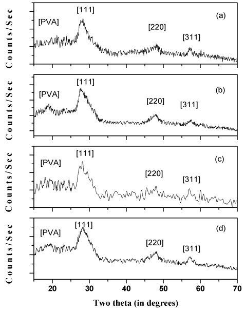
Figure 1. X-ray diffraction patterns of nanocrystalline ZnS thin films as a function of pH variation (a) 2.2, (b) 2.4, (c) 2.6, (d) 2.8.
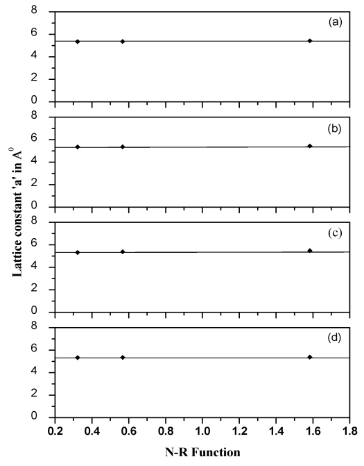
Figure 2. Nelson-Riley plots for ZnS nanocrystalline films, prepared at different pH values (a) 2.2, (b) 2.4, (c) 2.6, (d) 2.8.
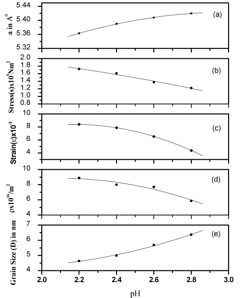
Figure 3. Variation of (a) Lattice constant, (b) Average stress, (c) Average strain, (d) Dislocation density and (e) Average grain size with different pH value of ZnS nancrystalline thin films.
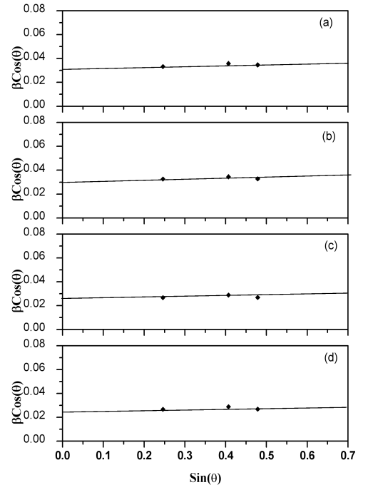
Figure 4. W-H plots for ZnS nanocrystalline thin films for different pH value (a) 2.2, (b) 2.4, (c) 2.6, (d) 2.8.
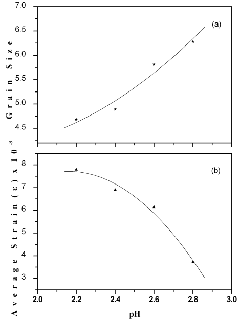
Figure 5. Change in (a) average grain size and (b) average strain from the W-H plot for ZnS nanocrystalline films at varying pH values.
Information

