Abstract
Objective: The pathological process of vascular dementia originates from functional impairment of brain microvascular endothelial cells under ischemia‑hypoxia conditions, and endothelial nitric oxide synthase (eNOS) uncoupling serves as a critical hallmark of endothelial dysfunction. This study aimed to explore whether the protective effect of Shenfu injection against oxygen‑glucose deprivation (OGD)‑induced injury in brain microvascular endothelial cells involves eNOS uncoupling. Methods: An OGD model of bEnd.3 cells was induced by sodium dithionite treatment. Experimental groups included control group, OGD group, low‑dose Shenfu group (0.1mg/mL), medium‑dose Shenfu group (0.5mg/mL), and high‑dose Shenfu group (1.0mg/mL). MTT assay and lactate dehydrogenase (LDH) detection were performed to evaluate cell viability and injury. Griess assay was adopted to measure nitric oxide (NO) concentration in cell supernatants. Enzyme‑linked immunosorbent assay (ELISA) was used to detect intracellular cyclic guanosine monophosphate (cGMP) levels. Western blot was applied to determine the protein expression of eNOS. Reactive oxygen species (ROS) content was quantified with DCFH‑DA fluorescent probe. Xanthine oxidase assay was utilized for superoxide dismutase (SOD) activity measurement, and DTNB method was used to detect reduced glutathione (GSH) content. Results: Compared with the control group, the OGD group exhibited marked cellular morphological damage, significantly reduced cell viability, elevated LDH release and ROS levels (P<0.01); the contents of NO and cGMP decreased significantly (P<0.01); eNOS protein expression showed no statistically significant difference (P>0.05); meanwhile, antioxidant indicators including SOD activity and GSH content declined remarkably (P<0.01). Compared with the OGD group, Shenfu injection produced dose‑dependent improvements in above‑mentioned parameters: cell viability was significantly increased while LDH activity was lowered (P<0.01); NO and cGMP contents were elevated (P<0.01), without significant alteration in eNOS protein expression (P>0.05); additionally, intracellular ROS level was reduced (P<0.01), accompanied by increases in SOD activity and GSH content (P<0.01). Conclusion: Shenfu injection mitigates OGD‑induced injury of brain microvascular endothelial cells. Its underlying mechanism may be associated with potentiating the antioxidant capacity of SOD and GSH, eliminating excessive ROS, inhibiting eNOS uncoupling, and further improving NO bioavailability.
Keywords
Shenfu Injection, Brain Microvascular Endothelial Cells, Oxygen‑Glucose Deprivation, eNOS Uncoupling, Oxidative Stress
1. Introduction
With global population aging accelerating, vascular dementia (VaD) has become an increasingly concerning public‑health issue.Ranked second only to Alzheimer's disease in prevalence, vascular dementia accounts for approximately 20% of the total dementia population.
. Endothelial cells exert vital functions in maintaining normal circulatory homeostasis. They synthesize and secrete multiple bioactive molecules participating in blood coagulation, vasomotor regulation, barrier permeability modulation and inflammatory responses, consequently governing cardiac and vascular functions
| [2] | Michiels C. Endothelial cell functions [J]. Journal of Cellular Physiology, 2003, 196(3): 430‑443. |
[2]
.
Nitric oxide (NO) acts as an essential messenger molecule maintaining endothelial integrity and cardiovascular health. It participates in modulating neural plasticity, regulating vascular tone and decreasing peripheral vascular resistance
| [3] | Zhang X, Chen Z, Xiong Y, et al. The emerging role of nitric oxide in the synaptic dysfunction of vascular dementia [J]. Neural Regeneration Research, 2025, 20(2): 402‑415.
https://doi.org/10.4103/NRR.NRR-D-23-01353 |
[3]
. As a pivotal enzyme generating vasodilatory NO, endothelial nitric oxide synthase (eNOS) exhibits dual biological properties. Under physiological circumstances, eNOS exists as homodimers and catalyzes the conversion of L‑arginine into NO and L‑citrulline, which is indispensable for stabilizing vascular tone and suppressing platelet aggregation as well as leukocyte adhesion
| [4] | Anna J, Agata P, Michał J W, et al. Endothelial dysfunction due to eNOS uncoupling: molecular mechanisms as potential therapeutic targets [J]. Cellular & Molecular Biology Letters, 2023, 28(1): 21. https://doi.org/10.1186/s11658-023-00423-2 |
[4]
. Nevertheless, hypoxia and severe oxidative stress trigger massive accumulation of reactive oxygen species (ROS), driving eNOS uncoupling. During uncoupling, electron flow is redirected, and eNOS ceases to produce protective NO but generates detrimental superoxide anion radicals (O
2•⁻)
| [5] | De Pascali F, Hemann C, Samons K, et al. Hypoxia and reoxygenation induce endothelial nitric oxide synthase uncoupling in endothelial cells through tetrahydrobiopterin depletion and S‑glutathionylation [J]. Biochemistry, 2014, 53(22): 3679‑3688. https://doi.org/10.1021/bi500076r |
[5]
. Superoxide anions directly damage biological macromolecules. Moreover, they rapidly react with the diminished endogenous NO pool to yield highly toxic peroxynitrite (ONOO⁻), which further amplifies oxidative stress and cellular injury and forms a vicious cycle that exacerbates eNOS uncoupling
| [6] | Zhao Y X, Ge R L. Pathogenic roles of endothelial nitric oxide synthase uncoupling in cardiovascular diseases [J]. Chinese Circulation Journal, 2014, 29(04): 315‑318. |
[6]
. Endothelial cells under eNOS‑uncoupled status suffer dual insults: attenuation of protective signaling cascades and persistent oxidative attack, which facilitates cell apoptosis, disrupts intercellular junctions and impairs blood‑brain barrier integrity
| [7] | Abdullah S, Karl A, Roland A, et al. Oxidative stress‑induced endothelial dysfunction in cardiovascular diseases [J]. Frontiers in Bioscience (Landmark Edition), 2022, 27(3): 105. https://doi.org/10.31083/j.fbl2703105 |
[7]
. Accordingly, eNOS uncoupling is not merely a biomarker of endothelial dysfunction but also a core pathogenic driver. It has been validated as a key contributor to microvascular malfunction and blood‑brain barrier disruption in cerebral ischemia‑reperfusion injury
| [8] | Katz P S, Wappler E A, Katakam P V, et al. eNOS Inhibition is Protective against Oxygen Glucose Deprivation in Brain Microvascular Endothelial Cells by Preempting eNOS Uncoupling [J]. 2012. |
[8]
.
Current clinical managements for vascular dementia mainly adopt cholinesterase inhibitors and glutamate receptor antagonists, which are occasionally combined with atypical antipsychotic agents; however, these therapies are associated with adverse effects
| [9] | Chu X Q, Qin Y K, Xie H Y, et al. Clinical evaluation of butylphthalide combined with galantamine for vascular dementia [J]. Chinese Journal of Drug Application and Monitoring, 2026, 23(03): 349‑352. |
[9]
. Traditional Chinese medicine accumulates abundant clinical experience in treating ischemic encephalopathy, featured by multi‑target and holistic regulatory effects in compound prescriptions
. Derived from the classical ancient formula Shenfu Decoction, Shenfu injection is manufactured from Radix Ginseng and Radix Aconiti Lateralis Preparata via modern purification procedures. Clinically, it is primarily applied for emergency interventions for shock, cardiac insufficiency and sepsis‑induced critical illnesses
| [11] | Tian R, Li R, Chen Y, et al. Shenfu injection ameliorates endotoxemia‑associated endothelial dysfunction and organ injury via inhibiting PI3K/Akt‑mediated glycolysis [J]. Journal of Ethnopharmacology, 2024, 335: 118634.
https://doi.org/10.1016/j.jep.2024.118634 |
[11]
. Accumulating evidence demonstrates diverse pharmacological activities of Shenfu injection, including cardiac function enhancement, vasodilation for microcirculation restoration, anti‑inflammation, anti‑oxidation and anti‑apoptosis
| [12] | Chen R J, Wu J, Tian F, et al. Effects of Shenfu injection on oxidative‑stress‑triggered vascular endothelial injury and related signal transduction pathways [J]. Journal of Nanjing University of Traditional Chinese Medicine, 2021, 37(4): 529‑534. https://doi.org/10.14148/j.issn.1672-0482.2021.0529 |
[12]
. In animal models of sepsis and hemorrhagic shock, Shenfu injection alleviates endothelial damage and reduces vascular permeability, possibly through suppressing pro‑inflammatory cytokine secretion and modulating apoptosis‑related gene expression
| [13] | Zhang M Q, Zhang Q, Yuan W, et al. Protective Effect of Shenfu Injection on Vascular Endothelial Damage in a Porcine Model of Hemorrhagic Shock [J]. Chinese Journal of Integrative Medicine, 2022, 28(9): 794‑801.
https://doi.org/10.1007/s11655-021-2876-x |
| [14] | Xing Y, Cheng D, Shi C. Protective effect of Shenfu injection on the endothelium of severe sepsis by inhibiting CD11b+ cell paralysis induced by high mobility group protein B1 [J]. Chinese Journal of Critical Care Medicine, 2020, 32(6): 696‑701. https://doi.org/10.3760/cma.j.cn121430-20191127-00066 |
[13, 14]
. Furthermore, in rat acute cerebral infarction models, Shenfu injection lowers blood‑brain barrier permeability and modulates cerebral SOD levels
| [15] | Chen J, Leng J. Pre‑treatment with Shenfu injection ameliorates oxidative stress following acute cerebral infarction in rats [J]. Journal of Emergency in Traditional Chinese Medicine, 2017, 26(05): 761‑763. |
[15]
. Nevertheless, systematic investigations regarding whether Shenfu injection intervenes in eNOS uncoupling under OGD stress in brain microvascular endothelial cells and its concrete molecular mechanisms remain scarce. Interpreting this clinically effective Chinese medicinal compound with modern molecular biology techniques not only validates its scientific rationale but also reveals its unique pharmacological modes, which may provide novel therapeutic paradigms for complicated diseases.
2. Materials and Methods
2.1. Cells
Rat brain microvascular endothelial cells (bEnd.3) were purchased from Zhongao Ruisai Biotechnology Co., Ltd.
2.2. Reagents
Table 1. Main Reagents.
No. | Reagent | Batch/Specification | Manufacturer |
1 | DMEM/F12 Medium | SW-CJ-2FD | Shanghai Fuheng Biotechnology Co., Ltd. |
2 | Fetal Bovine Serum | 20240801 | Umedium |
3 | Trypsin-EDTA Digestion Solution | BL512A | Hefei Baisai Biotechnology Co., Ltd. |
4 | Shenfu Injection | 241204AK01 | China Resources Sanjiu Medical & Pharmaceutical Co., Ltd. |
5 | Sodium Dithionite | LW449243 | Shanghai Yuanye Bio-Technology Co., Ltd. |
6 | MTT | No. 5651030002 | Beijing Solarbio Science & Technology Co., Ltd. |
7 | LDH Assay Kit | A020-2-2 | Nanjing Jiancheng Bioengineering Institute |
8 | cGMP ELISA Kit | 52017640 | ELK Biotechnology |
9 | Sodium Nitrite | No. M10097467 | Shanghai Meryer Chemical Technology Co., Ltd. |
10 | BCA Protein Quantification Kit | BF03031040 | Beijing Bioss Antibodies Co., Ltd. |
11 | eNOS Rabbit Polyclonal Antibody | bs-20609R | Beijing Bioss Antibodies Co., Ltd. |
12 | Β-Actin Recombinant Rabbit Monoclonal Antibody | bsm-63325R | Beijing Bioss Antibodies Co., Ltd. |
13 | HRP-Conjugated Goat Anti-Rabbit IgG H&L | bs-0295G-HRP | Beijing Bioss Antibodies Co., Ltd. |
14 | ECL Chemiluminescence Detection Kit | J18E01 | Absin Bioscience Inc. |
15 | ROS Fluorometric Assay Kit | WO10V0228366 | Wuhan Eleray Biotechnology Co., Ltd. |
16 | Total SOD Assay Kit | LR449975 | Shanghai Yuanye Bio Technology Co., Ltd. |
17 | Reduced GSH Assay Kit | No. 2604002001 | Beijing Solarbio Science & Technology Co., Ltd. |
2.3. Cell Culture
bEnd.3 cells were maintained in DMEM/F12 medium enriched with 10% fetal bovine serum and incubated at 37°C in a humidified atmosphere containing 5% CO2.Cells at the logarithmic growth stage were collected for further experiments.
2.4. OGD Model and Drug Treatments
Procedures were modified from published literature
. OGD groups received 200 μL of 2 mmol/L Na
2S
2O₄ prepared in low‑glucose medium. After 4‑h hypoxia in CO
2 incubator, supernatants were discarded, and cells were gently rinsed twice with PBS. The normal group received medium renewal. The OGD model group was replenished with complete medium, whereas intervention groups were supplemented with respective concentrations of Shenfu injection. All plates were incubated at 37 °C with 5 % CO
2 for 24‑h re‑oxygenation.
Prior to formal experiments, pre‑experiments screened proper concentrations of Shenfu injection (SF). SF was diluted with complete medium to 0.1 mg/mL, 0.5 mg/mL, 1.0 mg/mL and 2.0 mg/mL. After OGD insult, corresponding SF concentrations were administrated during re‑oxygenation for 24 h, followed by MTT assay. The 2.0 mg/mL group displayed significantly decreased cell viability, implying potential cytotoxicity. Therefore, three dosage gradients were adopted in formal experiments: low‑dose (0.1 mg/mL), medium‑dose (0.5 mg/mL) and high‑dose (1.0 mg/mL).
Normal control group: bEnd.3 cells incubated with 200 μL complete medium without OGD treatment.
OGD model group: Cells subjected to OGD, with equal‑volume complete medium added during re‑oxygenation.
SF‑L group: OGD‑treated cells intervened with 0.1 mg/mL Shenfu injection upon re‑oxygenation.
SF‑M group: OGD‑treated cells intervened with 0.5 mg/mL Shenfu injection upon re‑oxygenation.
SF‑H group: OGD‑treated cells intervened with 1.0 mg/mL Shenfu injection upon re‑oxygenation.
2.5. Cells Morphology
At 24 h post‑re‑oxygenation, cellular morphology including cell contour, adherence status and cytoplasmic transparency was observed under inverted microscope. Images were captured for morphological description.
2.6. Determination of bEnd.3 Cell Viability by MTT Assay
Upon completion of re‑oxygenation, old medium was discarded carefully. Twenty microliters of 5 mg/mL MTT solution were added into each well. Plates were incubated at 37 °C with 5 % CO
2 for 4 h in darkness. Supernatants were aspirated without disturbing formazan precipitates. A volume of 150 µL dimethyl sulfoxide was added into every well, and the plates were agitated horizontally for 10 min so that the formazan crystals dissolved thoroughly. Subsequently, absorbance values were recorded at 490 nm with a microplate reader. Cell viability (%) = (OD value of experimental group / OD value of normal control group) × 100 %
| [17] | Shen J, Li L, Jiang L, et al. Effect of dexmedetomidine hydrochloride on H2O2‑induced oxidative stress and inflammatory response in Kupffer cells [J]. Journal of Central South University (Medical Sciences), 2016, 41(5): 477‑481.
https://doi.org/10.11817/j.issn.1672-7347.2016.05.005 |
[17]
.
2.7. Evaluation of bEnd.3 Injury by LDH Colorimetric Assay
After 24‑h re‑oxygenation, cell culture supernatants were collected and stored temporarily at 4 °C. All procedures were carried out in strict accordance with the protocol provided by the LDH cytotoxicity kit manufacturer. Fifty microliters of supernatant were mixed with 50 μL substrate buffer and incubated at 37 °C for 30 min. Absorbance was measured immediately at 440 nm. LDH activity (U/L) = [(A_sample − A_control)/(A_standard − A_blank)] × C_standard × N × 1000, where A_sample = absorbance of sample; A_control = absorbance of control well; A_standard = absorbance of standard well; A_blank = absorbance of blank well; C_standard = standard solution concentration (0.2 μmol/mL); N = sample dilution multiple; 1000 stands for unit conversion coefficient
| [17] | Shen J, Li L, Jiang L, et al. Effect of dexmedetomidine hydrochloride on H2O2‑induced oxidative stress and inflammatory response in Kupffer cells [J]. Journal of Central South University (Medical Sciences), 2016, 41(5): 477‑481.
https://doi.org/10.11817/j.issn.1672-7347.2016.05.005 |
[17]
.
2.8. Measurement of NO Content in bEnd.3 Supernatants by Griess Assay
Prepare 10 mM NaNO2 stock solution: accurately weigh 6.90 mg NaNO2 and dilute to 10 mL with double‑distilled water, store at 4 °C away from light. Prepare serial diluted standards: label wells 1‑7 in 96‑well plate. Add 1 mL double‑distilled water to wells 2‑7. Add 2 mL 10 mM NaNO2 stock solution to well 1. Transfer 1 mL solution from well 1 to well 2, mix well and serially transfer 1 mL to subsequent wells for serial dilution (discard 1 mL from well 6). Final standard concentrations: 80, 40, 20, 10, 5, 2.5 μM. Well 7 served as blank control (double‑distilled water only).
Griess reagent preparation: Reagent A: mix 6 mL concentrated phosphoric acid with 70 mL deionized water, dissolve 1.0 g sulfanilic acid, make up volume to 100 mL. Reagent B: dissolve 0.1 g N‑1‑naphthylethylenediamine dihydrochloride with deionized water to 100 mL.
Color‑development reaction: add 50 μL Reagent A to each well, incubate at 37 °C for 10 min (or room temperature for 20 min). Then add 50 μL Reagent B and incubate for another 10‑20 min to generate pink‑rose colored products. Absorbance was detected at 540 nm or 546 nm. Establish standard curve with NaNO2 concentration as X‑axis and absorbance as Y‑axis (R² ≥ 0.99 to guarantee reliable linearity).
Sample pretreatment: collect cell supernatants, centrifuge at 12 000 rpm for 5 min to remove cell debris and retain supernatants. Load 50 μL sample supernatant into 96‑well plate, set blank controls and replicate wells (3‑6 replicates per sample to reduce experimental error). Perform the color‑development procedure described above. Sample nitrite concentration calculated from standard curve indirectly reflects NO level.
2.9. Detection of Intracellular cGMP in bEnd.3 by ELISA
Assays were conducted according to ELISA kit instructions. Lysed cell supernatants were loaded onto microplate wells, followed by incubation, washing, secondary antibody addition and chromogenic reaction. Optical density was read at 450 nm, and cGMP concentrations were quantified against standard curves.
2.10. Western Blot Analysis for eNOS Protein Expression
After re‑oxygenation, cells from each group were harvested and rinsed twice with PBS.Total cellular proteins were obtained by lysing cells in pre‑chilled RIPA lysis buffer supplemented with protease and phosphatase inhibitors, keeping the samples on ice for 30 min.The lysates were then spun down at 12,000×g for 15 min under 4°C, after which the clear supernatant was harvested.The protein concentration of each sample was measured via BCA assay, and the samples were subsequently adjusted to ensure uniform loading amounts.Samples were mixed with 5 × SDS loading buffer and denatured at 95°C for 5 min. SDS‑PAGE electrophoresis was performed, and proteins were transferred onto PVDF membranes. Membranes were blocked with 5 % BSA at room temperature for 1 h. Primary antibodies (eNOS 1:1000 dilution, β‑actin 1:5000 dilution) were incubated overnight at 4 °C. Membranes were washed three times with TBST (10 min each), and HRP‑conjugated secondary antibody (1:10 000 dilution) was incubated for 1 h at room temperature. After another three rounds of TBST washing (10 min each), ECL chemiluminescent substrate was applied. Band images were captured with chemiluminescence imaging system. Relative eNOS expression was calculated as the gray‑scale ratio of target band to β‑actin band.
2.11. Intracellular ROS Measurement Using DCFH‑DA Probe
After OGD treatment, supernatants were discarded and cells were gently washed twice with PBS to remove residual medium and drugs. Serum‑free medium containing DCFH‑DA working solution (final concentration 10 μM) was added, and cells were incubated at 37 °C for 30 min in darkness to allow probe uptake. Non‑specifically bound probes were eliminated by three gentle PBS washes. Fluorescence intensity was detected by microplate reader with excitation wavelength 470 nm and emission wavelength 525 nm. Fluorescence intensity positively correlates with intracellular ROS abundance.
2.12. Determination of SOD Activity via Xanthine Oxidase Method
At 24 h post‑re‑oxygenation, culture medium was removed, cells were washed twice with PBS and lysed in pre‑cooled RIPA buffer on ice for 30 min. After centrifugation of the lysates at 12,000 × g for 15 min at 4 °C, the resulting supernatants were collected, and their protein levels were determined using the BCA assay. Operations complied with SOD assay kit instructions: buffer solution, NBT and xanthine solution were sequentially added, and reactions were initiated by xanthine oxidase. Samples were incubated at 37 °C in darkness for precisely 20 min, absorbance was recorded at 560 nm.
2.13. Quantification of GSH Content Using DTNB Method
After 24‑h re‑oxygenation, cell lysates were centrifuged and supernatants collected. Assays were performed following GSH detection kit protocols. DTNB chromogenic reagent was added and incubated at 37 °C for 30 min in darkness. Absorbance was measured at 412 nm. Standard curve was plotted with standard GSH concentration as abscissa and OD value as ordinate to calculate sample GSH concentrations.
2.14. Statistical Analysis
GraphPad Prism 8.0 software was used for statistical processing. Measurement data were presented as mean ± standard deviation(±s). Normality test and homogeneity‑of‑variance test were conducted prior to statistical comparison. One‑way analysis of variance (ANOVA) was applied for multiple‑group comparison when data followed normal distribution with equal variance, and LSD‑t test was adopted for pairwise inter‑group comparisons. Kruskal‑Wallis H non‑parametric test was used for data violating normality or homogeneity of variance. P< 0.05 was defined as statistically significant difference.
3. Results
3.1. Morphological Changes of bEnd.3 Cells
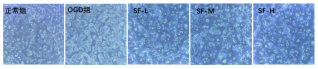
In normal control group, bEnd.3 cells adhered firmly and exhibited regular polygonal morphology with transparent cytoplasm without observable damage. In OGD model group, cells displayed conspicuous shrinkage and deformation, weakened adherence, partial cell detachment, increased cytoplasmic granularity and decreased cytoplasmic transparency, indicating severe morphological injury.
Shenfu injection intervention alleviated cellular morphological damage in a dose‑dependent manner. Mild shrinkage persisted in low‑dose group. Medium‑ and high‑dose groups showed improved adherence, restored regular shape and enhanced cytoplasmic transparency. The high‑dose group achieved the most prominent protective effect, whose morphology was closest to normal control group.
Figure 1. Morphological alterations of bEnd.3 cells in each experimental group (20 × magnification).
3.2. Effects of Shenfu Injection on Viability and LDH Activity of OGD‑Insulted bEnd.3 Cells
As summarized in
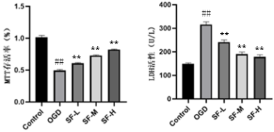
Table 3, compared with normal control group, the OGD model group manifested significantly reduced cell viability and elevated LDH release (P < 0.01), confirming successful establishment of OGD‑induced cellular injury. Compared with OGD model group, all Shenfu‑treated groups obtained elevated cell viability and reduced LDH release, in a dose‑dependent fashion. Medium‑ and high‑dose groups exerted more remarkable protective effects. These results demonstrated that Shenfu injection effectively ameliorates OGD‑triggered bEnd.3 injury.
Table 2. Cell viability and LDH activity of bEnd.3 cells in different groups (±s, n=6).
Group | Cell Viability (%) | LDH Activity (U/L) |
Normal Control | 100.00±2.86 | 149.3±3.8 |
OGD Model | 48.84±1.37## | 316.2±11.7## |
SF-L (0.1 mg/mL) | 59.91±0.99** | 241.6±9.1** |
SF-M (0.5 mg/mL) | 71.83±0.99** | 179.3±8.3** |
SF-H (1.0 mg/mL) | 81.13±0.59** | 190.8±8.6** |
Figure 2. Effects of different concentrations of Shenfu injection on cell viability and LDH activity of bEnd.3 cells.
3.3. Impacts of Shenfu Injection on NO and cGMP Levels in Supernatants of OGD‑Damaged bEnd.3 Cells
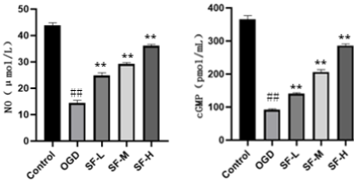
Further investigations focused on NO/cGMP signaling cascade, which governs endothelial function. Compared with normal control group, both NO and cGMP concentrations were markedly lowered in OGD model group (P < 0.01), suggesting impaired NO bioavailability, disrupted NO/cGMP pathway and aggravated eNOS uncoupling. Relative to OGD model group, Shenfu injection dose‑dependently raised NO and cGMP concentrations (P< 0.01), implying that Shenfu injection enhances NO bioavailability, restores NO/cGMP signaling and suppresses eNOS uncoupling.
Table 3. Comparison of NO and cGMP levels among experimental groups (±s,, n=6).
Group | NO(μmol/L) | cGMP(pmol/mL) |
Normal Control | 43.83±0.99 | 365.82±19.12 |
OGD Model | 14.50±0.97## | 91.90±4.78## |
SF L (0.1 mg/mL) | 24.84±1.06** | 140.00±5.93** |
SF M (0.5 mg/mL) | 29.20±0.59** | 208.29±7.21** |
SF H (1.0 mg/mL) | 36.14±0.50** | 285.91±10.45** |
Figure 3. Effects of Shenfu injection on NO and cGMP contents in bEnd.3 cells.
3.4. Influence of Shenfu Injection on eNOS Protein Expression in OGD‑Challenged bEnd.3 Cells
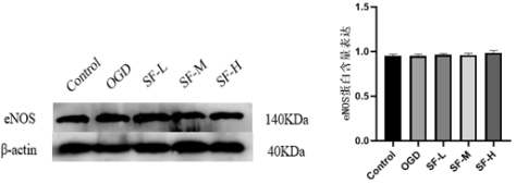
Western blot results revealed no significant difference in total eNOS protein abundance between OGD model group and normal control group (P > 0.05). No obvious changes in total eNOS protein expression were observed in all Shenfu‑intervened groups compared with OGD model group (P > 0.05). It indicated that the enhancement of NO bioavailability induced by Shenfu injection was not derived from up‑regulated eNOS protein abundance, but most likely resulted from inhibition of eNOS uncoupling.
Figure 4. Effect of Shenfu injection on eNOS protein expression in OGD‑treated bEnd.3 cells.
3.5. Effects of Shenfu Injection on ROS, SOD and GSH in OGD‑Stressed bEnd.3 Cells
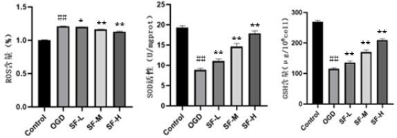
Oxidative stress serves as a key trigger for eNOS uncoupling. ROS, SOD and GSH were measured to assess the anti‑oxidative capacity of Shenfu injection. Compared with normal control group, OGD model group exhibited elevated ROS level and declined SOD activity as well as GSH content (P< 0.01). After Shenfu injection intervention, intracellular ROS level decreased, whereas SOD activity and GSH content increased significantly (P < 0.05 or P < 0.01), demonstrating that Shenfu injection ameliorates oxidative stress injury.
Table 4. Levels of ROS, SOD and GSH in each experimental group (±s, n=6).
Group | Relative ROS Fluorescence Intensity (%) | SOD Activity(U/mgprot) | GSH Content(μg/106cell) |
Normal Control | 1.00±0.00 | 21.07±0.69 | 269.15±4.71 |
OGD Model | 1.21±0.00## | 8.97±0.34## | 115.14±2.96## |
SF-L (0.1 mg/mL) | 1.20±0.00* | 12.01±0.41** | 136.10±4.61** |
SF-M (0.5 mg/mL) | 1.16±0.00** | 15.59±0.69** | 170.50±6.75** |
SF-H (1.0 mg/mL) | 1.13±0.01** | 17.61±0.49** | 209.62±4.23** |
Figure 5. Effects of Shenfu injection on ROS, SOD and GSH in bEnd.3 cells subjected to OGD.
4. Discussion
In this study, sodium dithionite was applied to successfully establish bEnd.3 OGD model simulating in‑vivo ischemia‑hypoxia endothelial damage. The protective effect and molecular mechanisms of Shenfu injection against OGD‑induced bEnd.3 injury were systematically explored from multiple dimensions: cell viability, membrane integrity, NO‑cGMP pathway, eNOS expression and oxidative stress status. Our results confirmed that Shenfu injection markedly attenuates OGD‑mediated bEnd.3 injury. Its core mechanisms involve restoring NO bioavailability, activating the protective NO‑cGMP signaling axis, suppressing eNOS uncoupling and counteracting oxidative stress.
OGD mimics ischemic‑hypoxic pathological status and provokes cellular energy metabolic disturbance, mitochondrial dysfunction and oxidative stress, leading to membrane lipid peroxidation, protein oxidative modification, loss of cell viability, membrane rupture and even cell death
| [18] | Islam R, Ahlfors J E, Siu R, et al. Inhibition of apoptosis in a model of ischemic stroke leads to enhanced cell survival, endogenous neural precursor cell activation and improved functional outcomes [J]. International Journal of Molecular Sciences, 2024, 25: 1786. https://doi.org/10.3390/ijms25031786 |
| [19] | Mo Y, Sun Y Y, Liu K Y. Autophagy and inflammation in ischemic stroke [J]. Neural Regeneration Research, 2020, 15(8): 1388‑1396. https://doi.org/10.4103/1673-5374.274331 |
| [20] | Ma C, Gao Q, Zhang L, et al. miR‑9a‑5p Protects Ischemic Stroke by Regulating Oxidative Stress and Mitochondrial Autophagy [J]. Disease Markers, 2023, 2023: 5146305.
https://doi.org/10.1155/2023/5146305 |
[18-20]
. In our experiments, OGD model group presented cell shrinkage, detachment, reduced viability and increased LDH release, verifying severe cellular damage and disrupted membrane integrity, which confirmed successful model construction. Shenfu injection intervention improved cell adherence and morphology, raised cell viability and reduced LDH leakage in a dose‑dependent pattern, suggesting that Shenfu injection stabilizes cellular structure and function. These findings are consistent with prior reports regarding endothelial‑protective, anti‑ischemia‑hypoxia and anti‑apoptotic properties of Shenfu injection
| [21] | Gao X M, Wu L D, Hu B. Research progress of Shenfu injection for acute lung injury [J]. Yunnan Journal of Traditional Chinese Medicine, 2025, 46(06): 108‑110.
https://doi.org/10.16254/j.cnki.53-1120/r.2025.06.016 |
| [22] | Chen C, Zhang Z H, Li J T. Research progress of Shenfu formula for cardiovascular diseases [J]. Lishizhen Medicine and Materia Medica Research, 1‑6. |
[21, 22]
.
NO‑cGMP cascade represents the major downstream signaling pathway mediating eNOS‑derived endothelial protection. NO stimulates soluble guanylate cyclase (sGC) to generate cGMP. As a secondary messenger, cGMP modulates vasodilation, inhibits platelet aggregation, mitigates inflammatory responses, stabilizes blood‑brain barrier and represses endothelial apoptosis
| [23] | Zhu S L. The Roles of Nitric Oxide Synthase/Nitric Oxide Pathway in the Pathology of Vascular Dementia and Related Therapeutic Approaches [J]. Current Drug Targets, 2021, 22(9). https://doi.org/10.3390/ijms22094540 |
[23]
. Therefore, cGMP level reflects NO bioavailability and eNOS functional status, and acts as an indicator for endothelial function evaluation
| [23] | Zhu S L. The Roles of Nitric Oxide Synthase/Nitric Oxide Pathway in the Pathology of Vascular Dementia and Related Therapeutic Approaches [J]. Current Drug Targets, 2021, 22(9). https://doi.org/10.3390/ijms22094540 |
[23]
. In the present research, cGMP was drastically decreased in OGD model group, which implied diminished NO bioavailability and suppressed endothelial protective signaling. Shenfu injection treatment significantly elevated cGMP concentration synchronously with NO increment, which illustrated that Shenfu injection restores NO‑cGMP protective signaling via inhibiting eNOS uncoupling.
eNOS uncoupling constitutes a central molecular mechanism for cerebral microvascular endothelial dysfunction under ischemic conditions, characterized by reduced NO synthesis, augmented ROS generation, without significant alteration in total eNOS protein abundance
| [24] | Zhang H P, Tang N, Bian K. Endothelial nitric oxide synthase uncoupling and oxidative stress [J]. Journal of Liaoning University of Traditional Chinese Medicine, 2009, 11(12): 36‑40. https://doi.org/10.13194/j.jlunivtcm.2009.12.38.zhanghp.092 |
| [25] | Zhang X F, Liu Y, Wang L C, et al. Xuebijing injection alleviates oxidative stress in sepsis‑induced vascular endothelial cells via inhibiting eNOS uncoupling [J]. Journal of Guangzhou Medical University, 2020, 48(06): 29‑32. |
[24, 25]
. Under physiological conditions, eNOS catalyzes the formation of beneficial NO. Under pathological stimuli, eNOS undergoes functional uncoupling, ceases NO synthesis and generates ROS, thereby initiating a vicious pathological cycle
| [5] | De Pascali F, Hemann C, Samons K, et al. Hypoxia and reoxygenation induce endothelial nitric oxide synthase uncoupling in endothelial cells through tetrahydrobiopterin depletion and S‑glutathionylation [J]. Biochemistry, 2014, 53(22): 3679‑3688. https://doi.org/10.1021/bi500076r |
[5]
. Our data demonstrated decreased NO and cGMP, unchanged total eNOS protein expression and elevated ROS in OGD model group, indicating that eNOS was functionally uncoupled rather than quantitatively down‑regulated. After Shenfu injection administration, NO and cGMP were restored while total eNOS protein level remained unchanged, accompanied by ROS reduction. It indicated that Shenfu injection exerts protective effects not by up‑regulating eNOS protein expression, but by alleviating oxidative stress, maintaining eNOS functional homeostasis and reversing eNOS uncoupling
| [23] | Zhu S L. The Roles of Nitric Oxide Synthase/Nitric Oxide Pathway in the Pathology of Vascular Dementia and Related Therapeutic Approaches [J]. Current Drug Targets, 2021, 22(9). https://doi.org/10.3390/ijms22094540 |
[23]
. This finding clarifies the key molecular mechanism underlying Shenfu injection‑mediated bEnd.3 protection.
Oxidative stress serves as the initiating driver of eNOS uncoupling as well as a fundamental pathological factor for ischemic endothelial injury
| [23] | Zhu S L. The Roles of Nitric Oxide Synthase/Nitric Oxide Pathway in the Pathology of Vascular Dementia and Related Therapeutic Approaches [J]. Current Drug Targets, 2021, 22(9). https://doi.org/10.3390/ijms22094540 |
[23]
. SOD, GSH and ROS are classic markers reflecting oxidative‑redox status. SOD specifically scavenges superoxide anions and interrupts oxidative chain reactions
| [26] | Dong L, He Y Z, Wang Y L, et al. Advances in application research of superoxide dismutase (SOD) [J]. Journal of Agricultural Science and Technology, 2013, 15(05): 53‑58. |
[26]
. GSH is the major non‑enzymatic intracellular antioxidant, directly neutralizing free radicals, sustaining cellular reductive milieu and preserving normal eNOS function
| [27] | Lian S P, Zhou Y, Lu C H. Study on protective effect of reduced glutathione against pristane‑induced diffuse alveolar hemorrhage in mice [J]. Chinese Journal of New Clinical Medicine, 2026, 19(02): 177‑183. |
[27]
. ROS level mirrors the severity of intracellular oxidative stress
. In OGD model group, ROS was accumulated while SOD activity and GSH content declined, manifesting impaired antioxidant defense and severe oxidative insult. Shenfu injection intervention lowered ROS accumulation, enhanced SOD activity and increased GSH content, revealing that Shenfu injection eliminates excessive free radicals through boosting enzymatic and non‑enzymatic antioxidant systems, mitigates oxidative damage and provides favorable redox microenvironment to maintain normal eNOS function
.
Limitations still exist in this investigation. bEnd.3 is an immortalized cell line; its gene expression profiles and functional characteristics may differ from primary brain microvascular endothelial cells. This study focused primarily on eNOS uncoupling. As a multi‑component Chinese medicinal compound, Shenfu injection may also modulate inflammatory factors (e.g. TNF‑α, IL‑6)
| [30] | Jin X H, Gao M Y, Zhu Z Y, et al. Experimental study of Shenfu injection combined with sivelestat sodium injection improving sublingual microcirculation and inflammatory response in septic rats [J]. Journal of Emergency in Traditional Chinese Medicine, 2026, 35(04): 405‑409. |
[30]
, pyroptosis, ferroptosis and other biological processes
| [31] | Wang Z Y, Zhang Q, Guo J, et al. Pathogenesis of chronic heart failure in rats mediated by ferroptosis‑dependent oxidative stress and intervention effect of Shenfu injection [J]. China Journal of Chinese Materia Medica, 2023, 48(19): 5285‑5293. https://doi.org/10.19540/j.cnki.cjcmm.20230601.702 |
[31]
. In complex in‑vivo neurovascular unit, crosstalk among astrocytic end‑feet, pericytes and endothelial cells also modulates eNOS function and drug efficacy, which requires further validation in animal models. In addition, Shenfu injection is an extract from Radix Ginseng and Radix Aconiti Lateralis Preparata with complicated chemical constituents. Whether the inhibitory effect against eNOS uncoupling is dominated by certain components such as ginsenosides
, or arises from synergistic interactions among multiple ingredients, awaits chemical separation and pharmacodynamic tracing experiments. Furthermore, upstream signaling pathways governing eNOS function modulated by Shenfu injection (e.g. Akt activation or SIRT1‑mediated eNOS deacetylation) remain to be elucidated.
In summary, this study illustrates the core protective mechanism of Shenfu injection against bEnd.3 OGD injury: OGD triggers oxidative stress, which induces eNOS uncoupling, reduces NO bioavailability and elevates ROS, ultimately aggravating endothelial injury. Shenfu injection suppresses oxidative stress, reverses eNOS uncoupling, restores physiological NO production, elevates cGMP level and improves NO bioavailability via enhancing SOD‑ and GSH‑dependent antioxidant defense, thereby alleviating ischemia‑hypoxia‑mediated brain microvascular endothelial injury.
5. Conclusions
OGD injury reduces brain microvascular endothelial cell viability, disrupts membrane integrity, impairs SOD and GSH‑dependent antioxidant defense, provokes excessive ROS generation and eNOS uncoupling, consequently decreasing cGMP level and NO bioavailability and exacerbating cerebrovascular endothelial damage.
Shenfu injection exerts dose‑dependent protective effects on OGD‑injured brain microvascular endothelial cells. It potentiates SOD and GSH antioxidant capacity, eliminates excessive ROS, inhibits eNOS uncoupling, restores physiological NO synthesis, elevates cGMP concentration and augments NO bioavailability.
Abbreviations
eNOS | Endothelial Nitric Oxide Synthase |
OGD | Oxygen-Glucose Deprivation |
MTT | 3-(4, 5-Dimethylthiazol-2-yl)-2, 5-Diphenyltetrazolium Bromide |
LDH | Lactate Dehydrogenase |
ELISA | Enzyme-Linked Immunosorbent Assay |
cGMP | Cyclic Guanosine Monophosphate |
DCFH-DA | 2′, 7′-Dichlorodihydrofluorescein Diacetate |
DTNB | 5, 5′-Dithiobis (2-Nitrobenzoic Acid) |
ROS | Reactive Oxygen Species |
SOD | Superoxide Dismutase |
GSH | Glutathione |
VaD | Vascular Dementia |
NO | Nitric Oxide |
DMEM/F12 | Dulbecco’s Modified Eagle Medium/Nutrient Mixture F-12 |
SF | Shenfu Injection |
DMSO | Dimethyl Sulfoxide |
BSA | Bovine Serum Albumin |
ANOVA | Analysis of Variance |
RIPA | Radioimmunoprecipitation Assay |
Acknowledgments
The authors would like to thank Ziyi Li for the technical support provided during this study.
Author Contributions
Meiwen Yang: Data curation, Formal Analysis, Investigation, Methodology, Writing – original draft
Jiazilin Hu: Data curation, Formal Analysis, Investigation, Methodology, Writing – original draft
Fenfang Hong: Conceptualization, Funding acquisition, Project administration, Supervision, Writing – review & editing
Shulong Yang: Conceptualization, Funding acquisition, Project administration, Supervision, Writing – review & editing
Funding
This work was supported by National Natural Science Foundation of China (№.82360880, 82060661, 81660751), Jiangxi Provincial Natural Science Foundation (№ 20232ACB206057, 20212BAB206092), and Educational Commission of Jiangxi Province of China (№. GJJ218104). Teaching reform research project of Jiangxi Province of China (№JXJG-22-130-1), and Science and Technology Key Project of Fuzhou Medical College (fykj202301).
Data Availability Statement
The data supporting the outcome of this research work has been reported in this manuscript.
Conflicts of Interest
The authors declare no conflicts of interest.
References
| [1] |
Iadecola C. The pathobiology of vascular dementia [J]. Neuron, 2013, 80(4): 844‑866.
https://doi.org/10.1016/j.neuron.2013.10.008
|
| [2] |
Michiels C. Endothelial cell functions [J]. Journal of Cellular Physiology, 2003, 196(3): 430‑443.
|
| [3] |
Zhang X, Chen Z, Xiong Y, et al. The emerging role of nitric oxide in the synaptic dysfunction of vascular dementia [J]. Neural Regeneration Research, 2025, 20(2): 402‑415.
https://doi.org/10.4103/NRR.NRR-D-23-01353
|
| [4] |
Anna J, Agata P, Michał J W, et al. Endothelial dysfunction due to eNOS uncoupling: molecular mechanisms as potential therapeutic targets [J]. Cellular & Molecular Biology Letters, 2023, 28(1): 21.
https://doi.org/10.1186/s11658-023-00423-2
|
| [5] |
De Pascali F, Hemann C, Samons K, et al. Hypoxia and reoxygenation induce endothelial nitric oxide synthase uncoupling in endothelial cells through tetrahydrobiopterin depletion and S‑glutathionylation [J]. Biochemistry, 2014, 53(22): 3679‑3688.
https://doi.org/10.1021/bi500076r
|
| [6] |
Zhao Y X, Ge R L. Pathogenic roles of endothelial nitric oxide synthase uncoupling in cardiovascular diseases [J]. Chinese Circulation Journal, 2014, 29(04): 315‑318.
|
| [7] |
Abdullah S, Karl A, Roland A, et al. Oxidative stress‑induced endothelial dysfunction in cardiovascular diseases [J]. Frontiers in Bioscience (Landmark Edition), 2022, 27(3): 105.
https://doi.org/10.31083/j.fbl2703105
|
| [8] |
Katz P S, Wappler E A, Katakam P V, et al. eNOS Inhibition is Protective against Oxygen Glucose Deprivation in Brain Microvascular Endothelial Cells by Preempting eNOS Uncoupling [J]. 2012.
|
| [9] |
Chu X Q, Qin Y K, Xie H Y, et al. Clinical evaluation of butylphthalide combined with galantamine for vascular dementia [J]. Chinese Journal of Drug Application and Monitoring, 2026, 23(03): 349‑352.
|
| [10] |
Cao S, Qi X, Zhang Y J, et al. Research progress of traditional Chinese medicine targeting inflammasomes for vascular dementia treatment [J]. Archives of Traditional Chinese Medicine, 2024, 42(11): 8‑13.
https://doi.org/10.13193/j.issn.1673-7717.2024.11.002
|
| [11] |
Tian R, Li R, Chen Y, et al. Shenfu injection ameliorates endotoxemia‑associated endothelial dysfunction and organ injury via inhibiting PI3K/Akt‑mediated glycolysis [J]. Journal of Ethnopharmacology, 2024, 335: 118634.
https://doi.org/10.1016/j.jep.2024.118634
|
| [12] |
Chen R J, Wu J, Tian F, et al. Effects of Shenfu injection on oxidative‑stress‑triggered vascular endothelial injury and related signal transduction pathways [J]. Journal of Nanjing University of Traditional Chinese Medicine, 2021, 37(4): 529‑534.
https://doi.org/10.14148/j.issn.1672-0482.2021.0529
|
| [13] |
Zhang M Q, Zhang Q, Yuan W, et al. Protective Effect of Shenfu Injection on Vascular Endothelial Damage in a Porcine Model of Hemorrhagic Shock [J]. Chinese Journal of Integrative Medicine, 2022, 28(9): 794‑801.
https://doi.org/10.1007/s11655-021-2876-x
|
| [14] |
Xing Y, Cheng D, Shi C. Protective effect of Shenfu injection on the endothelium of severe sepsis by inhibiting CD11b+ cell paralysis induced by high mobility group protein B1 [J]. Chinese Journal of Critical Care Medicine, 2020, 32(6): 696‑701.
https://doi.org/10.3760/cma.j.cn121430-20191127-00066
|
| [15] |
Chen J, Leng J. Pre‑treatment with Shenfu injection ameliorates oxidative stress following acute cerebral infarction in rats [J]. Journal of Emergency in Traditional Chinese Medicine, 2017, 26(05): 761‑763.
|
| [16] |
Wang D, Tian C Y, Chen G S. Preparation of HT22 cell hypoxia‑reoxygenation model induced by sodium dithionite [J]. Journal of Ningxia Medical University, 2020, 42(10): 983‑986.
https://doi.org/10.16050/j.cnki.issn1674-6309.2020.10.003
|
| [17] |
Shen J, Li L, Jiang L, et al. Effect of dexmedetomidine hydrochloride on H2O2‑induced oxidative stress and inflammatory response in Kupffer cells [J]. Journal of Central South University (Medical Sciences), 2016, 41(5): 477‑481.
https://doi.org/10.11817/j.issn.1672-7347.2016.05.005
|
| [18] |
Islam R, Ahlfors J E, Siu R, et al. Inhibition of apoptosis in a model of ischemic stroke leads to enhanced cell survival, endogenous neural precursor cell activation and improved functional outcomes [J]. International Journal of Molecular Sciences, 2024, 25: 1786.
https://doi.org/10.3390/ijms25031786
|
| [19] |
Mo Y, Sun Y Y, Liu K Y. Autophagy and inflammation in ischemic stroke [J]. Neural Regeneration Research, 2020, 15(8): 1388‑1396.
https://doi.org/10.4103/1673-5374.274331
|
| [20] |
Ma C, Gao Q, Zhang L, et al. miR‑9a‑5p Protects Ischemic Stroke by Regulating Oxidative Stress and Mitochondrial Autophagy [J]. Disease Markers, 2023, 2023: 5146305.
https://doi.org/10.1155/2023/5146305
|
| [21] |
Gao X M, Wu L D, Hu B. Research progress of Shenfu injection for acute lung injury [J]. Yunnan Journal of Traditional Chinese Medicine, 2025, 46(06): 108‑110.
https://doi.org/10.16254/j.cnki.53-1120/r.2025.06.016
|
| [22] |
Chen C, Zhang Z H, Li J T. Research progress of Shenfu formula for cardiovascular diseases [J]. Lishizhen Medicine and Materia Medica Research, 1‑6.
|
| [23] |
Zhu S L. The Roles of Nitric Oxide Synthase/Nitric Oxide Pathway in the Pathology of Vascular Dementia and Related Therapeutic Approaches [J]. Current Drug Targets, 2021, 22(9).
https://doi.org/10.3390/ijms22094540
|
| [24] |
Zhang H P, Tang N, Bian K. Endothelial nitric oxide synthase uncoupling and oxidative stress [J]. Journal of Liaoning University of Traditional Chinese Medicine, 2009, 11(12): 36‑40.
https://doi.org/10.13194/j.jlunivtcm.2009.12.38.zhanghp.092
|
| [25] |
Zhang X F, Liu Y, Wang L C, et al. Xuebijing injection alleviates oxidative stress in sepsis‑induced vascular endothelial cells via inhibiting eNOS uncoupling [J]. Journal of Guangzhou Medical University, 2020, 48(06): 29‑32.
|
| [26] |
Dong L, He Y Z, Wang Y L, et al. Advances in application research of superoxide dismutase (SOD) [J]. Journal of Agricultural Science and Technology, 2013, 15(05): 53‑58.
|
| [27] |
Lian S P, Zhou Y, Lu C H. Study on protective effect of reduced glutathione against pristane‑induced diffuse alveolar hemorrhage in mice [J]. Chinese Journal of New Clinical Medicine, 2026, 19(02): 177‑183.
|
| [28] |
Liu C G, Li L, Zou C. Roles of oxidative stress in the pathogenesis of acute brain injury [J]. Basic & Clinical Medicine, 2026, 46(05): 721‑725.
https://doi.org/10.16352/j.issn.1001-6325.2026.05.0721
|
| [29] |
Su W Y, Hong F F, Yang S L. Research progress on mechanism of Shenfu injection for vascular diseases [J]. Shanghai Journal of Traditional Chinese Medicine, 2018, 52(09): 92‑97.
https://doi.org/10.16305/j.1007-1334.2018.09.024
|
| [30] |
Jin X H, Gao M Y, Zhu Z Y, et al. Experimental study of Shenfu injection combined with sivelestat sodium injection improving sublingual microcirculation and inflammatory response in septic rats [J]. Journal of Emergency in Traditional Chinese Medicine, 2026, 35(04): 405‑409.
|
| [31] |
Wang Z Y, Zhang Q, Guo J, et al. Pathogenesis of chronic heart failure in rats mediated by ferroptosis‑dependent oxidative stress and intervention effect of Shenfu injection [J]. China Journal of Chinese Materia Medica, 2023, 48(19): 5285‑5293.
https://doi.org/10.19540/j.cnki.cjcmm.20230601.702
|
| [32] |
Dong X, Sun B, Luo C, et al. Ginsenoside Rb1 and skin flap ischemia‑reperfusion injury [J]. Chemistry of Life, 1‑7.
https://doi.org/10.13488/j.smhx.20250644
|
Cite This Article
-
APA Style
Yang, M., Hu, J., Hong, F., Yang, S. (2026). Shenfu Injection Alleviating OGD-Induced Brain Microvascular Endothelial Cell Injury via Inhibiting eNOS Uncoupling. American Journal of Clinical and Experimental Medicine, 14(6), 129-139. https://doi.org/10.11648/j.ajcem.20261406.11
 Copy
|
 Download
ACS Style
Yang, M.; Hu, J.; Hong, F.; Yang, S. Shenfu Injection Alleviating OGD-Induced Brain Microvascular Endothelial Cell Injury via Inhibiting eNOS Uncoupling. Am. J. Clin. Exp. Med. 2026, 14(6), 129-139. doi: 10.11648/j.ajcem.20261406.11
Copy
|
Download
AMA Style
Yang M, Hu J, Hong F, Yang S. Shenfu Injection Alleviating OGD-Induced Brain Microvascular Endothelial Cell Injury via Inhibiting eNOS Uncoupling. Am J Clin Exp Med. 2026;14(6):129-139. doi: 10.11648/j.ajcem.20261406.11
Copy
|
Download
-
@article{10.11648/j.ajcem.20261406.11,
  author = {Meiwen Yang and Jiazilin Hu and Fenfang Hong and Shulong Yang},
  title = {Shenfu Injection Alleviating OGD-Induced Brain Microvascular Endothelial Cell Injury via Inhibiting eNOS Uncoupling},
  journal = {American Journal of Clinical and Experimental Medicine},
  volume = {14},
  number = {6},
  pages = {129-139},
  doi = {10.11648/j.ajcem.20261406.11},
  url = {https://doi.org/10.11648/j.ajcem.20261406.11},
  eprint = {https://article.sciencepublishinggroup.com/pdf/10.11648.j.ajcem.20261406.11},
  abstract = {Objective: The pathological process of vascular dementia originates from functional impairment of brain microvascular endothelial cells under ischemia‑hypoxia conditions, and endothelial nitric oxide synthase (eNOS) uncoupling serves as a critical hallmark of endothelial dysfunction. This study aimed to explore whether the protective effect of Shenfu injection against oxygen‑glucose deprivation (OGD)‑induced injury in brain microvascular endothelial cells involves eNOS uncoupling. Methods: An OGD model of bEnd.3 cells was induced by sodium dithionite treatment. Experimental groups included control group, OGD group, low‑dose Shenfu group (0.1mg/mL), medium‑dose Shenfu group (0.5mg/mL), and high‑dose Shenfu group (1.0mg/mL). MTT assay and lactate dehydrogenase (LDH) detection were performed to evaluate cell viability and injury. Griess assay was adopted to measure nitric oxide (NO) concentration in cell supernatants. Enzyme‑linked immunosorbent assay (ELISA) was used to detect intracellular cyclic guanosine monophosphate (cGMP) levels. Western blot was applied to determine the protein expression of eNOS. Reactive oxygen species (ROS) content was quantified with DCFH‑DA fluorescent probe. Xanthine oxidase assay was utilized for superoxide dismutase (SOD) activity measurement, and DTNB method was used to detect reduced glutathione (GSH) content. Results: Compared with the control group, the OGD group exhibited marked cellular morphological damage, significantly reduced cell viability, elevated LDH release and ROS levels (P0.05); meanwhile, antioxidant indicators including SOD activity and GSH content declined remarkably (P0.05); additionally, intracellular ROS level was reduced (P<0.01), accompanied by increases in SOD activity and GSH content (P<0.01). Conclusion: Shenfu injection mitigates OGD‑induced injury of brain microvascular endothelial cells. Its underlying mechanism may be associated with potentiating the antioxidant capacity of SOD and GSH, eliminating excessive ROS, inhibiting eNOS uncoupling, and further improving NO bioavailability.},
 year = {2026}
}
Copy
|
Download
-
TY  - JOUR
T1  - Shenfu Injection Alleviating OGD-Induced Brain Microvascular Endothelial Cell Injury via Inhibiting eNOS Uncoupling
AU  - Meiwen Yang
AU  - Jiazilin Hu
AU  - Fenfang Hong
AU  - Shulong Yang
Y1  - 2026/11/10
PY  - 2026
N1  - https://doi.org/10.11648/j.ajcem.20261406.11
DO  - 10.11648/j.ajcem.20261406.11
T2  - American Journal of Clinical and Experimental Medicine
JF  - American Journal of Clinical and Experimental Medicine
JO  - American Journal of Clinical and Experimental Medicine
SP  - 129
EP  - 139
PB  - Science Publishing Group
SN  - 2330-8133
UR  - https://doi.org/10.11648/j.ajcem.20261406.11
AB  - Objective: The pathological process of vascular dementia originates from functional impairment of brain microvascular endothelial cells under ischemia‑hypoxia conditions, and endothelial nitric oxide synthase (eNOS) uncoupling serves as a critical hallmark of endothelial dysfunction. This study aimed to explore whether the protective effect of Shenfu injection against oxygen‑glucose deprivation (OGD)‑induced injury in brain microvascular endothelial cells involves eNOS uncoupling. Methods: An OGD model of bEnd.3 cells was induced by sodium dithionite treatment. Experimental groups included control group, OGD group, low‑dose Shenfu group (0.1mg/mL), medium‑dose Shenfu group (0.5mg/mL), and high‑dose Shenfu group (1.0mg/mL). MTT assay and lactate dehydrogenase (LDH) detection were performed to evaluate cell viability and injury. Griess assay was adopted to measure nitric oxide (NO) concentration in cell supernatants. Enzyme‑linked immunosorbent assay (ELISA) was used to detect intracellular cyclic guanosine monophosphate (cGMP) levels. Western blot was applied to determine the protein expression of eNOS. Reactive oxygen species (ROS) content was quantified with DCFH‑DA fluorescent probe. Xanthine oxidase assay was utilized for superoxide dismutase (SOD) activity measurement, and DTNB method was used to detect reduced glutathione (GSH) content. Results: Compared with the control group, the OGD group exhibited marked cellular morphological damage, significantly reduced cell viability, elevated LDH release and ROS levels (P0.05); meanwhile, antioxidant indicators including SOD activity and GSH content declined remarkably (P0.05); additionally, intracellular ROS level was reduced (P<0.01), accompanied by increases in SOD activity and GSH content (P<0.01). Conclusion: Shenfu injection mitigates OGD‑induced injury of brain microvascular endothelial cells. Its underlying mechanism may be associated with potentiating the antioxidant capacity of SOD and GSH, eliminating excessive ROS, inhibiting eNOS uncoupling, and further improving NO bioavailability.
VL  - 14
IS  - 6
ER  -
Copy
|
Download





 Share This Article
 Twitter
 Linked In
 Facebook
